Granuloma annulare 2.0
•Download as PPT, PDF•
6 likes•2,705 views

Granuloma annulare

Recommended
More Related Content
What's hot
What's hot (20)
Similar to Granuloma annulare 2.0
Similar to Granuloma annulare 2.0 (20)
More from Dr. Varughese George
More from Dr. Varughese George (20)
Recently uploaded
Recently uploaded (20)
Granuloma annulare 2.0
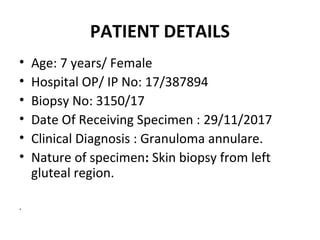
- 1. PATIENT DETAILS • Age: 7 years/ Female • Hospital OP/ IP No: 17/387894 • Biopsy No: 3150/17 • Date Of Receiving Specimen : 29/11/2017 • Clinical Diagnosis : Granuloma annulare. • Nature of specimen: Skin biopsy from left gluteal region. .
- 2. GROSS EXAMINATION Received single skin punch biopsy measuring 0.5x0.5x0.3cm. All embedded.
- 6. MICROSCOPIC EXAMINATION Sections studied shows • epidermis with hyperkeratosis, focal acanthosis, elongation of rete pegs and follicular keratotic plugging. • the papillary dermis shows dense perivascular and periadnexal lymphocytic infiltration along with focal collection of histiocytes and macrophages seen around degenerated collagen. • the inflammation is extending upto reticular dermis and subcutaneous plane.
- 7. IMPRESSION • Features consistent with granuloma annulare – left gluteal region.
- 8. DISCUSSION
- 9. Granuloma annulare • A benign inflammatory, self-limiting granulomatous dermatoses that is seen in both adults and children • Females are more commonly affected than males. • The lesions involves skin and/or subcutaneous tissue.
- 10. Granuloma annulare • The etiology of granuloma annulare is unknown. • Lesions could be related to insect bites, sun exposure, viral infections, diabetes, thyroiditis, immunoglobulin-mediated vasculitis, and certain medications such as antibiotics, antiinflammatory agents and oral contraceptives. • Cases have also been reported in patients with AIDS, sarcoidosis, hepatitis C infection, Hodgkin's and non Hodgkin's lymphoma, metastatic adenocarcinoma and granulomatous mycosis fungoides.
- 11. Histopathology Histological patterns in Granuloma Annulare: 1. Necrobiotic granuloma. 2. Interstitial or 'incomplete' form - Most common. 3. Granuloma of sarcoidal or tuberculoid type with epithelioid histiocytes or a type of giant cell - Rare
- 12. Differential Diagnosis • Necrobiosis lipodica • Annular elastolytic giant cell granuloma • Rheumatoid arthritis and rheumatic fever nodules.
- 14. Annular elastolytic giant cell granuloma
- 15. Necrobiosis Lipoidica • Predominantly involves the dermis. • Abundant mucin is distinctly uncommon. • Multilayered necrobiosis (stacks of plates) with open ends. • Thickened collagen bundles within palisaded granuloma. • Shows extensive deposits of lipids or nodular lymphocytic infiltrates in the deep dermis or subcutis. • Numerous deep dermal plasma cells.
- 16. Rheumatoid Nodule • Sharp irregular areas of necrobiosis (huge deposits of fibrin). • Located in the subcutis and deep reticular dermis. • Surrounded by a palisade of elongated histiocytes. • Stroma surrounding the nodules show perivascular lymphocytic infiltrate including plasma cells. • Some neutrophils and nuclear dusts of neutrophils may be present. • Acute or chronic thrombotic endoarteritis is observed in some cases around rheumatoid nodules. • Old lesions show dense fibrosis, clefts and cystic degeneration.
- 17. Special Stains • Mucin stains such as colloidal iron and alcian blue may be used to highlight the increased connective tissue mucins. • Histiocytes gives positivity to vimentin and CD68.
- 18. References Lever's histopathology of the skin. 10th ed.
- 19. Thank you
Editor's Notes
- delayed hypersensitivity reaction to some component of the dermis. Inflammation is mediated by tumour necrosis factor alpha (TNFα).