SlideShare a Scribd company logo
1 of 64
Dr. Jerriton, 1st Year PG, DVL,
SVMCH, Pondy.
11.09.18
SUBCUTANEOUS MYCOSIS
• Infections limited to skin & subcutaneous tissue
• Rarely spreading to internal organs
• Caused by fungi with saprophytic existence in nature
• Usual mode of transmission by inoculation
DEFINITION
EYMYCE
-TOMA
CHROMO-
MYCOSIS
PHAEOHYP
OMYCOSIS
SPOROTRI-
CHOSIS
LOBOMYC-
OSIS
SC
ZYGOMYC-
OSIS
RHINOSPO
-RIDIASIS
ETIOLOGY M.
mycetomat
is
C. carrionii,
F. pedrosoi
E.
jeanselmei
, P.
verrucosa
S. schenkii Lacazia
loboi
Basidiobol
us,
conidiobol
us spp.
R. seebri
CLINICAL
FEATURES
Painless SC
swelling +
sinus +
discharging
granules
Verrucous
plaques or
nodules +
ulceration
Subcutane
ous cysts
Ulcerated
nodules
along
lymphatics
Polymorph
ic; keloidal
& painless
Painless SC
masses –
bathsuit,
monstrous
face
Painless
friable
warty
outgrowth
with
ulceration
DIAGNOSIS Granules Sclerotic
bodies
Melanin Asteroid
bodies
Round
cells,
tubular
projections
, bifringent
memb.
Mixed
inflammat
ory cells
with fungal
elements
Sporangia
with
endospore
s
TREATMENT Surgery +
Antifungals
Antifungals Surgery +
Antifungals
SSKI +
Antifungals
Surgery +
Clofazimin
e +
Antifungals
SSKI +
Antifungals
Surgery
MYCETOMA
Definition
• Chronic, suppurative, granulomatous disease of subcutaneous (SC) tissue
& bones.
• It’s a triad of:
a) Painless SC Tumefaction
b) Sinuses
c) Discharge of granules
MYCETOMA
Etiology
• Bacteria (Actinomycotic Mycetoma) – M/C in India
 Actinomadura madurae (M/C)
• Fungi (Eumycotic Mycetoma)
 Madurella mycetomatis (M/C in South India)
Brown grain – M. mycetomatis White grain – A. madurae
MYCETOMA
HISTORY & TRANSMISSION
• John Gill in 1842 described “Madura Foot” first.
• Mycetoma means fungal tumor.
• The organisms are soil / plant saprophytes.
• Entry is by abrasion / implantation.
• More common in tropics.
MYCETOMA
EPIDEMIOLOGY
• 20 and 40 years, mostly in developing countries.
• Tropics and sub-tropics.
• Barefoot walkers.
• Low socioeconomic status.
MYCETOMA
CLINICAL FEATURES
• Begins as small, painless SC nodule at site of injury.
• Nodules increase in number, ulcerate & drain through sinuses.
• Discharge will have the characteristic granules.
• Surrounding skin becomes swollen, indurated & deformed.
• Spreads by direct contiguity along facial planes.
• Spares tendons till late stage.
• Becomes painful if bones are involved / 2° infection.
• Bones show punched out lytic lesions.
• Ankylosis can happen.
MYCETOMA
MACROSCOPIC COLOR OF GRAINS
• Black Grains – Eumycotic
• Red Grains – Actinomadura pelletierii
• White Grains – Eumycotic / Actinomycotic
DIAGNOSIS
MYCETOMA
• Granules microscopy (KOH / Gram stain) of discharge are characteristic:
1) Actinomycetomas – Fine, branching interlacing filaments with no
chlamydospores
2) Eumycetomas – Thick walled septate hyphae with chalmydospores
MICROSCOPY OF DISCHARGE
DIAGNOSIS
HPE
• Initially – Acute suppurative reaction
• Later – Suppurative granuloma (peripheral epithelioid histiocytes and
MNGs with central PMNs)
• Characteristic granules can be seen within central abscess of granuloma
MYCETOMA
EUMYCETOMA GRAIN
EUMYCETOMA
EUMYCETOMA GRAIN
A: Ulcerated, indurated plaque of
eumycotic mycetoma on the foot.
B: H&E staining shows a “sulfur granule”
in a purulent area of granulation tissue.
C: The sulfur granule is composed largely
of septate hyphae of P. boydii (GMS stain).
MYCETOMA
TREATMENT
ACTINOMYCETOMA
1. Modified Welsch Regimen (3-4 cycles)
Amikacin
15 mg/Kg IV x 21 days with 15 day intervals
PLUS
Co-trimoxazole
35 mg/Kg oral daily
PLUS
Rifampicin
10 mg/day oral daily
MYCETOMA
TREATMENT
ACTINOMYCETOMA
1. Modified Two – Step Regimen
Gentamycin
80 mg IV 12 hourly
PLUS
Co-trimoxazole
320 mg/1600 mg oral BD x 4 weeks
FOLLOWED BY
Maintenance doses
Co-trimoxazole & Doxy 100 mg oral BD
Continued 6 months till after healing
MYCETOMA
TREATMENT
EUMYCETOMA
• Ketaconazole
• Terbinafine
• Irraconazole 400 mg/day for 3 months f/b 200 mg/day for 9 months
• Voriconazole 300 mg BD doe 16 months
• Posaconazole for refractory cases
MYCETOMA
TREATMENT
ROLE OF SURGERY
• Exploration & drainage of sinus tracts
• Debridement of diseased tissue
• Removal of bone cysts
• They help healing faster
CHROMOBLASTOMYCOSIS
DEFINITION
• Chronic granulomatous infection, usually of exposed areas,
characterised by verrucous plaques or nodules that ulcerate.
CHROMOBLASTOMYCOSIS
ETIOLOGY
• Dematiaceous (brown-pigmented) fungi
1. Cladophialophora carrionii
2. Fonsecaea pedrosoi
• Saprophytes of soil, decaying vegetation and rotting wood
• Enter skin through abrasion
• Most common in tropics and subtropics
CHROMOBLASTOMYCOSIS
CLINICAL FEATURES
• 3 clinical forms exist:
1. Localized
2. Multiple with satellite lesions
3. Sporotrichoid
• Begins as a verrucous papule ->
verrucous plaque, which may have
central atrophy / scarring
• Seen mostly at exposed sites
• Complications include elephantiasis, 2° infection, ulceration &
malignant change
CHROMOBLASTOMYCOSIS
DIAGNOSIS
• KOH of skin scraping, crusts, aspiration, biopsy tissue
Sclerotic bodies / muriform cells
CHROMOBLASTOMYCOSIS
DIAGNOSIS
• H & E stain of biopsy specimen
Sclerotic bodies / muriform cells
CHROMOBLASTOMYCOSIS
DIAGNOSIS
• Culture for species identification
Sabouraud dextrose agar
• Serology if culture is not possible
CHROMOBLASTOMYCOSIS
TREATMENT
• Systemic antifungals
Itraconazole 200-400 mg / day
PLUS
Terbinafine 250-500 mg / day
X 6 – 12 months
CHROMOBLASTOMYCOSIS
TREATMENT
• Other treatment options
1. Iodides, Fluconazole, posiconazole
2. Surgical excision, cryotherapy, local heat
PHAEOHYPOMYCOSIS
DEFINITION
• Infections other than chromoblastomycosis and eumycetoma caused
by dematiaceous (melanized / phaeoid) fungi
• They do not have grains or sclerotic bodies that characterized
mycetoma and chromomycosis respectively.
• This entity is characterized by dark septate hyphae, pseudohyphae,
yeast or their combinations.
PHAEOHYPOMYCOSIS
ETIOLOGY
• Most common are:
1. Exophiala jeanselmei
2. Wangiella dermatitidis
3. Phialophora verrucosa
4. Bipolaris spp.
• They live in decaying vegetation, bird nests and soil
• They are seen mostly in tropics and subtropics
• Infection is by local trauma by abrasion or inhalation
PHAEOHYPOMYCOSIS
PATHOGENESIS
• DHN melanin – fungal armor in cell wall
1. Scavenges free radicals
2. Prevents action of hydrolytic enzymes
• Thermotolerance – can cause deep invasive cerebral lesions
PHAEOHYPOMYCOSIS
CLASSIFICATION
1. Superficial: black piedra and tinea nigra
2. Cutaneous: dermatomycosis and onychomycosis
3. Mycotic keratitis
4. Subcutaneous phaeohypomycosis
5. Invasive, systemic & cerebral
PHAEOHYPOMYCOSIS
CLINICAL FEATURES
• Classical presentation: SC cyst.
• Begins as small papule -> evolves into SC cyst
• Usually single
• M/C site: extremities
• Children: face
PHAEOHYPOMYCOSIS
DIAGNOSIS
• KOH of pus, drainage or skin scrapings
Pigmented yeasts, pseudohyphae and hyphae
• Biopsy
1. Foreign body granuloma
2. Pigmented fungi seen within granuloma
3. Splendore-Hoeppli reaction +/-
4. Fontana Masson stain for melanin is diagnostic
• FNAC
Pigmented fungi with inflammatory cells
PHAEOHYPOMYCOSIS
TREATMENT
• Triple antifungal combinations give best results for refractory cases
Amphotericin B, glucytosine and itraconazole
• Localised lesion: excision f/b pre & post op antifungal therapy
• Antifungals used:
• Flucytosine 150 mg/kg/day
• Itraconazole 200 mg/day
• Ketoconazole 200 mg/day
• IV / ILS Amphotericin B
SPOROTRICHOSIS
DEFINITION
• Subacute or chronic infection caused by dimorphic fungi, S. schenckii
• Characterized by nodular and ulcerative lesions along lymphatics
SPOROTRICHOSIS
ETIOLOGY
S. schenkii
• Dimorphic fungi
• Mycelia at 26°C and yeast at 37°C
• Mycelia bears conidia resembling flower
• Grows in common agar
• Produces creamy white colonies
• Turns black later
• It is a saprophyte in dead plants
• Introduced by trauma to skin
SPOROTRICHOSIS
HISTOLOGY
• HPE shows three granulomatous patterns observed
1. Sporotrichotic (central suppuration seen)
2. Tuberculoid (central suppuration seen)
3. Foreign body (no central suppuration)
• Asteroid bodies are characteristic
Round basophilic yeast-like body with surrounding
elongated radiating eosinophilic material (a type of
Splendore – Hoeppli reaction)
SPOROTRICHOSIS
CLINICAL FEATURES
• Cutaneous
1. Lymphocutaneous
2. Localized
• Extra cutaneous
1. Pulmonary
2. Disseminated
SPOROTRICHOSIS
CLINICAL FEATURES
Lymphocutaneous form
• M/C seen in exposed site of upper extremity
• Small nodule / pustule develops at site of trauma
• Nodule breaks to form ulcer
• New nodules form along lymphatics at few days interval
• Ulcerated nodules connect by cord-like swollen lymphatics
• Heals with scarring and new nodules develop at other sites
• These secondary lesions are gummatous and persist for years
SPOROTRICHOSIS
CLINICAL FEATURES
Lymphocutaneous form
SPOROTRICHOSIS
CLINICAL FEATURES
Localized cutaneous form
• Primary lesion is restricted to site of injury
• It can be ulcerative, verrucous, acneiform or scaly plaque
• Does not involve local lymphatics
• Mucous membrane can be involved
• Pain is predominant complaint
SPOROTRICHOSIS
INVESTIGATIONS
• Sporotrichin skin test
• 0.1 mL of intradermal sporotrichin M (mycelia) is injected
into their forearm and the reading is ascertained at 48
hours, using the same criterion as for the tuberculin skin
test.
• Induration ≥ 0.8 cm is positive.
• Serology – for extra-cutaneous forms
SPOROTRICHOSIS
INVESTIGATIONS
• Culture – definitive diagnosis
1. Sabouraud’s dextrose agar at 26°C and 37°C
2. Conversion to yeast form at 37°C is important
• Animal innoculation
1. Gram positive cigar bodies in pus
SPOROTRICHOSIS
TREATMENT
• SSKI
1. Given orally in milk
2. Initial dose: 5 drops (1 ml) TID after meals
3. Increased by 1 drop / dose till 40 drops TID
4. Continued till signs of active disease are gone
5. Then dose decreased by 1 drop / dose till 5 drops
6. Then discontinued
• Itraconazole 100-200 mg/day
• Terbinafine 250 mg/day
• Thermotherapy / pocket warmer
LOBOMYCOSIS
DEFINITION
Also known as keloidal blastomycosis, pseudoleprosy. It is
characterized by pleomorphic lesions.
LOBOMYCOSIS
ETIOLOGY
• Caused by fungi, Lacazia loboi
• Has a saprophytic phase in vegetation, soil & water
• Infection is acquired through trauma
• Farmers, gold miners, fishermen and hunters are affected
• Dolphin to human transmission is reported
• No person-to-person transmission
LOBOMYCOSIS
CLINICAL FEATURES
• Pleomorphic lesions
• Nodules or plaques (hypopigmented / hyperpigmented)
• Ulcers
• Sclerodermoid
• Keloidal
• Verrucous
• Legs, outer ears and arms are commonly affected
• Single or multiple, become confluent
• Generally painless, occasionally painless
LOBOMYCOSIS
INVESTIGATIONS
• Direct microscopy
• KOH shows round yeast-like organisms, singly or in chains
connected by short tubular projections.
• They have bifringngent membrane with central granules
• HPE
• Granulomatous infiltrate without suppuration
• Grenz zone +/-
• Asteroid bodies +/-
• Fungal forms seen at different levels of epidermis (TEE)
LOBOMYCOSIS
TREATMENT
• Medical:
1. Clofazimine 300 mg/day initial dose with maintainance
dose of 100 mg/day for 2 years
2. Ketoconazole, Itraconazole, Posaconazole
• Surgical excision
• Electrocautery
• Cryosurgery
SUBCUTANEOUS ZYGOMYCOSIS
DEFINITION
• Chronic subcutaneous infection characterized by woody
swelling of SC tissue
SUBCUTANEOUS ZYGOMYCOSIS
ETIOLOGY & CLASSIFICATION
FEATURES ENTOMOPHTHORALES
(Basidiobolus, conidiobolus)
MUCORALES
Host Immunocompetent Immunocompromised
Distribution Tropics and subtropics Worldwide
Transmission Traumatic implantation Inhalation of spores
Systems involved SC mycosis and sinusitis RS, CNS, GIT, skin
Histopathology Chronic inflammatory
response
Angioinvasion,
thrombosis,tissue necrosis
Splendore-Hoeppli Characteristic Rarely seen
Septation More common Less common
Dissemination Uncommon Common
SUBCUTANEOUS ZYGOMYCOSIS
EPIDEMIOLOGY & TRANSMISSION
BASIDIOBOLUS
• Children less than 20 years
• Male > Female
• Africa > India
• Transmitted by minor trauma / insect bite / contaminated toilet
leaves (bathing suit distribution)
• Also transmitted through soil and vegetation containing
contaminated animal faeces
SUBCUTANEOUS ZYGOMYCOSIS
EPIDEMIOLOGY & TRANSMISSION
CONIDIOBOLUS
• Young adults
• Male > female
• Africa > India
• Identified in soil & plant debris, M/C c. coronatus
• Transmitted by inhalation of fungal spores / frequent nose
pricking habits
• Leads to monstrous disfigurement of face
SUBCUTANEOUS ZYGOMYCOSIS
PATHOGENESIS
BASIDIOBOLUS
• Produces extracellular proteinases and lipases
 Phospholipase A hydrolizes lecithin, which destroyed
membranes of blood, skin & muscle cells
 Once lipase liberates cellular protein components,
proteinases digest them as its nutrients
• Thermotolerant – grows poorly at 37°C
SUBCUTANEOUS ZYGOMYCOSIS
PATHOGENESIS
CONIDIOBOLUS
• Produces elastase, esterase, collagenase and lipase
 Proteinase is secreted first – breaks down proteins to amino acids
 Lipase is produced later – hydrolizes fatty materials in SC tissue
• Thermophilic – grows readily at 37°C
SUBCUTANEOUS ZYGOMYCOSIS
CLINICAL FEATURES
BASIDIOBOLOMYCOSIS
• M/C site: limb girdles / proximal limbs
• Bathing suit distribution
• Painless well-circumcized, firm to hard, smooth, rounded SC masses
that can be raised by inserting fingers underneath it (freely mobile)
• Satellite lesions may be seen at advancing margins
• May encompass part / whole of limb
• Overlying skin may be tense, edematous, desquamating,
hyperpigmented or normal
• Non-pitting oedema +/-
• Underlying muscle / visceral involvement can occur
SUBCUTANEOUS ZYGOMYCOSIS
CLINICAL FEATURES
CONIDIOBOLOMYCOSIS
• Begins as swelling of inferior nasal turbinates
• Stuffiness, discharge, epistaxis, nasal obstruction are symptoms
• Diffuse erythematous infiltration with skin thickening on nose, cheeks,
forehead and lips – monstrous disfigurement, facial elephantiasis,
palatal perforation, orbital cellulitis, saddle nose deformity
• Phase 1 – nose, paranasal sinuses & pharynx
• Phase 2 – frontal region and lips
• Phase 3 – muscle, bones & viscera
SUBCUTANEOUS ZYGOMYCOSIS
DIAGNOSIS
HPE
• Mixed inflammatory infiltrate
• Fungal hyphal elements with surrounding dense eosinophilic granular
aterial (Splenndore-Hoeppli phenomenon)
MICROSCOPY
• KOH from scrapings show broad, septate branching hypahe
CULTURE
• Basidiobolus – flat & furrowed, yellowish grey color with musty odor
• Conidiobolus – white surface, becomes beige to brown, no odor
SUBCUTANEOUS ZYGOMYCOSIS
TREATMENT
• Itraconazole / SSKI – 1st line choices
• Treated continuously for 1-2 months after clinical cure
RHINOSPORIDIOSIS
DEFINITION
• Chronic granulomatous disease of mucocutaneous tissue
• Characterized by development of polypoid tumors or pedunculated /
sessile polyps
RHINOSPORIDIOSIS
ETIOLOGY & EPIDEMIOLOGY
• Caused by Rhinosporidium seeberi – protistan parasite of class
Mesomycetozoea
• Common pond bathing with buffalos is a risk factor
• Disease is endemic in Kerela, Tamil nadu and Chandigarh
RHINOSPORIDIOSIS
HISTOLOGY
• Thick walled sporangia with endospores
RHINOSPORIDIOSIS
CLINICAL FEATURES
CUTANEOUS RHINOSPORIDIOSIS
• Associated with mucosal disease
• Begin as tiny papules and enlarge to become wart like / tumorous
growth
• They are friable and have crenated surface, often ulcerated - & painless
RHINOSPORIDIOSIS
DIAGNOSIS
• KOH / HPE for sporangia with endospores
TREATMENT
• Surgical excision
• Electrocoagulation
REFERENCES
1. Rook’s Textbook of Surgery
2. IADVL Textbook of Dermatology
3. Fitzpatrick Textbook of Dermatology
4. Lever’s Histopathology
5. Weedon’s Histopathology
6. Online journals

More Related Content

What's hot

Opportunistic fungal infection
Opportunistic fungal infectionOpportunistic fungal infection
Opportunistic fungal infectionVishal Kulkarni
 
Opportunistic mycoses
Opportunistic mycosesOpportunistic mycoses
Opportunistic mycosesraghunathp
 
Laboratory diagnosis of fungal infections
Laboratory diagnosis of fungal infectionsLaboratory diagnosis of fungal infections
Laboratory diagnosis of fungal infectionsDr.Dinesh Jain
 
Laboratory diagnosis of mycology microscopy, staining techniques, culture me...
Laboratory diagnosis of mycology  microscopy, staining techniques, culture me...Laboratory diagnosis of mycology  microscopy, staining techniques, culture me...
Laboratory diagnosis of mycology microscopy, staining techniques, culture me...Prasad Gunjal
 
Tinea nigra presentation
Tinea nigra presentationTinea nigra presentation
Tinea nigra presentationSGowthamDhina
 
Lab diagnosis of fungal infections, Dr Naveen Reddy
Lab diagnosis of fungal infections, Dr Naveen ReddyLab diagnosis of fungal infections, Dr Naveen Reddy
Lab diagnosis of fungal infections, Dr Naveen ReddyNaveen Parvathareddy
 
Dematiaceous fungi.pptx
Dematiaceous fungi.pptxDematiaceous fungi.pptx
Dematiaceous fungi.pptxFarhanDar5
 
Chromoblastomycosis and phaeohyphomycosis
Chromoblastomycosis and phaeohyphomycosisChromoblastomycosis and phaeohyphomycosis
Chromoblastomycosis and phaeohyphomycosisadisutesfaye21
 
Overview of medical mycology
Overview of medical mycology Overview of medical mycology
Overview of medical mycology Dr.Dinesh Jain
 
Subcutaneous mycoses by Dr. Rakesh Prasad Sah
Subcutaneous mycoses by Dr. Rakesh Prasad SahSubcutaneous mycoses by Dr. Rakesh Prasad Sah
Subcutaneous mycoses by Dr. Rakesh Prasad SahDr. Rakesh Prasad Sah
 
Dimorphic fungi
Dimorphic  fungi Dimorphic  fungi
Dimorphic fungi Prbn Shah
 
Introduction to Medical mycology
Introduction to Medical mycologyIntroduction to Medical mycology
Introduction to Medical mycologyMuhammad Getso
 

What's hot (20)

Opportunistic fungal infection
Opportunistic fungal infectionOpportunistic fungal infection
Opportunistic fungal infection
 
Sporotrichosis
SporotrichosisSporotrichosis
Sporotrichosis
 
Mycetoma
MycetomaMycetoma
Mycetoma
 
Opportunistic mycoses
Opportunistic mycosesOpportunistic mycoses
Opportunistic mycoses
 
Pathology of fungal infection
Pathology of fungal infectionPathology of fungal infection
Pathology of fungal infection
 
Laboratory diagnosis of fungal infections
Laboratory diagnosis of fungal infectionsLaboratory diagnosis of fungal infections
Laboratory diagnosis of fungal infections
 
Laboratory diagnosis of mycology microscopy, staining techniques, culture me...
Laboratory diagnosis of mycology  microscopy, staining techniques, culture me...Laboratory diagnosis of mycology  microscopy, staining techniques, culture me...
Laboratory diagnosis of mycology microscopy, staining techniques, culture me...
 
Tinea nigra presentation
Tinea nigra presentationTinea nigra presentation
Tinea nigra presentation
 
Cutaneous mycoses.ppt
Cutaneous mycoses.pptCutaneous mycoses.ppt
Cutaneous mycoses.ppt
 
Lab diagnosis of fungal infections, Dr Naveen Reddy
Lab diagnosis of fungal infections, Dr Naveen ReddyLab diagnosis of fungal infections, Dr Naveen Reddy
Lab diagnosis of fungal infections, Dr Naveen Reddy
 
Dermatophytes
DermatophytesDermatophytes
Dermatophytes
 
Systemic mycosis
Systemic mycosisSystemic mycosis
Systemic mycosis
 
Dematiaceous fungi.pptx
Dematiaceous fungi.pptxDematiaceous fungi.pptx
Dematiaceous fungi.pptx
 
Chromoblastomycosis and phaeohyphomycosis
Chromoblastomycosis and phaeohyphomycosisChromoblastomycosis and phaeohyphomycosis
Chromoblastomycosis and phaeohyphomycosis
 
Chromoblastomycosis
ChromoblastomycosisChromoblastomycosis
Chromoblastomycosis
 
Overview of medical mycology
Overview of medical mycology Overview of medical mycology
Overview of medical mycology
 
Mycetoma
MycetomaMycetoma
Mycetoma
 
Subcutaneous mycoses by Dr. Rakesh Prasad Sah
Subcutaneous mycoses by Dr. Rakesh Prasad SahSubcutaneous mycoses by Dr. Rakesh Prasad Sah
Subcutaneous mycoses by Dr. Rakesh Prasad Sah
 
Dimorphic fungi
Dimorphic  fungi Dimorphic  fungi
Dimorphic fungi
 
Introduction to Medical mycology
Introduction to Medical mycologyIntroduction to Medical mycology
Introduction to Medical mycology
 

Similar to Subcutaneous Mycosis

1.DEEP FUNGAL INFECTIONS 10th feb 2022.pptx
1.DEEP FUNGAL INFECTIONS 10th feb 2022.pptx1.DEEP FUNGAL INFECTIONS 10th feb 2022.pptx
1.DEEP FUNGAL INFECTIONS 10th feb 2022.pptxAartiVinoj
 
SUBCUTANEOUS AND FUNGAL INFECTIONS
SUBCUTANEOUS AND FUNGAL INFECTIONSSUBCUTANEOUS AND FUNGAL INFECTIONS
SUBCUTANEOUS AND FUNGAL INFECTIONSAjeet Singh
 
Malignant skin lesions
Malignant skin lesionsMalignant skin lesions
Malignant skin lesionsAnkur Kajal
 
DEEP FUNGAL MYCOSIS_100307.pptx
DEEP FUNGAL MYCOSIS_100307.pptxDEEP FUNGAL MYCOSIS_100307.pptx
DEEP FUNGAL MYCOSIS_100307.pptxMehulChoudhary18
 
DEEP MYCOSES.pptx
DEEP MYCOSES.pptxDEEP MYCOSES.pptx
DEEP MYCOSES.pptxsriramck2
 
Subcutaneous Mycoses.pptx
Subcutaneous Mycoses.pptxSubcutaneous Mycoses.pptx
Subcutaneous Mycoses.pptxhabtamu biazin
 
Fungal allergies and infections
Fungal allergies and infections Fungal allergies and infections
Fungal allergies and infections SakethVuruma
 
NON ODONTOGENIC TUMORS OF ORAL CAVITY-I
NON ODONTOGENIC TUMORS OF ORAL CAVITY-I NON ODONTOGENIC TUMORS OF ORAL CAVITY-I
NON ODONTOGENIC TUMORS OF ORAL CAVITY-I Krupali Gandhi
 
Oral manifestations of bacterial infections
Oral manifestations of bacterial infectionsOral manifestations of bacterial infections
Oral manifestations of bacterial infectionsKarishma Sirimulla
 
Pigmented lesions of oral cavity (Oral Medicine and Radiology)
Pigmented lesions of oral cavity (Oral Medicine and Radiology)Pigmented lesions of oral cavity (Oral Medicine and Radiology)
Pigmented lesions of oral cavity (Oral Medicine and Radiology)RupaliBham
 
лекция.pptx for std dermatovenerology, cause and symptoms
лекция.pptx for std dermatovenerology, cause and symptomsлекция.pptx for std dermatovenerology, cause and symptoms
лекция.pptx for std dermatovenerology, cause and symptomsneestom1998
 
SYSTEMIC MYCOSIS
SYSTEMIC MYCOSISSYSTEMIC MYCOSIS
SYSTEMIC MYCOSISVAISHNAVI V
 
Medical mycology 2
Medical mycology 2Medical mycology 2
Medical mycology 2Gopi sankar
 

Similar to Subcutaneous Mycosis (20)

1.DEEP FUNGAL INFECTIONS 10th feb 2022.pptx
1.DEEP FUNGAL INFECTIONS 10th feb 2022.pptx1.DEEP FUNGAL INFECTIONS 10th feb 2022.pptx
1.DEEP FUNGAL INFECTIONS 10th feb 2022.pptx
 
cutaneous tuberculosis
cutaneous tuberculosiscutaneous tuberculosis
cutaneous tuberculosis
 
SUBCUTANEOUS AND FUNGAL INFECTIONS
SUBCUTANEOUS AND FUNGAL INFECTIONSSUBCUTANEOUS AND FUNGAL INFECTIONS
SUBCUTANEOUS AND FUNGAL INFECTIONS
 
Malignant skin lesions
Malignant skin lesionsMalignant skin lesions
Malignant skin lesions
 
DEEP FUNGAL MYCOSIS_100307.pptx
DEEP FUNGAL MYCOSIS_100307.pptxDEEP FUNGAL MYCOSIS_100307.pptx
DEEP FUNGAL MYCOSIS_100307.pptx
 
Mucocutaneous
Mucocutaneous Mucocutaneous
Mucocutaneous
 
DEEP MYCOSES.pptx
DEEP MYCOSES.pptxDEEP MYCOSES.pptx
DEEP MYCOSES.pptx
 
Subcutaneous Mycoses.pptx
Subcutaneous Mycoses.pptxSubcutaneous Mycoses.pptx
Subcutaneous Mycoses.pptx
 
Fungal allergies and infections
Fungal allergies and infections Fungal allergies and infections
Fungal allergies and infections
 
NON ODONTOGENIC TUMORS OF ORAL CAVITY-I
NON ODONTOGENIC TUMORS OF ORAL CAVITY-I NON ODONTOGENIC TUMORS OF ORAL CAVITY-I
NON ODONTOGENIC TUMORS OF ORAL CAVITY-I
 
Oral manifestations of bacterial infections
Oral manifestations of bacterial infectionsOral manifestations of bacterial infections
Oral manifestations of bacterial infections
 
Pigmented lesions of oral cavity (Oral Medicine and Radiology)
Pigmented lesions of oral cavity (Oral Medicine and Radiology)Pigmented lesions of oral cavity (Oral Medicine and Radiology)
Pigmented lesions of oral cavity (Oral Medicine and Radiology)
 
Deep fungal infection.pptx
Deep fungal infection.pptxDeep fungal infection.pptx
Deep fungal infection.pptx
 
Integumentary system
Integumentary systemIntegumentary system
Integumentary system
 
Tuberculosis mimics
Tuberculosis mimicsTuberculosis mimics
Tuberculosis mimics
 
лекция.pptx for std dermatovenerology, cause and symptoms
лекция.pptx for std dermatovenerology, cause and symptomsлекция.pptx for std dermatovenerology, cause and symptoms
лекция.pptx for std dermatovenerology, cause and symptoms
 
SYSTEMIC MYCOSIS
SYSTEMIC MYCOSISSYSTEMIC MYCOSIS
SYSTEMIC MYCOSIS
 
Medical mycology 2
Medical mycology 2Medical mycology 2
Medical mycology 2
 
Eumycetoma
EumycetomaEumycetoma
Eumycetoma
 
Black fungi
Black fungiBlack fungi
Black fungi
 

More from Jerriton Brewin

Superficial Fungal Infections
Superficial Fungal InfectionsSuperficial Fungal Infections
Superficial Fungal InfectionsJerriton Brewin
 
Cutaneous Vasculature and Thermoregulation
Cutaneous Vasculature and ThermoregulationCutaneous Vasculature and Thermoregulation
Cutaneous Vasculature and ThermoregulationJerriton Brewin
 
Severe Cutaneous Adverse Reactions to Drugs
Severe Cutaneous Adverse Reactions to DrugsSevere Cutaneous Adverse Reactions to Drugs
Severe Cutaneous Adverse Reactions to DrugsJerriton Brewin
 
Botulinum Toxin A (BOTOX)
Botulinum Toxin A (BOTOX)Botulinum Toxin A (BOTOX)
Botulinum Toxin A (BOTOX)Jerriton Brewin
 
Bacteriology & Immunology of Leprosy
Bacteriology & Immunology of LeprosyBacteriology & Immunology of Leprosy
Bacteriology & Immunology of LeprosyJerriton Brewin
 
Cutaneous T Cell Lymphomas
Cutaneous T Cell LymphomasCutaneous T Cell Lymphomas
Cutaneous T Cell LymphomasJerriton Brewin
 
Cutaneous Manifestations of Internal Malignancies
Cutaneous Manifestations of Internal MalignanciesCutaneous Manifestations of Internal Malignancies
Cutaneous Manifestations of Internal MalignanciesJerriton Brewin
 
Non Venereal Treponematosis
Non Venereal TreponematosisNon Venereal Treponematosis
Non Venereal TreponematosisJerriton Brewin
 
Mastocytosis (Cutaneous and Systemic)
Mastocytosis (Cutaneous and Systemic)Mastocytosis (Cutaneous and Systemic)
Mastocytosis (Cutaneous and Systemic)Jerriton Brewin
 
WHO ART Guidelines for HIV
WHO ART Guidelines for HIV WHO ART Guidelines for HIV
WHO ART Guidelines for HIV Jerriton Brewin
 
Direct Immunofluorescence in Dermatology
Direct Immunofluorescence in DermatologyDirect Immunofluorescence in Dermatology
Direct Immunofluorescence in DermatologyJerriton Brewin
 
Systemic Retinoids - Drug Review
Systemic Retinoids - Drug ReviewSystemic Retinoids - Drug Review
Systemic Retinoids - Drug ReviewJerriton Brewin
 
Treponemal Tests for Syphilis
Treponemal Tests for SyphilisTreponemal Tests for Syphilis
Treponemal Tests for SyphilisJerriton Brewin
 
Skin Keratinization & Its Disorders
Skin Keratinization & Its DisordersSkin Keratinization & Its Disorders
Skin Keratinization & Its DisordersJerriton Brewin
 
Methotrexate - A Comprehensive Drug Review in Dermatology
Methotrexate - A Comprehensive Drug Review in DermatologyMethotrexate - A Comprehensive Drug Review in Dermatology
Methotrexate - A Comprehensive Drug Review in DermatologyJerriton Brewin
 

More from Jerriton Brewin (20)

Vitiligo Surgeries
Vitiligo SurgeriesVitiligo Surgeries
Vitiligo Surgeries
 
Superficial Fungal Infections
Superficial Fungal InfectionsSuperficial Fungal Infections
Superficial Fungal Infections
 
Cutaneous Vasculature and Thermoregulation
Cutaneous Vasculature and ThermoregulationCutaneous Vasculature and Thermoregulation
Cutaneous Vasculature and Thermoregulation
 
Vitiligo Surgeries
Vitiligo SurgeriesVitiligo Surgeries
Vitiligo Surgeries
 
Severe Cutaneous Adverse Reactions to Drugs
Severe Cutaneous Adverse Reactions to DrugsSevere Cutaneous Adverse Reactions to Drugs
Severe Cutaneous Adverse Reactions to Drugs
 
Botulinum Toxin A (BOTOX)
Botulinum Toxin A (BOTOX)Botulinum Toxin A (BOTOX)
Botulinum Toxin A (BOTOX)
 
Bacteriology & Immunology of Leprosy
Bacteriology & Immunology of LeprosyBacteriology & Immunology of Leprosy
Bacteriology & Immunology of Leprosy
 
Cutaneous T Cell Lymphomas
Cutaneous T Cell LymphomasCutaneous T Cell Lymphomas
Cutaneous T Cell Lymphomas
 
Pregnancy Dermatoses
Pregnancy DermatosesPregnancy Dermatoses
Pregnancy Dermatoses
 
Cutaneous Manifestations of Internal Malignancies
Cutaneous Manifestations of Internal MalignanciesCutaneous Manifestations of Internal Malignancies
Cutaneous Manifestations of Internal Malignancies
 
Non Venereal Treponematosis
Non Venereal TreponematosisNon Venereal Treponematosis
Non Venereal Treponematosis
 
Mastocytosis (Cutaneous and Systemic)
Mastocytosis (Cutaneous and Systemic)Mastocytosis (Cutaneous and Systemic)
Mastocytosis (Cutaneous and Systemic)
 
WHO ART Guidelines for HIV
WHO ART Guidelines for HIV WHO ART Guidelines for HIV
WHO ART Guidelines for HIV
 
Direct Immunofluorescence in Dermatology
Direct Immunofluorescence in DermatologyDirect Immunofluorescence in Dermatology
Direct Immunofluorescence in Dermatology
 
Systemic Retinoids - Drug Review
Systemic Retinoids - Drug ReviewSystemic Retinoids - Drug Review
Systemic Retinoids - Drug Review
 
Treponemal Tests for Syphilis
Treponemal Tests for SyphilisTreponemal Tests for Syphilis
Treponemal Tests for Syphilis
 
ANA Negative Lupus
ANA Negative Lupus ANA Negative Lupus
ANA Negative Lupus
 
Skin Keratinization & Its Disorders
Skin Keratinization & Its DisordersSkin Keratinization & Its Disorders
Skin Keratinization & Its Disorders
 
Methotrexate - A Comprehensive Drug Review in Dermatology
Methotrexate - A Comprehensive Drug Review in DermatologyMethotrexate - A Comprehensive Drug Review in Dermatology
Methotrexate - A Comprehensive Drug Review in Dermatology
 
Normal Skin Flora
Normal Skin FloraNormal Skin Flora
Normal Skin Flora
 

Recently uploaded

College Call Girls Vyasarpadi Whatsapp 7001305949 Independent Escort Service
College Call Girls Vyasarpadi Whatsapp 7001305949 Independent Escort ServiceCollege Call Girls Vyasarpadi Whatsapp 7001305949 Independent Escort Service
College Call Girls Vyasarpadi Whatsapp 7001305949 Independent Escort ServiceNehru place Escorts
 
Low Rate Call Girls Pune Esha 9907093804 Short 1500 Night 6000 Best call girl...
Low Rate Call Girls Pune Esha 9907093804 Short 1500 Night 6000 Best call girl...Low Rate Call Girls Pune Esha 9907093804 Short 1500 Night 6000 Best call girl...
Low Rate Call Girls Pune Esha 9907093804 Short 1500 Night 6000 Best call girl...Miss joya
 
Call Girls Hosur Just Call 7001305949 Top Class Call Girl Service Available
Call Girls Hosur Just Call 7001305949 Top Class Call Girl Service AvailableCall Girls Hosur Just Call 7001305949 Top Class Call Girl Service Available
Call Girls Hosur Just Call 7001305949 Top Class Call Girl Service Availablenarwatsonia7
 
Russian Call Girl Brookfield - 7001305949 Escorts Service 50% Off with Cash O...
Russian Call Girl Brookfield - 7001305949 Escorts Service 50% Off with Cash O...Russian Call Girl Brookfield - 7001305949 Escorts Service 50% Off with Cash O...
Russian Call Girl Brookfield - 7001305949 Escorts Service 50% Off with Cash O...narwatsonia7
 
Call Girls Budhwar Peth 7001305949 All Area Service COD available Any Time
Call Girls Budhwar Peth 7001305949 All Area Service COD available Any TimeCall Girls Budhwar Peth 7001305949 All Area Service COD available Any Time
Call Girls Budhwar Peth 7001305949 All Area Service COD available Any Timevijaych2041
 
Book Call Girls in Kasavanahalli - 7001305949 with real photos and phone numbers
Book Call Girls in Kasavanahalli - 7001305949 with real photos and phone numbersBook Call Girls in Kasavanahalli - 7001305949 with real photos and phone numbers
Book Call Girls in Kasavanahalli - 7001305949 with real photos and phone numbersnarwatsonia7
 
Call Girls ITPL Just Call 7001305949 Top Class Call Girl Service Available
Call Girls ITPL Just Call 7001305949 Top Class Call Girl Service AvailableCall Girls ITPL Just Call 7001305949 Top Class Call Girl Service Available
Call Girls ITPL Just Call 7001305949 Top Class Call Girl Service Availablenarwatsonia7
 
Call Girls Frazer Town Just Call 7001305949 Top Class Call Girl Service Avail...
Call Girls Frazer Town Just Call 7001305949 Top Class Call Girl Service Avail...Call Girls Frazer Town Just Call 7001305949 Top Class Call Girl Service Avail...
Call Girls Frazer Town Just Call 7001305949 Top Class Call Girl Service Avail...narwatsonia7
 
Low Rate Call Girls Mumbai Suman 9910780858 Independent Escort Service Mumbai
Low Rate Call Girls Mumbai Suman 9910780858 Independent Escort Service MumbaiLow Rate Call Girls Mumbai Suman 9910780858 Independent Escort Service Mumbai
Low Rate Call Girls Mumbai Suman 9910780858 Independent Escort Service Mumbaisonalikaur4
 
Call Girls Thane Just Call 9910780858 Get High Class Call Girls Service
Call Girls Thane Just Call 9910780858 Get High Class Call Girls ServiceCall Girls Thane Just Call 9910780858 Get High Class Call Girls Service
Call Girls Thane Just Call 9910780858 Get High Class Call Girls Servicesonalikaur4
 
Call Girls Service Chennai Jiya 7001305949 Independent Escort Service Chennai
Call Girls Service Chennai Jiya 7001305949 Independent Escort Service ChennaiCall Girls Service Chennai Jiya 7001305949 Independent Escort Service Chennai
Call Girls Service Chennai Jiya 7001305949 Independent Escort Service ChennaiNehru place Escorts
 
Housewife Call Girls Hoskote | 7001305949 At Low Cost Cash Payment Booking
Housewife Call Girls Hoskote | 7001305949 At Low Cost Cash Payment BookingHousewife Call Girls Hoskote | 7001305949 At Low Cost Cash Payment Booking
Housewife Call Girls Hoskote | 7001305949 At Low Cost Cash Payment Bookingnarwatsonia7
 
High Profile Call Girls Jaipur Vani 8445551418 Independent Escort Service Jaipur
High Profile Call Girls Jaipur Vani 8445551418 Independent Escort Service JaipurHigh Profile Call Girls Jaipur Vani 8445551418 Independent Escort Service Jaipur
High Profile Call Girls Jaipur Vani 8445551418 Independent Escort Service Jaipurparulsinha
 
Bangalore Call Girls Marathahalli 📞 9907093804 High Profile Service 100% Safe
Bangalore Call Girls Marathahalli 📞 9907093804 High Profile Service 100% SafeBangalore Call Girls Marathahalli 📞 9907093804 High Profile Service 100% Safe
Bangalore Call Girls Marathahalli 📞 9907093804 High Profile Service 100% Safenarwatsonia7
 
Call Girls Service In Shyam Nagar Whatsapp 8445551418 Independent Escort Service
Call Girls Service In Shyam Nagar Whatsapp 8445551418 Independent Escort ServiceCall Girls Service In Shyam Nagar Whatsapp 8445551418 Independent Escort Service
Call Girls Service In Shyam Nagar Whatsapp 8445551418 Independent Escort Serviceparulsinha
 
Aspirin presentation slides by Dr. Rewas Ali
Aspirin presentation slides by Dr. Rewas AliAspirin presentation slides by Dr. Rewas Ali
Aspirin presentation slides by Dr. Rewas AliRewAs ALI
 
Call Girls Electronic City Just Call 7001305949 Top Class Call Girl Service A...
Call Girls Electronic City Just Call 7001305949 Top Class Call Girl Service A...Call Girls Electronic City Just Call 7001305949 Top Class Call Girl Service A...
Call Girls Electronic City Just Call 7001305949 Top Class Call Girl Service A...narwatsonia7
 
Call Girl Surat Madhuri 7001305949 Independent Escort Service Surat
Call Girl Surat Madhuri 7001305949 Independent Escort Service SuratCall Girl Surat Madhuri 7001305949 Independent Escort Service Surat
Call Girl Surat Madhuri 7001305949 Independent Escort Service Suratnarwatsonia7
 

Recently uploaded (20)

College Call Girls Vyasarpadi Whatsapp 7001305949 Independent Escort Service
College Call Girls Vyasarpadi Whatsapp 7001305949 Independent Escort ServiceCollege Call Girls Vyasarpadi Whatsapp 7001305949 Independent Escort Service
College Call Girls Vyasarpadi Whatsapp 7001305949 Independent Escort Service
 
Low Rate Call Girls Pune Esha 9907093804 Short 1500 Night 6000 Best call girl...
Low Rate Call Girls Pune Esha 9907093804 Short 1500 Night 6000 Best call girl...Low Rate Call Girls Pune Esha 9907093804 Short 1500 Night 6000 Best call girl...
Low Rate Call Girls Pune Esha 9907093804 Short 1500 Night 6000 Best call girl...
 
Call Girls Hosur Just Call 7001305949 Top Class Call Girl Service Available
Call Girls Hosur Just Call 7001305949 Top Class Call Girl Service AvailableCall Girls Hosur Just Call 7001305949 Top Class Call Girl Service Available
Call Girls Hosur Just Call 7001305949 Top Class Call Girl Service Available
 
Russian Call Girl Brookfield - 7001305949 Escorts Service 50% Off with Cash O...
Russian Call Girl Brookfield - 7001305949 Escorts Service 50% Off with Cash O...Russian Call Girl Brookfield - 7001305949 Escorts Service 50% Off with Cash O...
Russian Call Girl Brookfield - 7001305949 Escorts Service 50% Off with Cash O...
 
Call Girls Budhwar Peth 7001305949 All Area Service COD available Any Time
Call Girls Budhwar Peth 7001305949 All Area Service COD available Any TimeCall Girls Budhwar Peth 7001305949 All Area Service COD available Any Time
Call Girls Budhwar Peth 7001305949 All Area Service COD available Any Time
 
Book Call Girls in Kasavanahalli - 7001305949 with real photos and phone numbers
Book Call Girls in Kasavanahalli - 7001305949 with real photos and phone numbersBook Call Girls in Kasavanahalli - 7001305949 with real photos and phone numbers
Book Call Girls in Kasavanahalli - 7001305949 with real photos and phone numbers
 
Call Girls ITPL Just Call 7001305949 Top Class Call Girl Service Available
Call Girls ITPL Just Call 7001305949 Top Class Call Girl Service AvailableCall Girls ITPL Just Call 7001305949 Top Class Call Girl Service Available
Call Girls ITPL Just Call 7001305949 Top Class Call Girl Service Available
 
Call Girls Frazer Town Just Call 7001305949 Top Class Call Girl Service Avail...
Call Girls Frazer Town Just Call 7001305949 Top Class Call Girl Service Avail...Call Girls Frazer Town Just Call 7001305949 Top Class Call Girl Service Avail...
Call Girls Frazer Town Just Call 7001305949 Top Class Call Girl Service Avail...
 
Low Rate Call Girls Mumbai Suman 9910780858 Independent Escort Service Mumbai
Low Rate Call Girls Mumbai Suman 9910780858 Independent Escort Service MumbaiLow Rate Call Girls Mumbai Suman 9910780858 Independent Escort Service Mumbai
Low Rate Call Girls Mumbai Suman 9910780858 Independent Escort Service Mumbai
 
Call Girls Thane Just Call 9910780858 Get High Class Call Girls Service
Call Girls Thane Just Call 9910780858 Get High Class Call Girls ServiceCall Girls Thane Just Call 9910780858 Get High Class Call Girls Service
Call Girls Thane Just Call 9910780858 Get High Class Call Girls Service
 
Call Girls Service Chennai Jiya 7001305949 Independent Escort Service Chennai
Call Girls Service Chennai Jiya 7001305949 Independent Escort Service ChennaiCall Girls Service Chennai Jiya 7001305949 Independent Escort Service Chennai
Call Girls Service Chennai Jiya 7001305949 Independent Escort Service Chennai
 
Housewife Call Girls Hoskote | 7001305949 At Low Cost Cash Payment Booking
Housewife Call Girls Hoskote | 7001305949 At Low Cost Cash Payment BookingHousewife Call Girls Hoskote | 7001305949 At Low Cost Cash Payment Booking
Housewife Call Girls Hoskote | 7001305949 At Low Cost Cash Payment Booking
 
Russian Call Girls in Delhi Tanvi ➡️ 9711199012 💋📞 Independent Escort Service...
Russian Call Girls in Delhi Tanvi ➡️ 9711199012 💋📞 Independent Escort Service...Russian Call Girls in Delhi Tanvi ➡️ 9711199012 💋📞 Independent Escort Service...
Russian Call Girls in Delhi Tanvi ➡️ 9711199012 💋📞 Independent Escort Service...
 
High Profile Call Girls Jaipur Vani 8445551418 Independent Escort Service Jaipur
High Profile Call Girls Jaipur Vani 8445551418 Independent Escort Service JaipurHigh Profile Call Girls Jaipur Vani 8445551418 Independent Escort Service Jaipur
High Profile Call Girls Jaipur Vani 8445551418 Independent Escort Service Jaipur
 
Bangalore Call Girls Marathahalli 📞 9907093804 High Profile Service 100% Safe
Bangalore Call Girls Marathahalli 📞 9907093804 High Profile Service 100% SafeBangalore Call Girls Marathahalli 📞 9907093804 High Profile Service 100% Safe
Bangalore Call Girls Marathahalli 📞 9907093804 High Profile Service 100% Safe
 
Call Girls Service In Shyam Nagar Whatsapp 8445551418 Independent Escort Service
Call Girls Service In Shyam Nagar Whatsapp 8445551418 Independent Escort ServiceCall Girls Service In Shyam Nagar Whatsapp 8445551418 Independent Escort Service
Call Girls Service In Shyam Nagar Whatsapp 8445551418 Independent Escort Service
 
Aspirin presentation slides by Dr. Rewas Ali
Aspirin presentation slides by Dr. Rewas AliAspirin presentation slides by Dr. Rewas Ali
Aspirin presentation slides by Dr. Rewas Ali
 
Call Girls Electronic City Just Call 7001305949 Top Class Call Girl Service A...
Call Girls Electronic City Just Call 7001305949 Top Class Call Girl Service A...Call Girls Electronic City Just Call 7001305949 Top Class Call Girl Service A...
Call Girls Electronic City Just Call 7001305949 Top Class Call Girl Service A...
 
Escort Service Call Girls In Sarita Vihar,, 99530°56974 Delhi NCR
Escort Service Call Girls In Sarita Vihar,, 99530°56974 Delhi NCREscort Service Call Girls In Sarita Vihar,, 99530°56974 Delhi NCR
Escort Service Call Girls In Sarita Vihar,, 99530°56974 Delhi NCR
 
Call Girl Surat Madhuri 7001305949 Independent Escort Service Surat
Call Girl Surat Madhuri 7001305949 Independent Escort Service SuratCall Girl Surat Madhuri 7001305949 Independent Escort Service Surat
Call Girl Surat Madhuri 7001305949 Independent Escort Service Surat
 

Subcutaneous Mycosis

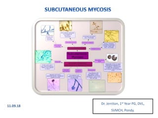
  • 1. Dr. Jerriton, 1st Year PG, DVL, SVMCH, Pondy. 11.09.18
  • 2. SUBCUTANEOUS MYCOSIS • Infections limited to skin & subcutaneous tissue • Rarely spreading to internal organs • Caused by fungi with saprophytic existence in nature • Usual mode of transmission by inoculation DEFINITION
  • 3.
  • 4. EYMYCE -TOMA CHROMO- MYCOSIS PHAEOHYP OMYCOSIS SPOROTRI- CHOSIS LOBOMYC- OSIS SC ZYGOMYC- OSIS RHINOSPO -RIDIASIS ETIOLOGY M. mycetomat is C. carrionii, F. pedrosoi E. jeanselmei , P. verrucosa S. schenkii Lacazia loboi Basidiobol us, conidiobol us spp. R. seebri CLINICAL FEATURES Painless SC swelling + sinus + discharging granules Verrucous plaques or nodules + ulceration Subcutane ous cysts Ulcerated nodules along lymphatics Polymorph ic; keloidal & painless Painless SC masses – bathsuit, monstrous face Painless friable warty outgrowth with ulceration DIAGNOSIS Granules Sclerotic bodies Melanin Asteroid bodies Round cells, tubular projections , bifringent memb. Mixed inflammat ory cells with fungal elements Sporangia with endospore s TREATMENT Surgery + Antifungals Antifungals Surgery + Antifungals SSKI + Antifungals Surgery + Clofazimin e + Antifungals SSKI + Antifungals Surgery
  • 5. MYCETOMA Definition • Chronic, suppurative, granulomatous disease of subcutaneous (SC) tissue & bones. • It’s a triad of: a) Painless SC Tumefaction b) Sinuses c) Discharge of granules
  • 6. MYCETOMA Etiology • Bacteria (Actinomycotic Mycetoma) – M/C in India  Actinomadura madurae (M/C) • Fungi (Eumycotic Mycetoma)  Madurella mycetomatis (M/C in South India) Brown grain – M. mycetomatis White grain – A. madurae
  • 7. MYCETOMA HISTORY & TRANSMISSION • John Gill in 1842 described “Madura Foot” first. • Mycetoma means fungal tumor. • The organisms are soil / plant saprophytes. • Entry is by abrasion / implantation. • More common in tropics.
  • 8. MYCETOMA EPIDEMIOLOGY • 20 and 40 years, mostly in developing countries. • Tropics and sub-tropics. • Barefoot walkers. • Low socioeconomic status.
  • 9. MYCETOMA CLINICAL FEATURES • Begins as small, painless SC nodule at site of injury. • Nodules increase in number, ulcerate & drain through sinuses. • Discharge will have the characteristic granules. • Surrounding skin becomes swollen, indurated & deformed. • Spreads by direct contiguity along facial planes. • Spares tendons till late stage. • Becomes painful if bones are involved / 2° infection. • Bones show punched out lytic lesions. • Ankylosis can happen.
  • 10. MYCETOMA MACROSCOPIC COLOR OF GRAINS • Black Grains – Eumycotic • Red Grains – Actinomadura pelletierii • White Grains – Eumycotic / Actinomycotic DIAGNOSIS
  • 11. MYCETOMA • Granules microscopy (KOH / Gram stain) of discharge are characteristic: 1) Actinomycetomas – Fine, branching interlacing filaments with no chlamydospores 2) Eumycetomas – Thick walled septate hyphae with chalmydospores MICROSCOPY OF DISCHARGE DIAGNOSIS HPE • Initially – Acute suppurative reaction • Later – Suppurative granuloma (peripheral epithelioid histiocytes and MNGs with central PMNs) • Characteristic granules can be seen within central abscess of granuloma
  • 13. EUMYCETOMA EUMYCETOMA GRAIN A: Ulcerated, indurated plaque of eumycotic mycetoma on the foot. B: H&E staining shows a “sulfur granule” in a purulent area of granulation tissue. C: The sulfur granule is composed largely of septate hyphae of P. boydii (GMS stain).
  • 14. MYCETOMA TREATMENT ACTINOMYCETOMA 1. Modified Welsch Regimen (3-4 cycles) Amikacin 15 mg/Kg IV x 21 days with 15 day intervals PLUS Co-trimoxazole 35 mg/Kg oral daily PLUS Rifampicin 10 mg/day oral daily
  • 15. MYCETOMA TREATMENT ACTINOMYCETOMA 1. Modified Two – Step Regimen Gentamycin 80 mg IV 12 hourly PLUS Co-trimoxazole 320 mg/1600 mg oral BD x 4 weeks FOLLOWED BY Maintenance doses Co-trimoxazole & Doxy 100 mg oral BD Continued 6 months till after healing
  • 16. MYCETOMA TREATMENT EUMYCETOMA • Ketaconazole • Terbinafine • Irraconazole 400 mg/day for 3 months f/b 200 mg/day for 9 months • Voriconazole 300 mg BD doe 16 months • Posaconazole for refractory cases
  • 17. MYCETOMA TREATMENT ROLE OF SURGERY • Exploration & drainage of sinus tracts • Debridement of diseased tissue • Removal of bone cysts • They help healing faster
  • 18. CHROMOBLASTOMYCOSIS DEFINITION • Chronic granulomatous infection, usually of exposed areas, characterised by verrucous plaques or nodules that ulcerate.
  • 19. CHROMOBLASTOMYCOSIS ETIOLOGY • Dematiaceous (brown-pigmented) fungi 1. Cladophialophora carrionii 2. Fonsecaea pedrosoi • Saprophytes of soil, decaying vegetation and rotting wood • Enter skin through abrasion • Most common in tropics and subtropics
  • 20. CHROMOBLASTOMYCOSIS CLINICAL FEATURES • 3 clinical forms exist: 1. Localized 2. Multiple with satellite lesions 3. Sporotrichoid • Begins as a verrucous papule -> verrucous plaque, which may have central atrophy / scarring • Seen mostly at exposed sites • Complications include elephantiasis, 2° infection, ulceration & malignant change
  • 21. CHROMOBLASTOMYCOSIS DIAGNOSIS • KOH of skin scraping, crusts, aspiration, biopsy tissue Sclerotic bodies / muriform cells
  • 22. CHROMOBLASTOMYCOSIS DIAGNOSIS • H & E stain of biopsy specimen Sclerotic bodies / muriform cells
  • 23. CHROMOBLASTOMYCOSIS DIAGNOSIS • Culture for species identification Sabouraud dextrose agar • Serology if culture is not possible
  • 24. CHROMOBLASTOMYCOSIS TREATMENT • Systemic antifungals Itraconazole 200-400 mg / day PLUS Terbinafine 250-500 mg / day X 6 – 12 months
  • 25. CHROMOBLASTOMYCOSIS TREATMENT • Other treatment options 1. Iodides, Fluconazole, posiconazole 2. Surgical excision, cryotherapy, local heat
  • 26. PHAEOHYPOMYCOSIS DEFINITION • Infections other than chromoblastomycosis and eumycetoma caused by dematiaceous (melanized / phaeoid) fungi • They do not have grains or sclerotic bodies that characterized mycetoma and chromomycosis respectively. • This entity is characterized by dark septate hyphae, pseudohyphae, yeast or their combinations.
  • 27. PHAEOHYPOMYCOSIS ETIOLOGY • Most common are: 1. Exophiala jeanselmei 2. Wangiella dermatitidis 3. Phialophora verrucosa 4. Bipolaris spp. • They live in decaying vegetation, bird nests and soil • They are seen mostly in tropics and subtropics • Infection is by local trauma by abrasion or inhalation
  • 28. PHAEOHYPOMYCOSIS PATHOGENESIS • DHN melanin – fungal armor in cell wall 1. Scavenges free radicals 2. Prevents action of hydrolytic enzymes • Thermotolerance – can cause deep invasive cerebral lesions
  • 29. PHAEOHYPOMYCOSIS CLASSIFICATION 1. Superficial: black piedra and tinea nigra 2. Cutaneous: dermatomycosis and onychomycosis 3. Mycotic keratitis 4. Subcutaneous phaeohypomycosis 5. Invasive, systemic & cerebral
  • 30. PHAEOHYPOMYCOSIS CLINICAL FEATURES • Classical presentation: SC cyst. • Begins as small papule -> evolves into SC cyst • Usually single • M/C site: extremities • Children: face
  • 31. PHAEOHYPOMYCOSIS DIAGNOSIS • KOH of pus, drainage or skin scrapings Pigmented yeasts, pseudohyphae and hyphae • Biopsy 1. Foreign body granuloma 2. Pigmented fungi seen within granuloma 3. Splendore-Hoeppli reaction +/- 4. Fontana Masson stain for melanin is diagnostic • FNAC Pigmented fungi with inflammatory cells
  • 32. PHAEOHYPOMYCOSIS TREATMENT • Triple antifungal combinations give best results for refractory cases Amphotericin B, glucytosine and itraconazole • Localised lesion: excision f/b pre & post op antifungal therapy • Antifungals used: • Flucytosine 150 mg/kg/day • Itraconazole 200 mg/day • Ketoconazole 200 mg/day • IV / ILS Amphotericin B
  • 33. SPOROTRICHOSIS DEFINITION • Subacute or chronic infection caused by dimorphic fungi, S. schenckii • Characterized by nodular and ulcerative lesions along lymphatics
  • 34. SPOROTRICHOSIS ETIOLOGY S. schenkii • Dimorphic fungi • Mycelia at 26°C and yeast at 37°C • Mycelia bears conidia resembling flower • Grows in common agar • Produces creamy white colonies • Turns black later • It is a saprophyte in dead plants • Introduced by trauma to skin
  • 35. SPOROTRICHOSIS HISTOLOGY • HPE shows three granulomatous patterns observed 1. Sporotrichotic (central suppuration seen) 2. Tuberculoid (central suppuration seen) 3. Foreign body (no central suppuration) • Asteroid bodies are characteristic Round basophilic yeast-like body with surrounding elongated radiating eosinophilic material (a type of Splendore – Hoeppli reaction)
  • 36.
  • 37. SPOROTRICHOSIS CLINICAL FEATURES • Cutaneous 1. Lymphocutaneous 2. Localized • Extra cutaneous 1. Pulmonary 2. Disseminated
  • 38. SPOROTRICHOSIS CLINICAL FEATURES Lymphocutaneous form • M/C seen in exposed site of upper extremity • Small nodule / pustule develops at site of trauma • Nodule breaks to form ulcer • New nodules form along lymphatics at few days interval • Ulcerated nodules connect by cord-like swollen lymphatics • Heals with scarring and new nodules develop at other sites • These secondary lesions are gummatous and persist for years
  • 40. SPOROTRICHOSIS CLINICAL FEATURES Localized cutaneous form • Primary lesion is restricted to site of injury • It can be ulcerative, verrucous, acneiform or scaly plaque • Does not involve local lymphatics • Mucous membrane can be involved • Pain is predominant complaint
  • 41. SPOROTRICHOSIS INVESTIGATIONS • Sporotrichin skin test • 0.1 mL of intradermal sporotrichin M (mycelia) is injected into their forearm and the reading is ascertained at 48 hours, using the same criterion as for the tuberculin skin test. • Induration ≥ 0.8 cm is positive. • Serology – for extra-cutaneous forms
  • 42. SPOROTRICHOSIS INVESTIGATIONS • Culture – definitive diagnosis 1. Sabouraud’s dextrose agar at 26°C and 37°C 2. Conversion to yeast form at 37°C is important • Animal innoculation 1. Gram positive cigar bodies in pus
  • 43. SPOROTRICHOSIS TREATMENT • SSKI 1. Given orally in milk 2. Initial dose: 5 drops (1 ml) TID after meals 3. Increased by 1 drop / dose till 40 drops TID 4. Continued till signs of active disease are gone 5. Then dose decreased by 1 drop / dose till 5 drops 6. Then discontinued • Itraconazole 100-200 mg/day • Terbinafine 250 mg/day • Thermotherapy / pocket warmer
  • 44. LOBOMYCOSIS DEFINITION Also known as keloidal blastomycosis, pseudoleprosy. It is characterized by pleomorphic lesions.
  • 45. LOBOMYCOSIS ETIOLOGY • Caused by fungi, Lacazia loboi • Has a saprophytic phase in vegetation, soil & water • Infection is acquired through trauma • Farmers, gold miners, fishermen and hunters are affected • Dolphin to human transmission is reported • No person-to-person transmission
  • 46. LOBOMYCOSIS CLINICAL FEATURES • Pleomorphic lesions • Nodules or plaques (hypopigmented / hyperpigmented) • Ulcers • Sclerodermoid • Keloidal • Verrucous • Legs, outer ears and arms are commonly affected • Single or multiple, become confluent • Generally painless, occasionally painless
  • 47. LOBOMYCOSIS INVESTIGATIONS • Direct microscopy • KOH shows round yeast-like organisms, singly or in chains connected by short tubular projections. • They have bifringngent membrane with central granules • HPE • Granulomatous infiltrate without suppuration • Grenz zone +/- • Asteroid bodies +/- • Fungal forms seen at different levels of epidermis (TEE)
  • 48. LOBOMYCOSIS TREATMENT • Medical: 1. Clofazimine 300 mg/day initial dose with maintainance dose of 100 mg/day for 2 years 2. Ketoconazole, Itraconazole, Posaconazole • Surgical excision • Electrocautery • Cryosurgery
  • 49. SUBCUTANEOUS ZYGOMYCOSIS DEFINITION • Chronic subcutaneous infection characterized by woody swelling of SC tissue
  • 50. SUBCUTANEOUS ZYGOMYCOSIS ETIOLOGY & CLASSIFICATION FEATURES ENTOMOPHTHORALES (Basidiobolus, conidiobolus) MUCORALES Host Immunocompetent Immunocompromised Distribution Tropics and subtropics Worldwide Transmission Traumatic implantation Inhalation of spores Systems involved SC mycosis and sinusitis RS, CNS, GIT, skin Histopathology Chronic inflammatory response Angioinvasion, thrombosis,tissue necrosis Splendore-Hoeppli Characteristic Rarely seen Septation More common Less common Dissemination Uncommon Common
  • 51. SUBCUTANEOUS ZYGOMYCOSIS EPIDEMIOLOGY & TRANSMISSION BASIDIOBOLUS • Children less than 20 years • Male > Female • Africa > India • Transmitted by minor trauma / insect bite / contaminated toilet leaves (bathing suit distribution) • Also transmitted through soil and vegetation containing contaminated animal faeces
  • 52. SUBCUTANEOUS ZYGOMYCOSIS EPIDEMIOLOGY & TRANSMISSION CONIDIOBOLUS • Young adults • Male > female • Africa > India • Identified in soil & plant debris, M/C c. coronatus • Transmitted by inhalation of fungal spores / frequent nose pricking habits • Leads to monstrous disfigurement of face
  • 53. SUBCUTANEOUS ZYGOMYCOSIS PATHOGENESIS BASIDIOBOLUS • Produces extracellular proteinases and lipases  Phospholipase A hydrolizes lecithin, which destroyed membranes of blood, skin & muscle cells  Once lipase liberates cellular protein components, proteinases digest them as its nutrients • Thermotolerant – grows poorly at 37°C
  • 54. SUBCUTANEOUS ZYGOMYCOSIS PATHOGENESIS CONIDIOBOLUS • Produces elastase, esterase, collagenase and lipase  Proteinase is secreted first – breaks down proteins to amino acids  Lipase is produced later – hydrolizes fatty materials in SC tissue • Thermophilic – grows readily at 37°C
  • 55. SUBCUTANEOUS ZYGOMYCOSIS CLINICAL FEATURES BASIDIOBOLOMYCOSIS • M/C site: limb girdles / proximal limbs • Bathing suit distribution • Painless well-circumcized, firm to hard, smooth, rounded SC masses that can be raised by inserting fingers underneath it (freely mobile) • Satellite lesions may be seen at advancing margins • May encompass part / whole of limb • Overlying skin may be tense, edematous, desquamating, hyperpigmented or normal • Non-pitting oedema +/- • Underlying muscle / visceral involvement can occur
  • 56. SUBCUTANEOUS ZYGOMYCOSIS CLINICAL FEATURES CONIDIOBOLOMYCOSIS • Begins as swelling of inferior nasal turbinates • Stuffiness, discharge, epistaxis, nasal obstruction are symptoms • Diffuse erythematous infiltration with skin thickening on nose, cheeks, forehead and lips – monstrous disfigurement, facial elephantiasis, palatal perforation, orbital cellulitis, saddle nose deformity • Phase 1 – nose, paranasal sinuses & pharynx • Phase 2 – frontal region and lips • Phase 3 – muscle, bones & viscera
  • 57. SUBCUTANEOUS ZYGOMYCOSIS DIAGNOSIS HPE • Mixed inflammatory infiltrate • Fungal hyphal elements with surrounding dense eosinophilic granular aterial (Splenndore-Hoeppli phenomenon) MICROSCOPY • KOH from scrapings show broad, septate branching hypahe CULTURE • Basidiobolus – flat & furrowed, yellowish grey color with musty odor • Conidiobolus – white surface, becomes beige to brown, no odor
  • 58. SUBCUTANEOUS ZYGOMYCOSIS TREATMENT • Itraconazole / SSKI – 1st line choices • Treated continuously for 1-2 months after clinical cure
  • 59. RHINOSPORIDIOSIS DEFINITION • Chronic granulomatous disease of mucocutaneous tissue • Characterized by development of polypoid tumors or pedunculated / sessile polyps
  • 60. RHINOSPORIDIOSIS ETIOLOGY & EPIDEMIOLOGY • Caused by Rhinosporidium seeberi – protistan parasite of class Mesomycetozoea • Common pond bathing with buffalos is a risk factor • Disease is endemic in Kerela, Tamil nadu and Chandigarh
  • 61. RHINOSPORIDIOSIS HISTOLOGY • Thick walled sporangia with endospores
  • 62. RHINOSPORIDIOSIS CLINICAL FEATURES CUTANEOUS RHINOSPORIDIOSIS • Associated with mucosal disease • Begin as tiny papules and enlarge to become wart like / tumorous growth • They are friable and have crenated surface, often ulcerated - & painless
  • 63. RHINOSPORIDIOSIS DIAGNOSIS • KOH / HPE for sporangia with endospores TREATMENT • Surgical excision • Electrocoagulation
  • 64. REFERENCES 1. Rook’s Textbook of Surgery 2. IADVL Textbook of Dermatology 3. Fitzpatrick Textbook of Dermatology 4. Lever’s Histopathology 5. Weedon’s Histopathology 6. Online journals

Editor's Notes

  1. M = Madurella mycetomatis C = Cla-do-phia-lo-phora carrionii F = Fon-se-caea pedrosoi E = Exophiala jeanselmei P = Phialophora verrucosa
  2. Other causes: Actinomycotic = Nocardia & Streptomyces spp. Actinomadura pelletieri alone has red granules. Others are white. Eumycotic = Leptosphaeria, Exophiala, Acremonium, etc.
  3. The fungal elements (hyphae with chlamydospores) comprising this grain are easily seen inside the central suppurative regio.
  4. Other tests: Culture and Serology
  5. Co-trimoxazole – Trimethoprim - Sulphomethoxazole
  6. Co-trimoxazole – Trimethoprim - Sulphomethoxazole
  7. Co-trimoxazole – Trimethoprim - Sulphomethoxazole
  8. Co-trimoxazole – Trimethoprim - Sulphomethoxazole
  9. Co-trimoxazole – Trimethoprim - Sulphomethoxazole
  10. Co-trimoxazole – Trimethoprim - Sulphomethoxazole
  11. Co-trimoxazole – Trimethoprim - Sulphomethoxazole
  12. Sclerotic bodies are thick walled dark brown bodies that are fungal elements
  13. Sclerotic bodies are thick walled dark brown bodies that are fungal elements
  14. Sclerotic bodies are thick walled dark brown bodies that are fungal elements
  15. Sclerotic bodies are thick walled dark brown bodies that are fungal elements
  16. Sclerotic bodies are thick walled dark brown bodies that are fungal elements
  17. Sclerotic bodies are thick walled dark brown bodies that are fungal elements
  18. Sclerotic bodies are thick walled dark brown bodies that are fungal elements
  19. Two virulence factors
  20. Two virulence factors
  21. Two virulence factors
  22. Two virulence factors
  23. Two virulence factors
  24. Two virulence factors
  25. Two virulence factors
  26. Two virulence factors
  27. Two virulence factors
  28. Two virulence factors
  29. Two virulence factors
  30. Two virulence factors
  31. Two virulence factors
  32. Two virulence factors
  33. SSKI = Saturated Solution of Potassium Iodide Thermotherapy = Immersion in hot water
  34. SSKI = Saturated Solution of Potassium Iodide Thermotherapy = Immersion in hot water
  35. SSKI = Saturated Solution of Potassium Iodide Thermotherapy = Immersion in hot water
  36. SSKI = Saturated Solution of Potassium Iodide Thermotherapy = Immersion in hot water
  37. SSKI = Saturated Solution of Potassium Iodide Thermotherapy = Immersion in hot water
  38. SSKI = Saturated Solution of Potassium Iodide Thermotherapy = Immersion in hot water
  39. SSKI = Saturated Solution of Potassium Iodide Thermotherapy = Immersion in hot water
  40. SSKI = Saturated Solution of Potassium Iodide Thermotherapy = Immersion in hot water
  41. SSKI = Saturated Solution of Potassium Iodide Thermotherapy = Immersion in hot water
  42. SSKI = Saturated Solution of Potassium Iodide Thermotherapy = Immersion in hot water
  43. SSKI = Saturated Solution of Potassium Iodide Thermotherapy = Immersion in hot water
  44. SSKI = Saturated Solution of Potassium Iodide Thermotherapy = Immersion in hot water
  45. SSKI = Saturated Solution of Potassium Iodide Thermotherapy = Immersion in hot water
  46. SSKI = Saturated Solution of Potassium Iodide Thermotherapy = Immersion in hot water
  47. SSKI = Saturated Solution of Potassium Iodide Thermotherapy = Immersion in hot water
  48. SSKI = Saturated Solution of Potassium Iodide Thermotherapy = Immersion in hot water
  49. SSKI = Saturated Solution of Potassium Iodide Thermotherapy = Immersion in hot water
  50. SSKI = Saturated Solution of Potassium Iodide Thermotherapy = Immersion in hot water
  51. SSKI = Saturated Solution of Potassium Iodide Thermotherapy = Immersion in hot water
  52. SSKI = Saturated Solution of Potassium Iodide Thermotherapy = Immersion in hot water