
Recommended
More Related Content
What's hot
What's hot (20)
Similar to Tight junctions workshop - By Vaishnavi Charanya Sundar - Physiology
Similar to Tight junctions workshop - By Vaishnavi Charanya Sundar - Physiology (20)
Recently uploaded
Recently uploaded (20)
Tight junctions workshop - By Vaishnavi Charanya Sundar - Physiology
- 2. What are Tight Junctions Tight junctions form the continuous intercellular barrier between epithelial cells, which is required to separate tissue spaces and regulate selective movement of solutes across the epithelium.
- 3. Structure of Tight junctions The portion of the cell exposed to the lumen is called its apical surface. The rest of the cell (i.e., its sides and base) make up the basolateral surface. Tight junctions seal adjacent epithelial cells in a narrow band just beneath their apical surface. They consist of a network of claudins and other proteins.
- 4. Functions of Tight Junctions Tight junctions perform two vital functions: ● They limit the passage of molecules and ions through the space between cells. So most materials must actually enter the cells (by diffusion or active transport) in order to pass through the tissue. This pathway provides tighter control over what substances are allowed through.
- 5. ● They block the movement of integral membrane proteins (red and green ovals) between the apical and basolateral surfaces of the cell. Thus the special functions of each surface, for example ○ receptor-mediated endocytosis at the apical surface ○ exocytosis at the basolateral surface can be preserved. Exocytosis is the reverse of endocytosis. And that is just as well. In 30 minutes an active cell like a macrophage (right) can endocytose an amount of plasma membrane equal to its complete plasma membrane.
- 6. Receptor mediated Endocytosis Some of the integral membrane proteins that a cell displays at its surface are receptors for particular components of the ECF (extracellular Fluid). For example, iron is transported in the blood complexed to a protein called transferrin. Cells have receptors for transferrin on their surface. When these receptors encounter a molecule of transferrin, they bind tightly to it. The complex of transferrin and its receptor is then engulfed by endocytosis. Ultimately, the iron is released into the cytosol. The strong affinity of the transferrin receptor for transferrin (its ligand) ensures that the cell will get all the iron it needs even if transferrin represents only a small fraction of the protein molecules present in the ECF. (Extracellular Fluid)
- 7. Pathway of tight junctions Transcellular transport involves the transportation of solutes by a cell through a cell.In contrast, paracellular transport is the transfer of substances across an epithelium by passing through an intercellular space between the cells. . Paracellular permeability can be divided into two distinct pathways, the Pore Pathway mediating the movement of small ions and solutes and the Leak Pathway mediating the movement of large solutes. Paracellular permeability barrier- Claudin protein. Leak permeability barrier-Occludin, Zo protein, Tricellulin and actin.
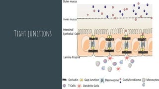
- 8. where would we find these tight junctions Tight junctions are located within our body's epithelia. Epithelia is the plural of epithelium. Epithelium is a word that refers to the covering of the body's internal and external surfaces. This includes organs (such as skin), blood vessels and body cavities.
- 9. How this information might be relevant to a future healthcare practitioner Tight junctions is crucial to protect the body against stress stimuli related to inflammation and infection. In Skin under homeostatic conditions efficiently protects and/or minimizes damage from both environmental (e.g. microorganisms, physical trauma, ultraviolet radiation) and endogenous (e.g., cancers, inflammation) factors. And in Intestinal epithelia the tight junctions (TJs) are essential to the function of the physical intestinal barrier, regulating the paracellular movement of various substances including ions, solutes, and water across the intestinal epithelium. Tight junctions contains number of protein like claudins , occludin etc which play a critical role in regulating TJ barrier function. So the tight junctions dysfunction may interfere with the protein expression causing pathogenesis of a number of intestinal and common cutaneous inflammatory and neoplastic conditions. So understanding the role of tight junctions may help to play a vital role in diagnosing many intestinal and skin conditions in the future.
- 10. What diseases or conditions can affect tight junctions? Barrier dysfunction includes increased paracellular permeability resulting from enhanced flux across the tight junction, but may also be caused by epithelial damage, including apoptosis, erosion, and ulceration. Studies reveal that TJ dysfunction is closely related to inflammatory and metabolic disorders including Inflammatory Bowel Disease, Non alcoholic fatty liver,Non-alcoholic Steatohepatitis, and obesity via the disruption of TJ barrier functions. Thus, the maintenance of TJ integrity is likely a good strategy to prevent and/or treat these diseases. And in Crohn's disease (CD) and ulcerative colitis (UC) share common features such as epithelial breaks, a reduction in tight junction strands, and glandular atrophy. Then Mutations in the claudin14 and the claudin16 genes (proteins of tight junctions) result in hereditary deafness and hereditary hypomagnesemia.
- 11. References Anderson, J. M., & Van Itallie, C. M. (2009). Physiology and function of the tight junction. Cold Spring Harbor perspectives in biology, 1(2), a002584. https://doi.org/10.1101/cshperspect.a002584 Brandner, J. M., Zorn-Kruppa, M., Yoshida, T., Moll, I., Beck, L. A., & De Benedetto, A. (2015). Epidermal tight junctions in health and disease. Tissue barriers, 3(1-2), e974451. https://doi.org/10.4161/21688370.2014.974451 Lee, B., Moon, K. M., & Kim, C. Y. (2018). Tight Junction in the Intestinal Epithelium: Its Association with Diseases and Regulation by Phytochemicals. Journal of immunology research, 2018, 2645465. https://doi.org/10.1155/2018/2645465 Sawada N. (2013). Tight junction-related human diseases. Pathology international, 63(1), 1–12. https://doi.org/10.1111/pin.12021 Shen, L., Weber, C. R., Raleigh, D. R., Yu, D., & Turner, J. R. (2011). Tight junction pore and leak pathways: a dynamic duo. Annual review of physiology, 73, 283–309. https://doi.org/10.1146/annurev-physiol-012110-142150