Parkinsonism syndromes with differential diagnosis
•Download as PPTX, PDF•
3 likes•76 views

Different parkinsonism syndromes compared with Idiopathic Parkinson's disease

Recommended
More Related Content
What's hot
What's hot (20)
Similar to Parkinsonism syndromes with differential diagnosis
Similar to Parkinsonism syndromes with differential diagnosis (20)
Recently uploaded
Recently uploaded (20)
Parkinsonism syndromes with differential diagnosis
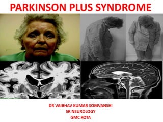
- 1. PARKINSON PLUS SYNDROME DR VAIBHAV KUMAR SOMVANSHI SR NEUROLOGY GMC KOTA
- 2. OUTLINE • Classification • Red flag signs • Diagnostic criteria • Phenotypic spectrum • Investigations • Novel biomarkers • Treatment • Future trends
- 3. INTRODUCTION • Common problem in neurology OPD • Wide variety of sporadic / heredodegenerative syndromes • 80-85% -IPD • Differentiation from other syndromes • Important in prognostication and management
- 5. PARKINSONISM PLUS • Progressive Supranuclear Palsy • Multiple System Atrophy [(Shy-Dragger syn.), SND (MSA P), OPCA (MSA C)] • Corticobasal Degeneration • Dementia with Lewy Body Disease
- 7. PROGRESSIVE SUPRANUCLEAR PALSY • Steele et al—1964 • 5% of parkinsonian pts • Male-to-female ratio is 1.5:1 • Commonly misdiagnosed as PD • Diagnosis is purely clinical • Always sporadic, few familial cases
- 8. • The usual interval from initial symptom occurrence to the need for a cane or a walker is 3.1 years, • Confinement to a chair or bed is 8.2 years. • Median disease duration of 9.7 years
- 9. • Postural Instability & EP Features : • Falls—backward • Rigidity –axial • Hypophonic • Widely based ataxic • Frontal release signs • Pseudobulbar palsy • L-DOPA UNRESPONSIVENESS
- 10. • Early signs- Slow vertical saccades and square wave jerks • Reduced blink rate and apraxia of eyelid opening • On doll’s eye maneuver, there is improved range • Subcortical-type dementia • Typical facies- “surprised look” • Advanced PSP - Complete ophthalmoparesis
- 12. NINDS PSP DIAGNOSTIC CRITERIA • Possible PSP(highly sensitive) • Mandatory inclusion criteria: • Gradually progressive disorder • Onset age 40 or later • Either vertical supranuclear palsy or both slowing of vertical saccades • Postural instability with falls within a year of disease onset • No evidence of other diseases that could explain the foregoing features, as Indicated by exclusion criteria
- 13. • Mandatory exclusion criteria: • Recent history of encephalitis • Alien limb syndrome • Cortical sensory deficits • Focal frontal or temporoparietal atrophy • Hallucinations or delusions unrelated to dopaminergic therapy • Cortical dementia of Alzheimer type • Prominent, early cerebellar symptoms • Unexplained dysautonomia
- 14. • Supportive features: • Symmetrical akinesia or rigidity • Proximal more than distal • Abnormal neck posture especially retrocollis • Poor or absent response of parkinsonism to levodopa • Early dysphagia and dysarthria • Early onset of cognitive impairment including two or more of: apathy, Impairment in abstract thought, decreased verbal fluency, utilisation or Imitation behaviour , or frontal release signs
- 15. • Probable PSP(highly specific) • Mandatory inclusion criteria • Gradually progressive disorder • Onset age 40 or later • Vertical supranuclear palsy • Prominent postural instability with falls within a year of disease onset • No evidence of other diseases that could explain the foregoing features, • As indicated by exclusion criteria
- 16. • Definite PSP • Mandatory inclusion criteria: • Clinically probable or possible PSP and • Histopathological evidence of typical PSP
- 18. INVESTIGATIONS • Clinical diagnosis • MRI midbrain atrophy (appearance of a flat or concave profile -68% sensitivity and an 89% Specificity • Superior cerebellar peduncle atrophy. • “Morning Glory Flower Sign” and the “Hummingbird Sign” – Highly specific(100%) low Sensitivity (50% and 68.4%) • Magnetic resonance parkinsonism index (MRPI) - sensitivity of 100% and specificity of 99·2–100·0% for PSP-RS. • Pons : Midbrain ratio
- 19. MAGNETIC RESONANCE PARKINSONISM INDEX (MRPI) P = area of pons in midsagittal plane MCP = width of middle cerebellar peduncle (P / M) x (MCP / SCP) M = area of midbrain in midsagittal plane SCP = width of superior cerebellar peduncle value more than 13.55 abnormal , strongly suggests will develop PSP.
- 20. • PET – lowered glucose metabolism in the midbrain ,caudate, thalamus of PSP • MIBG is abnormal in PD because of postganglionic sympathetic denervation, but is typically normal in PSP . • IBZM SPECT assessing the postsynaptic receptors is abnormal in PSP and normal in PD • IBZM SPECT is abnormal in all APS • DAT scan is abnormal in PD and all AP syndromes
- 23. PATHOLOGY • Spares the cortex and involves the basal ganglia,dentate, pontine, and oculomotor nuclei. • Abnormal tau hyperphosphorylation and deposition. Tau is encoded by MAPT and normally functions to stabilize microtubules. • Neurofibrillary tangles are present in reticular formation and ocular motor nuclei. • Tufted astrocytes -feature of PSP that differentiates other tauopathies such as CBD (astrocytic plaques,colloid bodies)
- 25. NOVEL DIAGNOSTIC APPROACH AND BIOMARKERS • CSF tau protein- CSF phospho-tau and total tau concentrations lower than AD • 2–5 times increased neurofilament light chain concentrations in PSP
- 26. TREATMENT • No effective symptomatic or neuroprotective treatments • A trial of levodopa (up to 1 g/d) and amantadine (up to 450 mg/d) • Botulinum toxin injections can be used to treat levator inhibition,rigidity , dystonia • Serotonin reuptake inhibitors (SSRIs) may be used for apathy with no clear benefit. • Supportive measures such as physiotherapy, walking aids, speech therapy and PEG
- 27. • A small study with Coenzyme Q10- no RCT study • Recent large, double-blind studies with (glycogen synthase kinase)GSK-3b inhibitors (Tideglusib,Davunetide) ,prevent hyperphosphorylation of tau- failed. • Tideglusib reduced the rate of brain atrophy in one study.
- 28. MULTIPLE SYSTEM ATROPHY • Sporadic neurodegenerative disorder clinically any combination of parkinsonian, autonomic, cerebellar, or pyramidal signs. • MSA is an alpha-synucleinopathy. • Usually a sporadic disease; however, rarely, familial cases - mutations in COQ2 gene. • Prevalence of MSA - ranged from 1·9 to 4·9 cases per 100 000 people
- 29. CLINICAL PRESENTATION : • Affects both men and women • Sixth decade of life • Mean survival of 6–9 years.(Upto 15yrs) Main features • Autonomic failure • Parkinsonism • Cerebellar ataxia • Pyramidal signs in any combination
- 30. TWO MAJOR MOTOR MANIFESTATIONS Distinguished clinically– • 1. Parkinsonian features predominate in 80% of patients (MSA-P subtype), • 2. Cerebellar ataxia is the main motor feature in 20% of patients (MSA-C subtype). • Both similar survival times. • MSA-P - more rapid functional deterioration
- 31. MSA-P • Progressive akinesia and rigidity • Jerky postural tremor and tremor at rest. • Orofacial or craniocervical dystonia • Recurrent falls at disease onset are unusual . • 90% of the MSA-P pts- unresponsive to levodopa in the long term.
- 32. MSA-C • Gait ataxia • Scanning dysarthria • Cerebellar oculomotor disturbances. • May be indistinguishable from other patients with idiopathic late onset cerebellar Ataxia
- 33. • Dysautonomia • Urogenital and orthostatic dysfunction. • Early erectile dysfunction is nearly universal in men with MSA • Female- genital insensitivity • Urinary incontinence or retention are common
- 34. CONSENSUS STATEMENT FOR CLINICAL DIAGNOSIS OF MSA • Autonomic and urinary dysfunction • Features • 1. Orthostatic hypotension(68% of patients) • 2. Urinary incontinence or incomplete bladder emptying • Criteria • Reduction of least 30mmhg or in diastolic blood pressure by at least 15 mm hg after 3 min of standing • Urinary incontinence (persistent, involuntary partial or total bladder emptying, • Accompanied by erectile dysfunction in men or both
- 35. • Parkinsonism : initial feature in 46% of patients with MSA-P • A. Features • 1. Bradykinesia • 2. Rigidity • 3. Postural instability (not caused by primary visual, vestibular, cerebellar, or proprioceptive dysfunction) • 4. Tremor (postural, resting or both) • B. Criteria • Bradykinesia plus at least one of features 2–4
- 36. • Cerebellar dysfunction :initial feature in 5% • A. Features • 1. Gait ataxia • 2. Ataxic dysarthria • 3. Limb ataxia • 4. Sustained gaze-evoked nystagmus • Criteria • Gait ataxia plus at least one of features 2–4
- 37. • Corticospinal tract dysfunction • A. Features • 1. Extensor plantar responses with hyper-reflexia • Criteria • No corticospinal tract features are used in defining the diagnosis of MSA • Prominent and severe spasticity should raise suspicion for an alternative diagnosis
- 38. • Exclusion criteria: • Symptomatic onset <30 years/>75YRS of age • Family history of a similar disorder • Systemic disease or other identifiable causes • Hallucinations unrelated to medication • Dementia
- 39. • Exclusion criteria: • Prominent slowing of vertical saccades or vertical supranuclear gaze palsy • Evidence of focal cortical dysfunction • Laboratory investigation- metabolic, molecular genetic and imaging evidence of an alternative cause of features
- 40. • Possible MSA • A sporadic, progressive, adult (>30y) with onset disease characterized by the following: • Parkinsonism or cerebellar syndrome • At least 1 feature of autonomic or urogenital dysfunction • At least 1 additional feature
- 41. • Probable MSA • A sporadic, progressive, adult (>30y) with onset disease characterized by the following: • Autonomic failure involving urinary dysfunction • Poorly levodopa-responsive parkinsonism or cerebellar dysfunction
- 42. Definitive MSA • A sporadic, progressive, adult (>30y) with onset disease pathologically confirmed by Presence of high density GCIS in association with degenerative changes in Striatonigral and olivopontocerebellar pathways
- 43. MSA ADDITIONAL FEATURES • Pyramidal signs • Orofacial dystonia or dyskinesias • Dyskinesia mainly affecting orofacial muscles • Axial dystonia -PISA syndrome (subacute axial dystonia with a severe tonic lateral flexion of the trunk, head, and neck) early severe camptocormia
- 44. • Jerky tremor • Dysarthria- Atypical, irregular and severely hypophonic • Dysphagia within 5 years of motor onset • Neuropsychiatric features – Depression (41%) , Hallucinations (5·5%),Dementia (4·5%) , Insomnia (19%) ,Daytime sleepiness (17%) , Restless legs (10%)
- 46. Investigations • Autonomic function tests (table tilt,24 hr ambulatory bp,heart rate monitoring,baroreflex sensitivity,qsart,gastric emptying study,psg) • Cardiovascular function • Standard urine analysis will exclude infection. • The residual volume –USG,Cystometry ,UDS
- 47. IMAGING • MRI • Hot cross burn sign- mcp/pons- MSA-c • Putaminal rim- MSA-p • The slight hyperintensity of the lateral margin of the putamen on T2-weighted MRI is a characteristic finding in patients with MSA involving the extrapyramidal system • MSA-C-cerebellum and middle cerebellar peduncle
- 49. INVESTIGATIONS • DAT scan abnormal in all MSA, PSP, and PD • MIBG scintigraphy abnormal in PD, normal in MSA • IBZM SPECT is normal in PD,abnormal in MSA (but also in PSP and CBD • PET- The caudate putamen index- lower in patients with MSA than in PD
- 50. TREATMENT • Symptomatic • PD- l-dopa/ dopa agonists- cranio cervical dystonia postural hypotension • Amantidine- gait disturbances • Orthostatic hypotension- high salt, fludrocortisone, midodrine,droxidopa,pyridostigmine • Urinary dysfunction- oxybutinin • Erectile dysfunction– sildenafil, Intracavernosal inj. Or penile implants
- 51. Depression • SSRI/TCA Promising studies • Rasagiline • Intrarterial/IV - autologous stem cells Future trials • Alpha synuclein targeting antibodies
- 52. CORTICOBASAL DEGENERATION • Sixth to eighth decades of life - mean age 63 years • Sporadic disease , 4–6% of parkinsonism.
- 53. • Clinical presentations • The most common presentation (55%) -“useless arm” (ie, a rigid, dystonic, akinetic, or apraxic arm), • Gait disorder (27%) • Prominent sensory symptoms • Isolated speech disturbance • Behavioural disturbance
- 54. CLINICAL FEATURES • Motor (asymmetric) • Limb clumsiness • Bradykinesia/ Akinesia • Rigidity • Tremor (action/postural) • Myoclonus • Limb dystonia • Blepharospasm • Choreoathetoid movements • Speech abnormalities • Gait disorder
- 55. • Higher cortical functions • Apraxia • Dementia • Alien-limb phenomenon • Aphasia • Frontal-lobe-release signs • Cortical sensory abnormalities • Depression • Apathy • Anxiety irritability • Disinhibition, delusions,obsessive compulsive disorde
- 56. DIAGNOSTIC CRITERIA • Inclusion criteria (one of A or B) • A) Rigidity (easily detectable without reinforcement) and one cortical sign: Apraxia, Cortical sensory loss, Alien-limb phenomenon • B) Asymmetric rigidity, dystonia (focal in limb; present at rest at onset),Focal reflex myoclonus (spreads beyond stimulated digits)
- 57. • Exclusion criteria • Early dementia (will exclude some patients) • Early vertical gaze palsy • Rest tremor • Severe autonomic disturbances • Sustained responsiveness to levodopa • Lesions on imaging studies indicate another pathological process
- 59. • Imaging • MRI • Asymmetric frontal, and parietal cortical atrophy becomes evident with dilatation of the lateral ventricle (temporal/parietal cortex (the later pattern is seen in dementia of the alzheimer type)
- 60. • Dopamine transporter SPECT- abnormal,differentiate them from those with alzheimer’s and pick’s diseases (in whom this scan is typically normal) early in the course of the disease. FDG-PET • Asymmetric reduction in fronto parietal regions
- 61. • The R2 component of the blink reflex recovery cycle (R2 BRRC) appears to be a useful tool to distinguish progressive supranuclear palsy (PSP) from corticobasal degeneration (CBD) • 4-repeat-tau aggregates - neocortex in CBD, brainstem in PSP Eur J Neurol.sciacca g et al, 2018 Aug;25(8):1100-e85. doi: 10.1111/ene.13673. Epub 2018 Jun 12.
- 62. TREATMENT • L-dopa trial (upto 1 gm/d) • Amantadine(450 mg/d) • Valproate, levetiracetam- myoclonus • Botox inj- dystonic hand • Antioxidants or vitamin E if the patient has memory loss • Palliative rx
- 63. DEMENTIA WITH LEWY BODY • Dementia -not occur in the early stages ,usually evident with progression. • Deficits on tests of attention, executive function, and visuospatial ability may be especially prominent.
- 64. CORE FEATURES • Two core features are sufficient for a diagnosis of probable, one for possible DLB • Fluctuating cognition with pronounced variations in attention and alertness • Recurrent visual hallucinations that are typically well formed and detailed features of parkinsonism
- 65. SUGGESTIVE FEATURES • REM sleep behavior disorder • Severe neuroleptic sensitivity • Low dopamine transporter uptake in basal ganglia demonstrated by SPECT or PET imaging • One or more of these + one or more core features (probable DLB ) • In the absence of any core features, one or more suggestive features - possible DLB. • Probable DLB should not be diagnosed on the basis of suggestive features alone
- 66. SUPPORTIVE FEATURES • Repeated falls and syncope, Transient unexplained loss of consciousness • Severe autonomic dysfunction • Hallucinations in other modalities • Depression • Relative preservation of medial temporal lobe structures on CT/MRI scan • Generalized low uptake on SPECT/PET perfusion scan with reduced occipital activity • Prominent slow wave activity on EEG with temporal lobe transient sharp waves
- 67. TEMPORAL SEQUENCE OF SYMPTOMS • Diagnosed when dementia occurs before or concurrently with parkinsonism (if it is present). • Parkinson disease dementia (PDD) - dementia that occurs in the context of well established parkinson disease. • The 1-year rule between the onset of dementia and parkinsonism – DLB .
- 68. INVESTIGATIONS • MRI BRAIN -diffuse cerebral atrophy with relative preservation of occipital and mesial temporal lobes compared to alzheimer disease. • SPECT & PET -decreased occipital lobe blood flow – DLB > AD -relative preservation of the posterior cingulate gyrus (cingulate island sign) - DLB > AD
- 69. • SPECT scanning studies in DLB patients : • Visual hallucinations - Were related to hypoperfusion of the parietal and occipital association cortices • Misidentifications - Were related to hypoperfusion of the limbic-paralimbic structures • Delusions - Were related to hyperperfusion of the frontal cortices
- 70. • CSF • Tau – DLB < AD • Beta amyloid are lower than normal in DLB, AD • LBCRS - lewy body composite risk score - help determine whether lewy body pathology is contributing to dementia.
- 72. CLINICAL MANAGEMENT • Motor parkinsonism-mild hallucinations and agitation may not require medical treatment. • Levodopa at low doses & titrate up. • Anticholinergics should be avoided,worsen cognition,psychosis • Neuropsychiatric symptoms.--Cholinesterase inhibitors atypical antipsychotic • Memantine improves cognitive function and neuropsychiatric features in patients with DLB. • Recently Pimavenserin selective 5 HT2 inverse agonist phase 3 trial promisi ng conrolling psychosis
- 76. RECENT TRIAL • Davunetide , Tideglusib - failed • Droxidopa - orthostatic hypotension –FDA approved • Losartan - supine hypertension - failed
- 77. CLINICAL FEATURES TAUPATHY SYNUCLEOPATHY Age of onset 7th 6th Initial symptoms Postural &gait disorder Tremor & bradykinesia Family history - +/- Multi infarct state +/- - Dementia +/- +/- Downgaze ophthalmoparesis + - Eyelid abnormalities + +/- Pseudobulbar palsy + +/- Gait Wide,stiff,unsteady Slow shuffling,narrow,festinating Rigidity Axial(neck) Generalised Facial expression Astonished,worried Hypomimia Tremor at rest - +/- Dystonia + +/-
- 78. Corticobulbar signs +/- - Symmetry of findings + - Weight loss - + Improvement with DA drugs _ + Levodopa induced dyskinesias _ +
- 86. SUMMARY • Careful clinical examination • AP mimickers • There are currently no biomarkers available. • There are currently no neuroprotective treatments available. • Symptomatic and supportive treatments with usually no sustained effect. • Further research required
- 87. REFERENCES • Eur J Neurol.sciacca g et al, 2018 Aug;25(8):1100-e85. doi:10.1111/ene.13673. Epub 2018 Jun 12. • Litvan I, Hauw JJ, Bartko JJ, et al. Validity and reliability of the preliminary NINDS neuropathologic criteria for progressive supranuclear palsy and related disorders.JNeuropathol Exp Neurol 1996;55(1):97Y105 • Strowd RE, Cartwright MS, Okun MS, et al. • Pseudobulbar affect: prevalence and quality of life impact in movement disorders.J Neurol 2010;257(8):1382Y1387. doi:10.1007/s00415-010-5550-3 • Bradley's Neurology in Clinical Practice,7th ed • Uptodate.com
- 88. Thank you