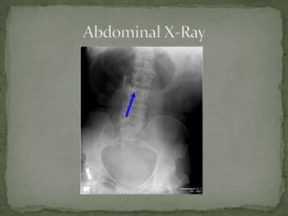
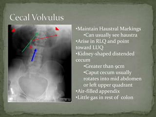
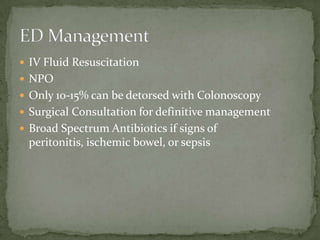
 56 year old female with     T 98.6 P 100 R 16 BP 120/80
                              Gen: normal appearing female who
 acute onset of severe        appears to be in pain
 abdominal pain and           HEENT: PERRLA, oropharynx
                              clear, NC/AT head
 nausea. Also complaining     CV: RRR no m/r/g
 of distended right sided     Pulm: CTAB
                              Abd: distended in Right abd > Left
 abdomen. She has no          abdomen, TTP diffusely, greater in
 medical history, otherwise   Right abdomen,
                              Ext: 2+ pulses, no c/c/e
 healthy female.
•Maintain Haustral Markings
    •Can usually see haustra
•Arise in RLQ and point
toward LUQ
•Kidney-shaped distended
cecum
    •Greater than 9cm
    •Caput cecum usually
    rotates into mid abdomen
    or left upper quadrant
•Air-filled appendix
•Little gas in rest of colon
 IV Fluid Resuscitation
 NPO
 Only 10-15% can be detorsed with Colonoscopy
 Surgical Consultation for definitive management
 Broad Spectrum Antibiotics if signs of
 peritonitis, ischemic bowel, or sepsis
 Misnomer: It does not just involve the cecum. the entire
  ascending colon lacks normal fusion of mesentery to
  posterior abdominal wall and you get cecocolonic twisting
  around the ileocolic vascular pedicle.
 Depending on the initial position and the length of mobile
  right colon, the cecum can be found anywhere in the
  abdomen but often found in LUQ.
 >9 cm is considered a dilated cecum. >12cm concern for
  impending perforation.
 Conservative management (No surgery) approaches 100%
  mortality rate; vascular compromise occurs early.
Cecal Volvulus   Point of torsion may be
                 identified by an area of cone
                 like narrowing, outlined by gas.
 http://radiopaedia.org/articles/caecal_volvulus
 http://www.learningradiology.com/archives05/COW%
  20152-Cecal%20Volvulus/cecalvolvuluscorrect.htm
 Schwartz, David. Emergency Radiology. 2000. pg 527-
  529.
 Tintanelli’s Emergency Medicine: A Comprehensive
  Study Guide. Chapter 79 Intestinal Obstruction

Cecal volvulus

  • 2.
     56 yearold female with T 98.6 P 100 R 16 BP 120/80 Gen: normal appearing female who acute onset of severe appears to be in pain abdominal pain and HEENT: PERRLA, oropharynx clear, NC/AT head nausea. Also complaining CV: RRR no m/r/g of distended right sided Pulm: CTAB Abd: distended in Right abd > Left abdomen. She has no abdomen, TTP diffusely, greater in medical history, otherwise Right abdomen, Ext: 2+ pulses, no c/c/e healthy female.
  • 4.
    •Maintain Haustral Markings •Can usually see haustra •Arise in RLQ and point toward LUQ •Kidney-shaped distended cecum •Greater than 9cm •Caput cecum usually rotates into mid abdomen or left upper quadrant •Air-filled appendix •Little gas in rest of colon
  • 5.
     IV FluidResuscitation  NPO  Only 10-15% can be detorsed with Colonoscopy  Surgical Consultation for definitive management  Broad Spectrum Antibiotics if signs of peritonitis, ischemic bowel, or sepsis
  • 6.
     Misnomer: Itdoes not just involve the cecum. the entire ascending colon lacks normal fusion of mesentery to posterior abdominal wall and you get cecocolonic twisting around the ileocolic vascular pedicle.  Depending on the initial position and the length of mobile right colon, the cecum can be found anywhere in the abdomen but often found in LUQ.  >9 cm is considered a dilated cecum. >12cm concern for impending perforation.  Conservative management (No surgery) approaches 100% mortality rate; vascular compromise occurs early.
  • 7.
    Cecal Volvulus Point of torsion may be identified by an area of cone like narrowing, outlined by gas.
  • 8.
     http://radiopaedia.org/articles/caecal_volvulus  http://www.learningradiology.com/archives05/COW% 20152-Cecal%20Volvulus/cecalvolvuluscorrect.htm  Schwartz, David. Emergency Radiology. 2000. pg 527- 529.  Tintanelli’s Emergency Medicine: A Comprehensive Study Guide. Chapter 79 Intestinal Obstruction