Download as PDF, PPTX

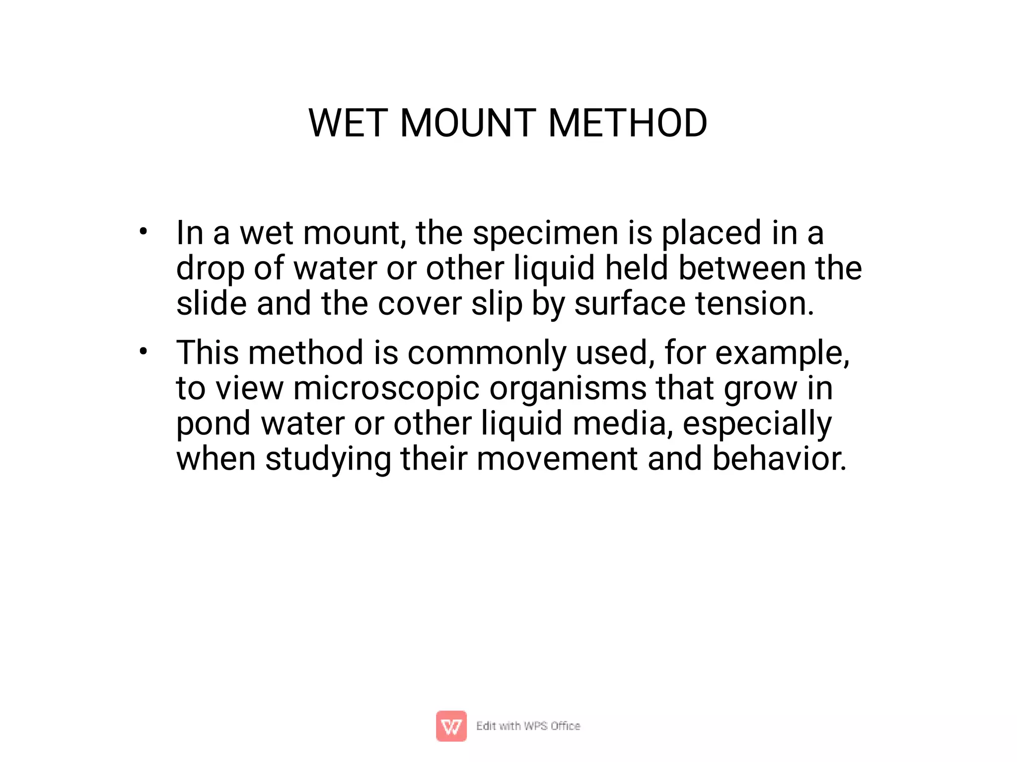
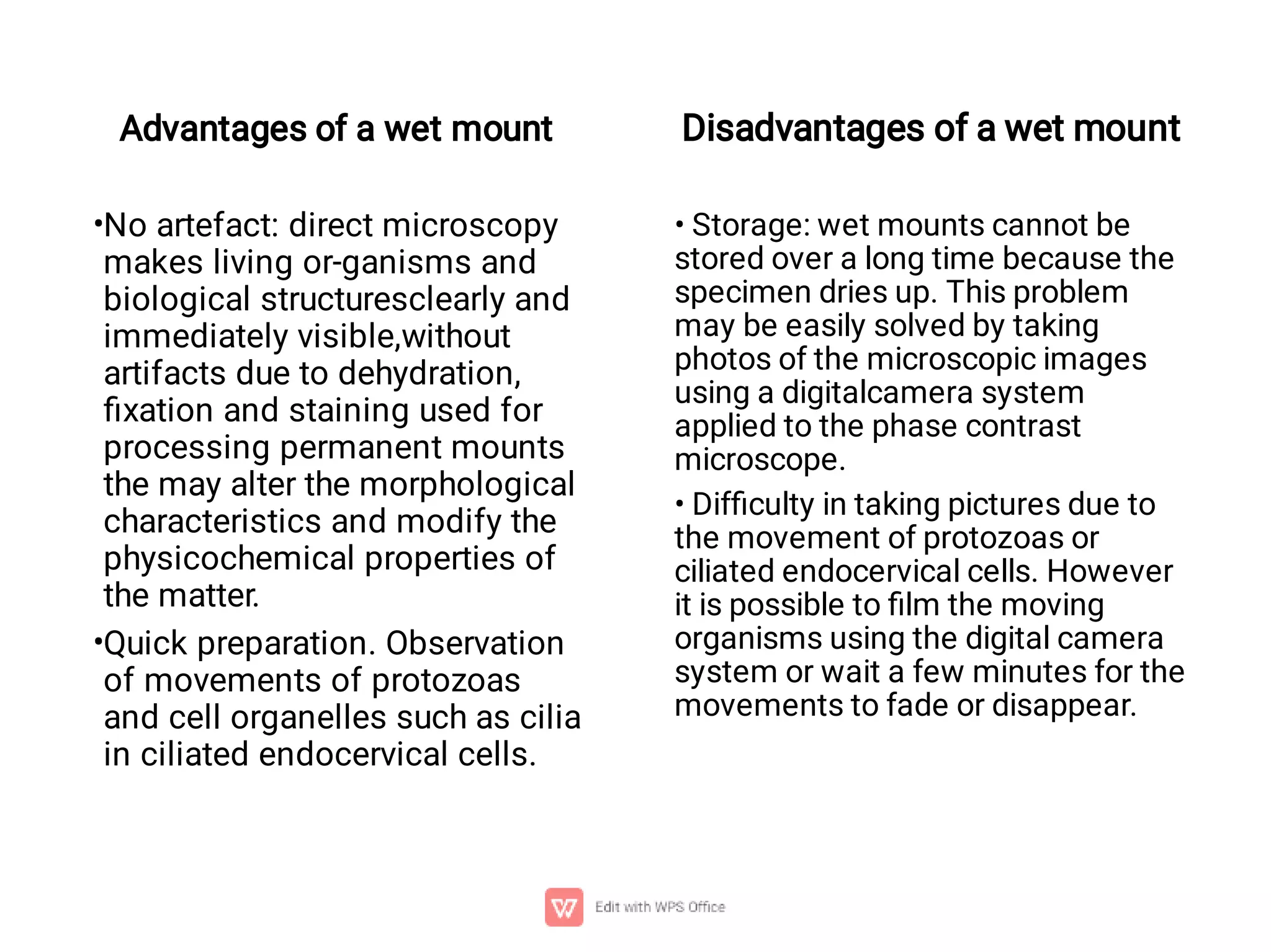
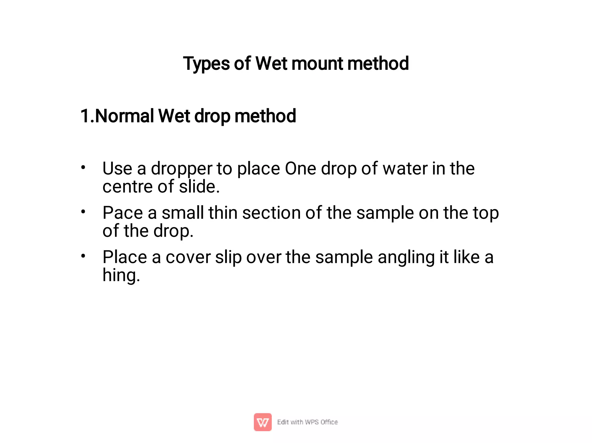
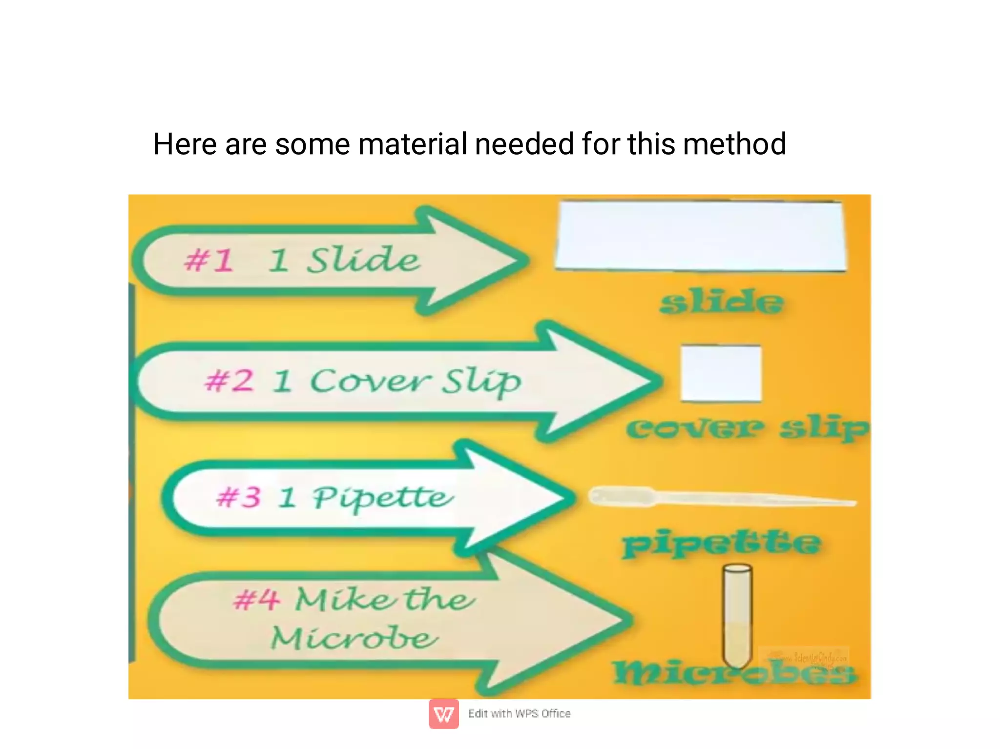
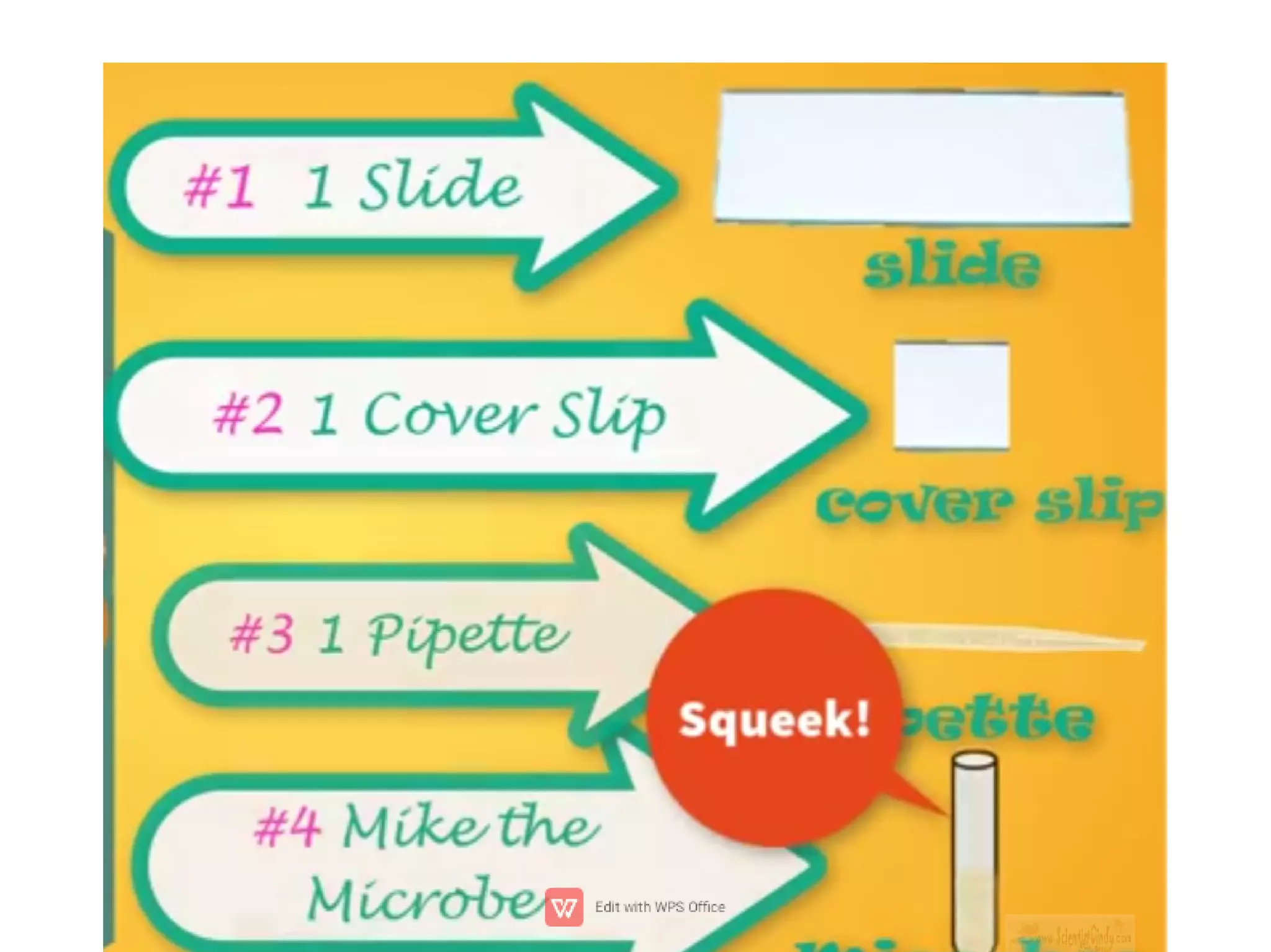

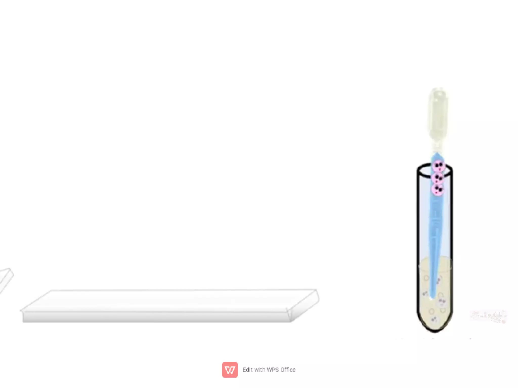

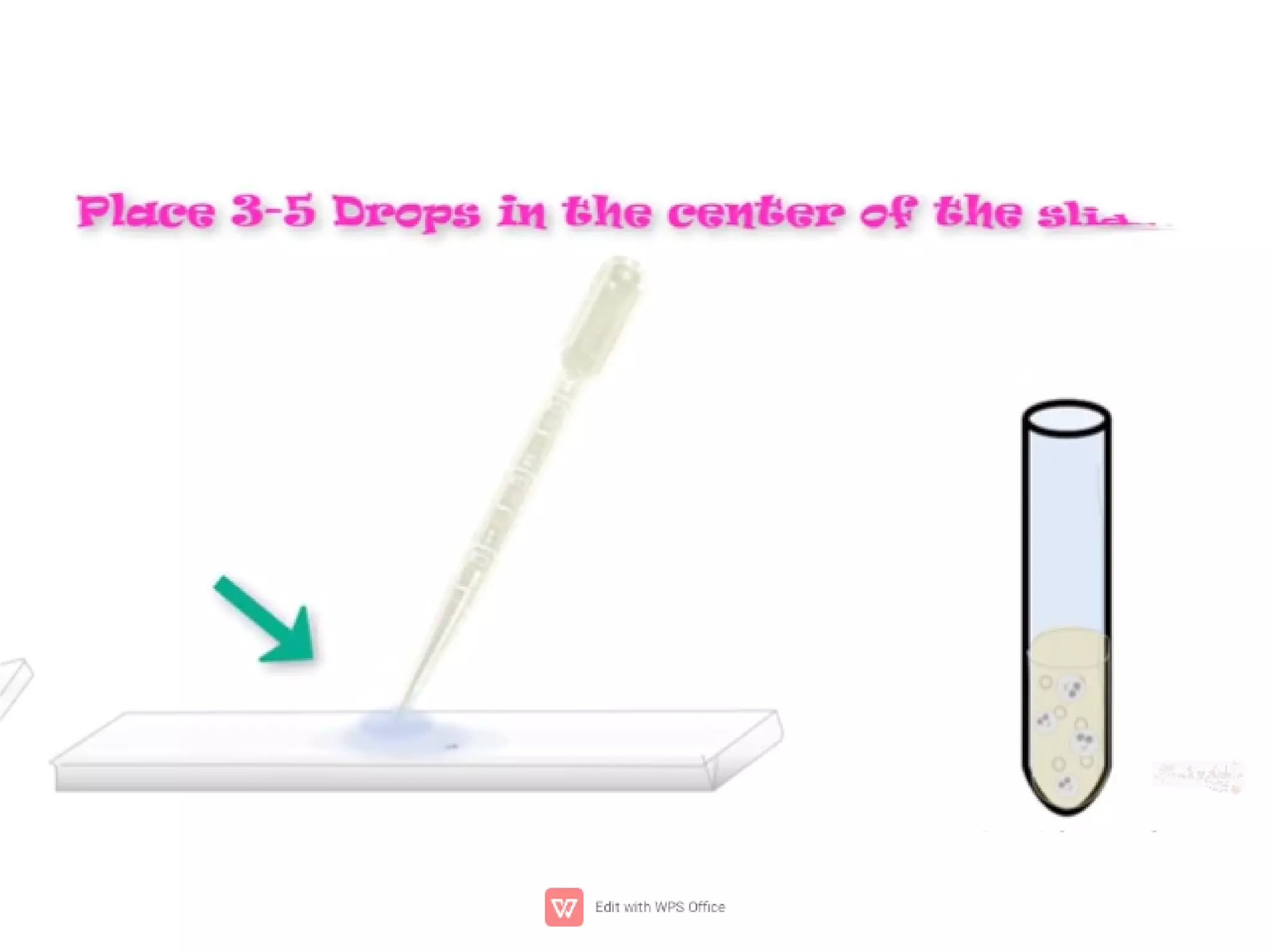
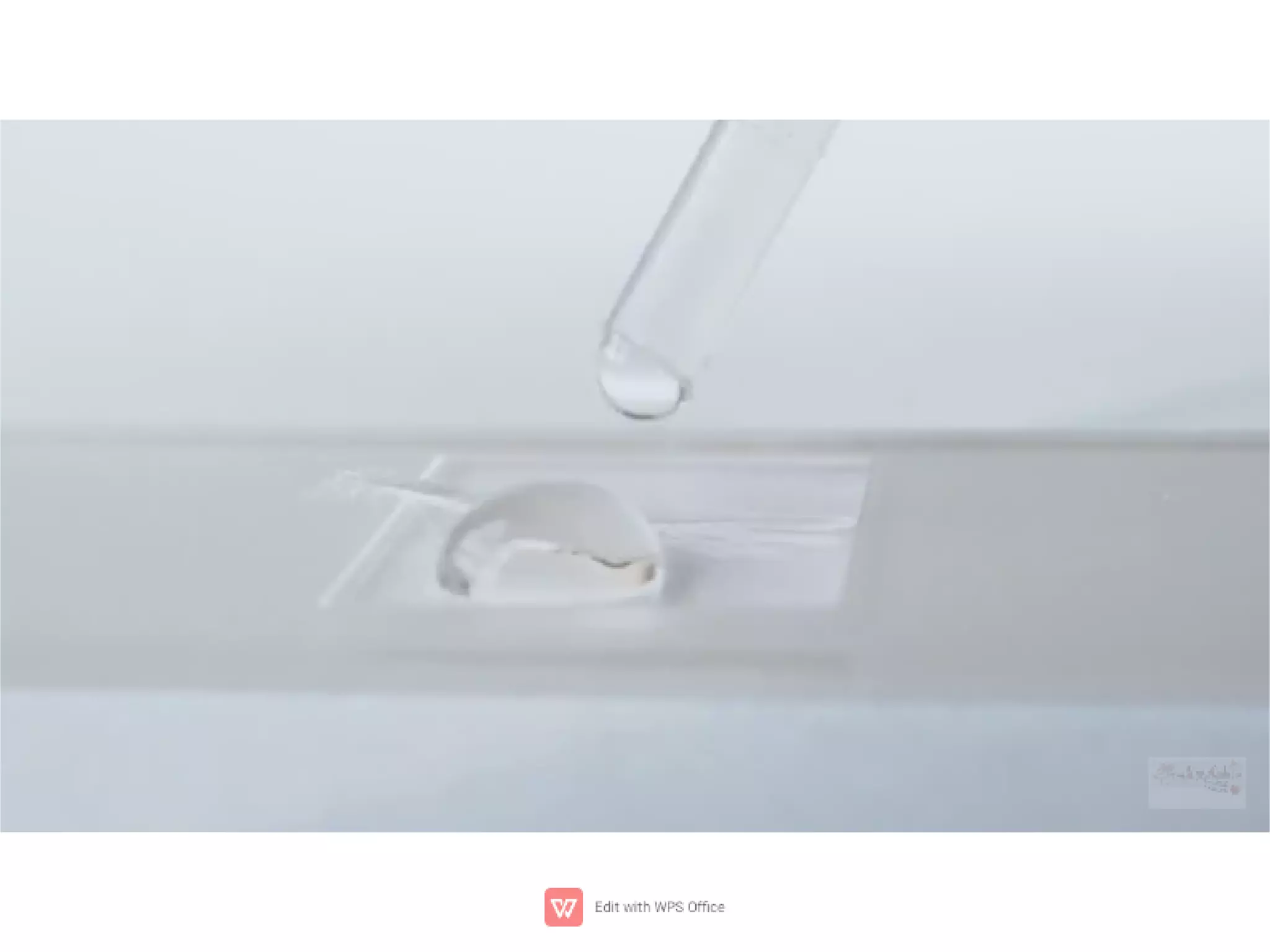

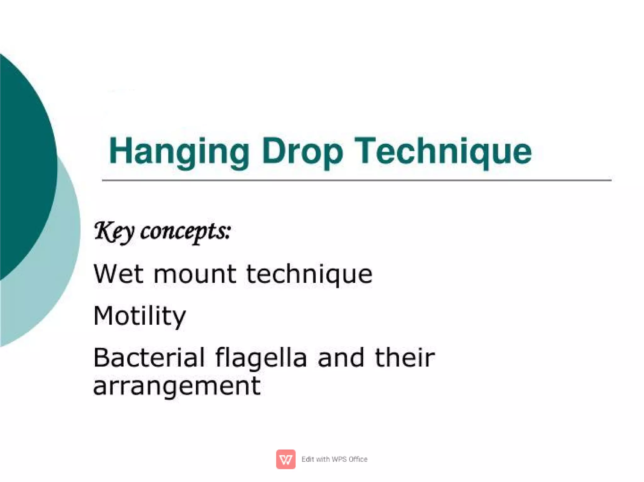
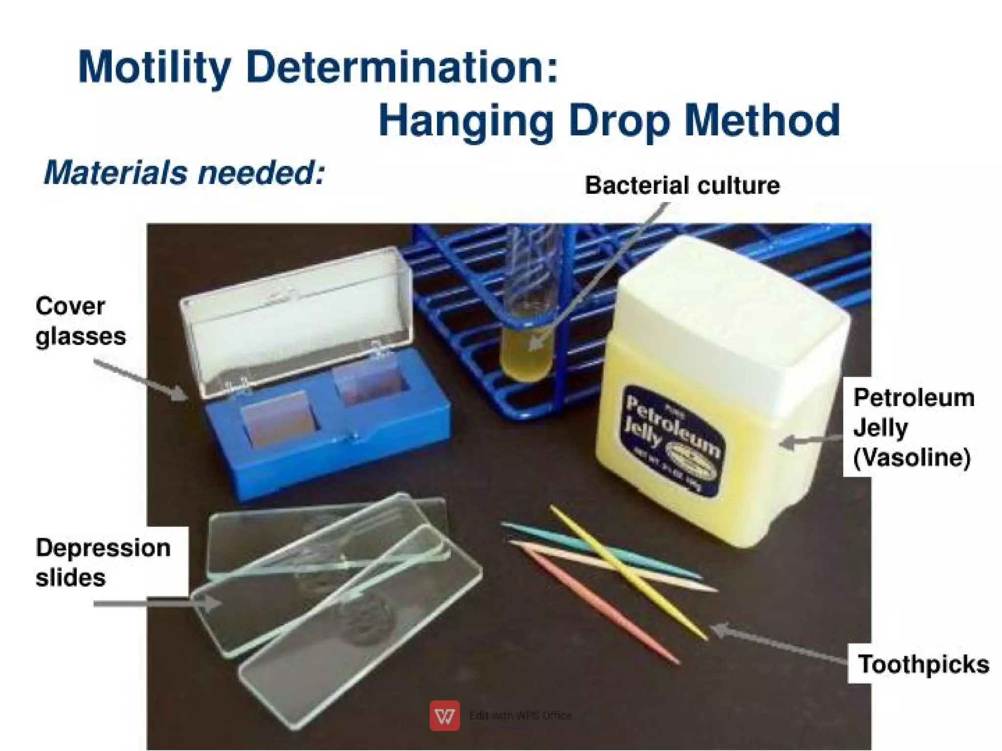
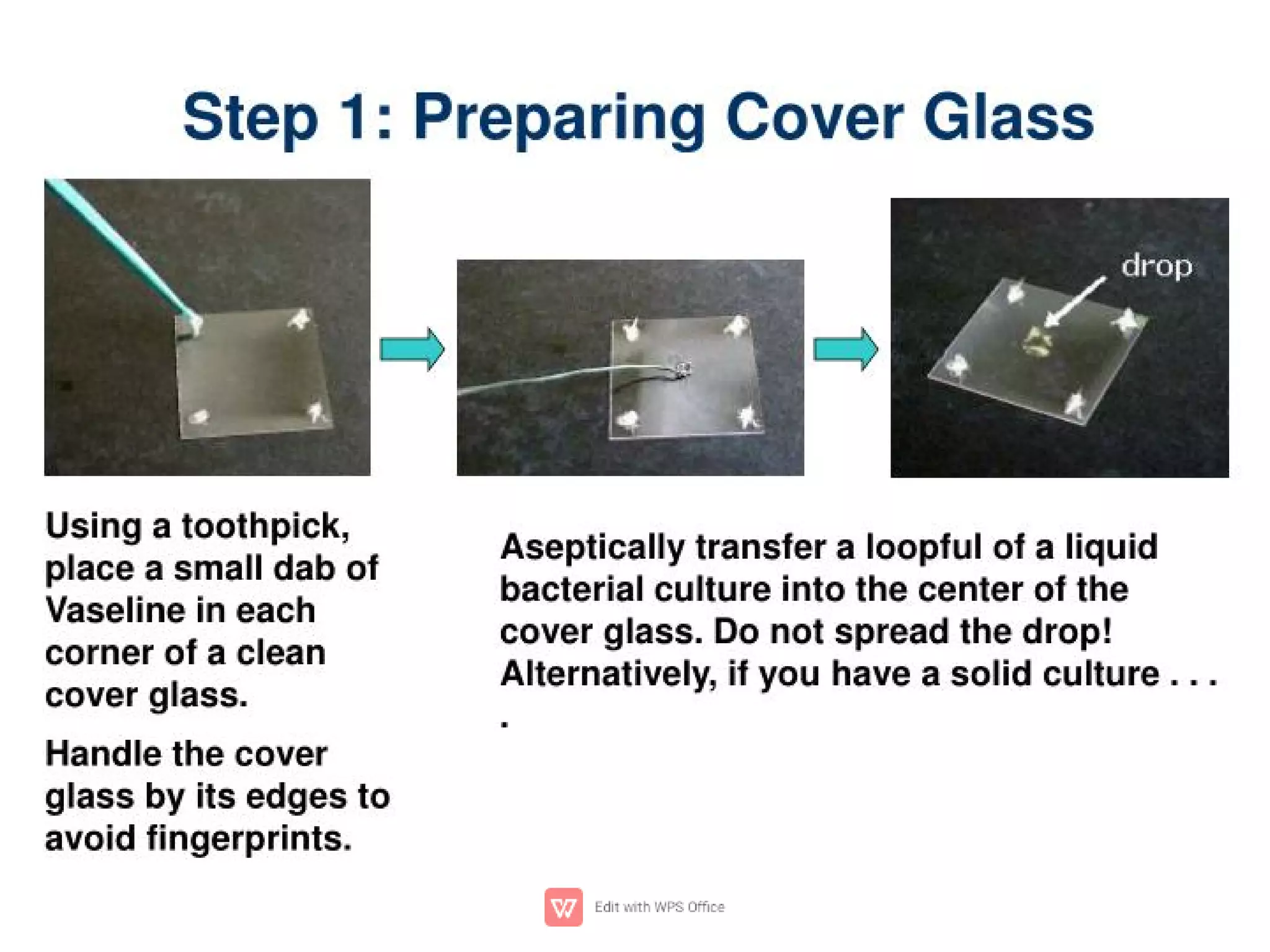

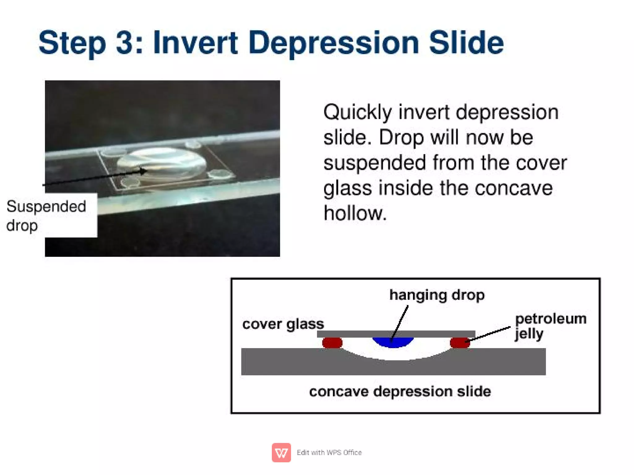
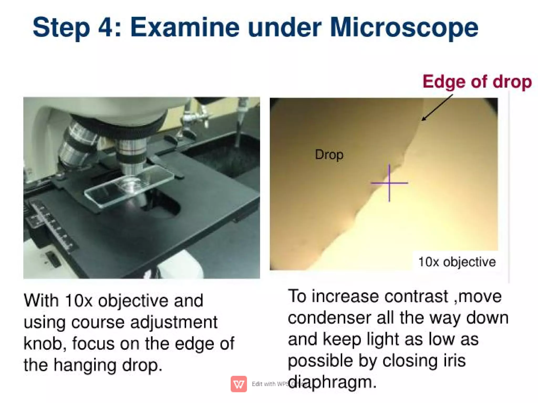
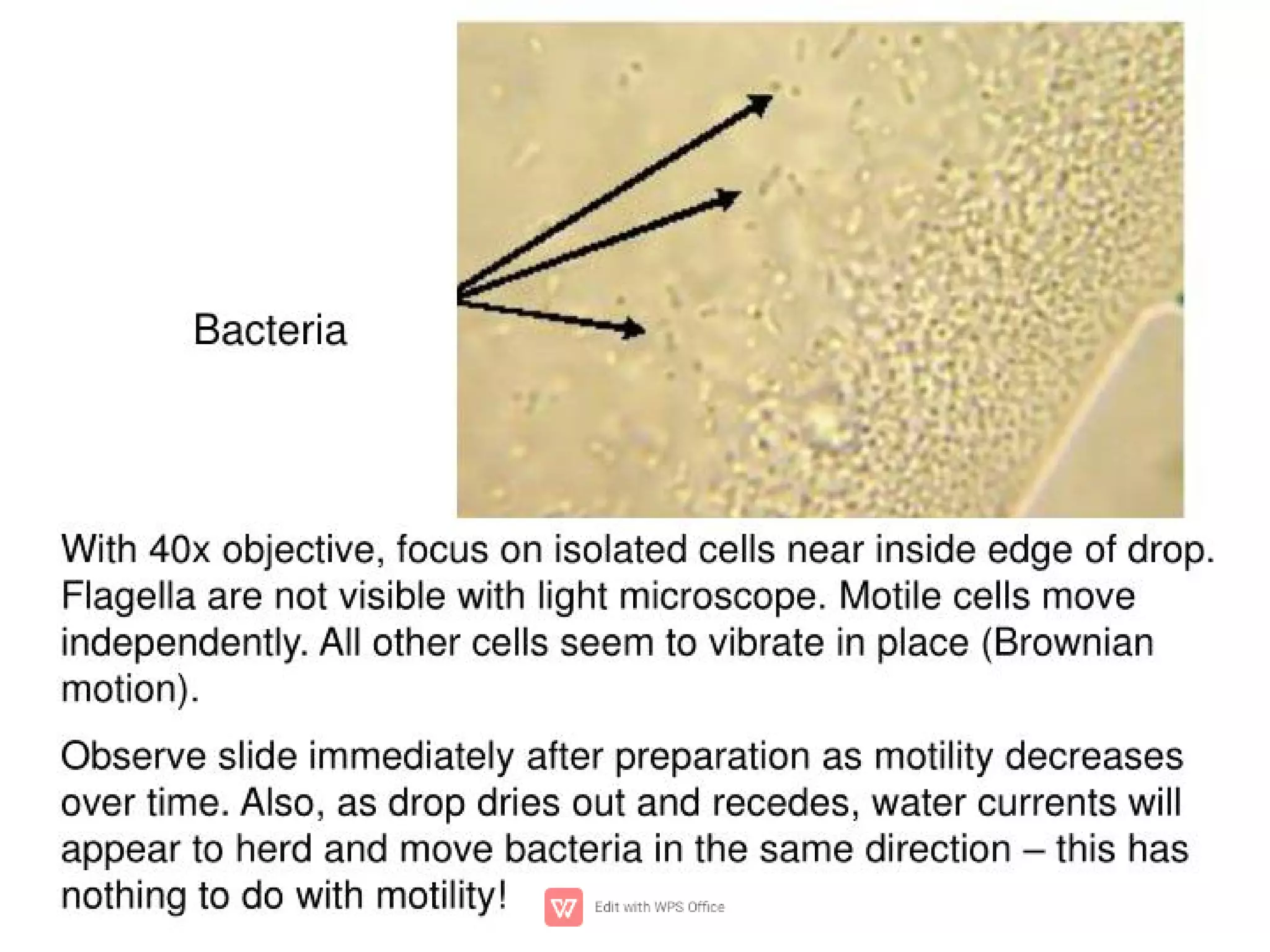
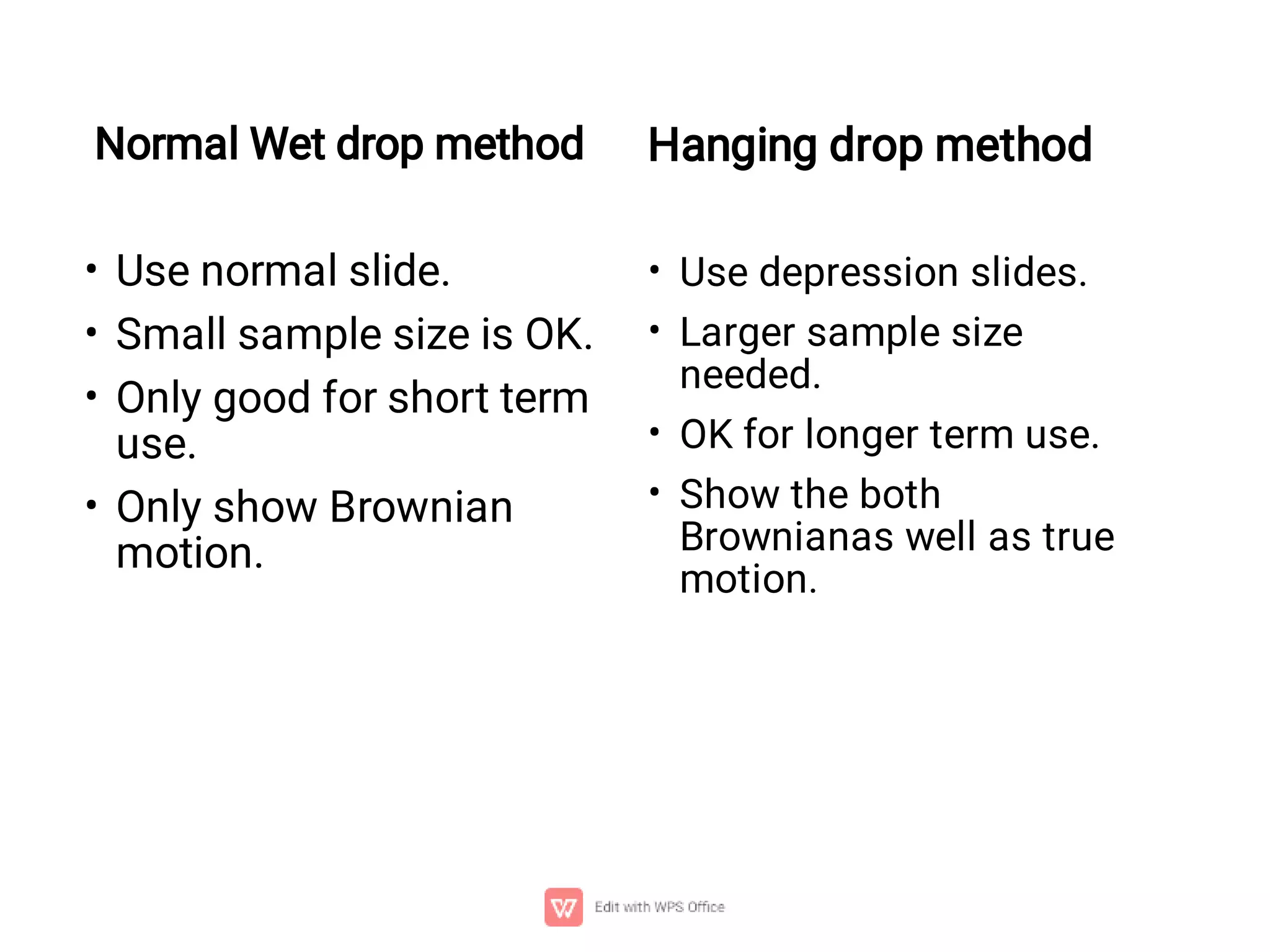


Wet mount and hanging drop methods are used to view microscopic organisms and structures in liquid. For a wet mount, a specimen is placed in a drop of liquid between a slide and cover slip. This allows viewing of movement and behavior. It has advantages of quick preparation and clear viewing without artifacts but specimens dry out over time. A hanging drop method uses a depression slide to suspend a larger sample in liquid for longer term observation of both Brownian and true motion.