SlideShare a Scribd company logo
1 of 14
Download to read offline
Caenorhabditis elegans
“A Transparent Window into Biology”
Guided by,
Ms. Sanjana Shajan
Guest Faculty
Dept. Molecular Biology,
Yuvaraja's College (Autonomous),
Mysuru
Presented by,
SHRYLI K S
VIIIth Semester
YMB17118 Molecular
Biology, Yuvaraja's
College (Autonomous),
Mysuru
11 June, 2021
Contents
 Classification
 History
 Basic Introduction
 Life Cycle
 Genetics and Genomics
 Why C. elegans?
 Epidermis: a model for extracellular matrix production, wound healing, and cell fusion
 Muscles—a model for controlling animal movement
 The digestive system—a model for organogenesis and pathogenesis
 The nervous system—a model for neurobiology
 Reproductive tissue—a model for sex-specific anatomy
 Key Discoveries
 Conclusion
 References
Classification
The binomial name fo C. elegans was first given in 1900, by Émile Maupas a French librarian, zoologist and
botanist.
Kingdom: Animalia
Phylum: Nematoda
Class: Chromadorea
Order: Rhabditida
Family: Rhabditidae
Genus: Caenorhabditis
Species: C. elegans
History
In the 1970s, Sydney Brenner and his students sought an organism wherein it might be possible to identify
each gene involved in development as well as to trace the lineage of each and every cell. Nematode
roundworms seemed like a good group to start with because embryologists such as Richard Goldschmidt
and Theodor Boveri had already shown that several nematode species have a relatively small number of
chromosomes and a small number of cells with invariant cell lineages. Brenner and his colleagues
eventually settled on C. elegans, a small (1 mm long), free-living (i.e., nonparasitic) soil nematode with
relatively few cell types. In Brenner's original vision, detailed elucidation of the development and anatomy
of C. elegans would serve as the foundation for the subsequent analysis of mutants. Both of these efforts
were completed primarily at the LMB (Laboratory of Molecular Biology) Cambridge. The transparency of
the animal allowed John Sulston, Robert Horvitz, Judith Kimble, David Hirsh, and Einhard Schierenberg to
describe every cell division starting with the single-celled zygote and ending with the adult male and
hermaphrodite. These efforts produced the first and only entire cell lineage of any multicellular organism.
During this same time, John White, Sydney Brenner, Donna Albertson, Eileen Southgate, Sam Ward, and
Nichol Thomson described the anatomy and connectivity of all 302 neurons of the adult hermaphrodite.
These projects set a standard for completeness in the understanding of the animal that has been a hallmark
of C. elegans research. Sydney Brenner, Robert Horvitz, and John Sulston were awarded the 2002 Nobel
Prize in Physiology or Medicine in part for the significance of the lineage project as a platform for
discovery of genes that orchestrated developmental decisions.
Introduction
Caenorhabditis elegans is a tiny, free-living nematode found worldwide. Newly hatched larvae are 0.25
millimeters long and adults are 1 millimeter long. Their small size means that the animals are usually
observed with either dissecting microscopes, which generally allow up to 100X magnification, or
compound microscopes, which allow up to 1000X magnification. Because C. elegans is transparent,
individual cells and subcellular details are easily visualized using Nomarski (differential interference
contrast, DIC) optics.
C. elegans has a rapid life cycle and exists primarily as a self-fertilizing hermaphrodite, although males
arise at a frequency of <0.2%. These features have helped to make C. elegans a powerful model of choice for
Fig 01: Émile Maupas
eukaryotic genetic studies. In addition, because the animal has an invariant numbers of somatic cells,
researchers have been able to track the fate of every cell between fertilization and adulthood in live animals
and to generate a complete cell lineage. Researchers have also reconstructed the shape of all C. elegans cells
from electron micrographs, including each of the 302 neurons of the adult hermaphrodite. Moreover,
because of the invariant wild-type cell lineage and neuroanatomy of C. elegans, mutations that give rise to
developmental and behavioral defects are readily identified in genetic screens. Finally, because C.
elegans was the first multicellular organism with a complete genome sequence, forward and reverse
genetics have led to the molecular identification of many key genes in developmental and cell biological
processes.
The experimental strengths and the similarities between the cellular and molecular processes present in C.
elegans and other animals across evolutionary time (metabolism, organelle structure and function, gene
regulation, protein biology, etc.) have made C. elegans an excellent organism with which to study general
metazoan biology. At least 38% of the C. elegans protein-coding genes have predicted orthologs in the
human genome, 60-80% of human genes have an ortholog in the C. elegans genome, and 40% of genes
known to be associated with human diseases have clear orthologs in the C. elegans genome. Thus, many
discoveries in C. elegans have relevance to the study of human health and disease.
Anatomy
Similar to other nematodes, C. elegans has an unsegmented, cylindrical body shape that is tapered at the
ends. It shows the typical nematode body plan with an outer tube and an inner tube separated from each
other by the pseudocoelomic space. The outer tube (body wall) consists of cuticle, hypodermis, excretory
system, neurons and muscles, and the inner tube, the pharynx, intestine and, in the adult, gonad. All of
these tissues are under an internal hydrostatic pressure, regulated by an osmoregulatory system
Life cycle
C. elegans embryogenesis takes approximately 16 hours at 20° (all of the subsequent times are also for
development at 20°). A virtually impermeable eggshell is made after fertilization, allowing the embryo to
develop completely independent of the mother. However, embryos are usually retained within the
hermaphrodite until about the 24-cell stage at which time they are laid. The hermaphrodite embryo hatches
with 558 nuclei (some nuclei are in multi-nuclear syncytia, so the cell count is lower) and becomes a first
Fig 02: C. elegans anatomy. Major anatomical
features of a hermaphrodite (A) and male (B)
viewed laterally. (A) The dorsal nerve cord
(DNC) and ventral nerve cord (VNC) run
along the entire length of the animal from
the nerve ring. Two of the four quadrants of
body wall muscles are shown. (B) The
nervous system and muscles are omitted in
this view, more clearly revealing the
pharynx and intestine. (C) Cross-section
through the anterior region of the C.
elegans hermaphrodite (location marked with
a black line in A) showing the four muscle
quadrants surrounded by the epidermis and
cuticle with the intestine and gonad housed
within the pseudocoelomic cavity.
stage (L1) larva (PAR-3, PAR-6, PKC-3, PAR-1, PAR-2, SKN-1, PAL-1, PIE-1, MED-1, MED-2, POP-1, TCF,
(pha-4) and muscles (hlh-1), MOM-2, MOM-5, GLP-1 and APX-1). The animals begin to eat and develop
through four larval stages (L1-L4). The L1 stage is ~16 hr long; the other stages are ~12 hr long. Each stage
ends with a sleep-like period of inactivity called lethargus in which a new cuticle (outer collagenous layer)
is made. Lethargus ends with the molting of the old cuticle. Approximately 12 hr after the L4 molt, adult
hermaphrodites begin producing progeny for a period of 2-3 days until they have utilized all of their self-
produced sperm; additional progeny can be generated if the sperm-depleted hermaphrodite mates with a
male. After the reproductive period, hermaphrodites can live several more weeks before dying of
senescence.
When bacteria are depleted and the animals are crowded, L2 larvae activate an alternative life cycle and
molt into an alternative L3 larval stage called the “dauer” larva (“dauer” in German means “lasting"; the
signal is actually processed by L1 animals, but its results are not seen until the so-called “L2d” stage). The
dauer larva cuticle completely surrounds the animal and plugs the mouth preventing the animal from
eating and thereby arresting development. The dauer cuticle has enhanced resistance to chemicals, so it
provides the dauer with greater protection against environmental stresses and caustic agents. Dauer larvae
can survive for many months and are the dispersal form most commonly encountered in the wild. When
the dauer larvae are transferred onto plates with bacteria, they shed their mouth plugs, molt, and continue
their development as slightly different L4 larvae.
Life span regulating genes: DAF-2, AGE-1, DAF-16, math-33, JNK-1, CST-1, HSF-1, SKN-1 and PQM-1.
Genetics and Genomics
A major reason Sydney Brenner chose to study C. elegans was the ease of genetic manipulation. C. elegans
is a well-established and powerful genetic system. Self-fertilization means that after hermaphrodites (P0s)
are mutagenized, any mutant alleles (except dominant lethals) can be maintained through self-propagation
in first-generation (F1) progeny, second-generation (F2) progeny, etc. without mating. This property makes
obtaining these progeny easy. A second genetic advantage of C. elegans is that it grows quickly. Since
animals take ~3 days at 25° (~3.5 days at 20°) to develop from fertilized eggs to adults producing their own
fertilized eggs, mutant homozygotes can be detected two generations (~1 week) after mutagenesis. In
addition, the ability to freeze and recover C. elegans makes it possible to preserve mutant strains without
Fig 06: Life Cycle of C. elegans
worrying that they have acquired unwanted suppressors, other modifiers, or additional background
mutations or have lost important mutations, particularly if they are maintained as heterozygotes.
Therefore, much less effort needs to be devoted to strain maintenance.
The use of genetics in C. elegans is in forward genetics reverse genetics and Mutant-like phenotypes can
also be obtained using RNA interference (RNAi), the use of double-stranded RNA (dsRNA) to reduce gene
activity.
C. elegans was the first multicellular eukaryotic organism to have its genome sequenced. The entire C.
elegans genome is 100 Mb and has 20,444 protein-coding genes. Both C. elegans sexes contain five autosomal
chromosomes named linkage group (LG ) I, II, III, IV, and V and the X chromosome. Compared to
vertebrate genes, C. elegans genes are relatively small with the average gene size of 3 kb due primarily to
the presence of very small introns. The chromosomes do not contain traditional centromeres; during
mitosis the microtubule spindle attaches to more than one position along the chromosome.
Why choose C. elegans?
Sharing large amounts of genetic and cellular information has been central to the success of C.
elegans research. In addition to being a powerful system for genetic studies, C. elegans has many inherent
advantages as a model for eukaryotic biology. These features include its small size, large brood size, ease of
cultivation, low maintenance expense, long-term cryopreservation, quick generation time, transparency,
invariant cell number and development, and the ability to reduce gene activity using feeding RNAi.
Although not usually mentioned, another favorable feature of C. elegans is the organisms are quite benign
to humans. In fact, because they cannot grow at body temperatures, they cannot grow in humans. Some
nematodes, e.g., Ascaris suum, induce a debilitating allergic reaction and must be studied in ventilated
hoods. As far as we are aware, allergic reactions to C. elegans have not been documented.
Studies of cell and developmental biology that use C. elegans are greatly aided by the transparency of the
animal, which allows researchers to examine development and changes due to mutations or altered
environments at the level of a single, identified cell within the context of the entire living organism. Thus,
many biological problems can be studied “in miniature” at the single-cell level, instead of large numbers of
cells in heterogeneous tissues. Transparency also enables a wealth of studies in living animals utilizing
fluorescent protein reporters. By labelling cells and proteins in living cells, fluorescent proteins enable
genetic screens to identify mutants defective in various cellular processes. In addition, fluorescent protein-
based reporters, which fluoresce in response to calcium flux, provide neuron-specific detection of calcium
flux under a fluorescent microscope, and therefore allow researchers to measure electrophysiological
activity in vivo. Furthermore, mapping of cell-cell and synaptic contacts can be accomplished by expressing
complementary fragments of GFP in different cells. Transparency also means that optogenetic tools, which
alter the activity of individual neurons, are particularly effective in C. elegans. In all of these experiments,
greater control of the animal's position and environment can be accomplished by microfluidic devices in
which individual worms are mounted in custom-designed channels allowing the application of various
compounds or other agents while simultaneously monitoring fluorescent readout of gene regulation or
electrophysiological activity by microscopy.
1. Epidermis: a model for extracellular matrix production, wound healing, and cell fusion
The outer epithelial layer, the epidermis, of the embryo undergoes a series of cell fusions to make large
multinucleate, or syncytial, epidermal cells. These cells secrete the cuticle, a protective layer of specialized
extracellular matrix (ECM) consisting primarily of collagen, lipids, and glycoproteins. The cuticle
determines the shape of the body and, through connection from the epidermis to muscle, provides
anchoring points for muscle contraction. The cuticle also serves as a model for ECM formation and function
with molecules and pathways involved in cuticle biogenesis conserved in vertebrates.
Mutations in several genes needed for cuticle formation produce visible phenotypes. Mutations in
collagen genes can result in animals that move in a corkscrew fashion [the Roller (Rol) phenotype] or that
have normal width but reduced length [the Dumpy (Dpy) phenotype]. Other mutations affect the struts
formed between layers of the adult cuticle, resulting in fluid-filled blisters [the Blister (Bli) phenotype]. Still
other mutations make the animals longer than normal [the Long (Lon) phenotype]. At the end of each
larval stage, C. elegans sheds its cuticle and secretes a new one to accommodate the growing organism.
Genes involved in cuticle formation are regulated so that the cuticle is re-established after each molt.
Studying the epidermis has led to insights in early cell movements, wound healing, cell-cell fusions, and
the establishment of epithelial layers in developing embryos. As the “skin” of C. elegans, the epidermis is a
model for the innate immune response to pathogens and for repair after a physical wound such as a needle
puncture. The wounded epidermis upregulates both Ca++ signalling to direct actin polymerization for
repair and innate immune signalling pathways to help promote survival after injury. The cell-cell fusion
events leading to the multinucleate epidermis and genes important for this process have been studied. This
work has supported the idea that repression of fusion in some cells may be just as important for proper
development as activation of cell fusion in other cells.
2. Muscles—a model for controlling animal movement
Just interior and connected to the epidermis are four quadrants (95 cells) of body-wall muscles that run
along the length of the body. The regular contraction and relaxation of the muscle cells leads to the
“elegant” sinusoidal movement of the animal. These somatic muscles are striated (although, unusually,
they are obliquely striated) and mononucleate (muscle cells do not fuse as they do in vertebrates) with
multiple sarcomeres per cell.
Genetic studies of muscle led to the first cloning and sequencing of a myosin gene (unc-54), and this
finding provided major insights into the structure of all myosins. unc-54 and many
other unc (uncoordinated) genes encoding proteins needed for muscle activity produce a “floppy
paralytic” phenotype. The study of the assembly of sarcomeres into functional muscles and, in particular,
the proteins mediating attachment to the plasma membrane has revealed many molecules in common with
vertebrate focal adhesion complexes. Genetic screens designed to understand molecules involved in muscle
contraction have also led to insights regarding muscle-wasting diseases such as Duchenne's Muscular
Dystrophy and cardiomyopathies. In addition to the body-wall muscle, C. elegans has muscles that control
eating (pharyngeal muscles), egg-laying (vulval and uterine muscles and the contractile gonad sheath),
mating (male-specific tail muscles), and defecation (enteric muscles).
3. The digestive system—a model for organogenesis and pathogenesis
Food (bacteria) enters the anterior of the animals and passes through the pharynx, a two-lobed
neuromuscular pump that grinds the food before it is passed on to the intestine for digestion. The pumping
behavior of the animals depends on the availability and the quality of the food; for example, animals pump
more when hungry and less when full. Studying pharyngeal development has been a model for
organogenesis, including how epithelial morphogenesis and cell-fate specification occur during
development. For example, the transcription factor PHA-4 plays a major regulatory role in the organ
identity of the pharynx. Animals defective in PHA-4 function do not contain a pharynx and embryos that
overexpress PHA-4 have more pharyngeal cells. The vertebrate FoxA transcription factors are homologous
to PHA-4 and are involved in gut development in many species. Intestinal development has been studied
in detail. C. elegans has served as a model to study infection and response to infection by several different
bacterial pathogens, microsporidia, and viruses that colonize the digestive system.
4. The nervous system—a model for neurobiology
C. elegans has become an important model for the study of neurobiological questions. Researchers have
identified genes and mechanisms needed for neuronal generation and specification, cell death, precursor
migration, synapse formation, chemosensory and mechanosensory transduction, neuronal degeneration,
neurite regeneration, and glial function. Researchers also have investigated a variety of behaviors, both
simple and complex, including chemotaxis, thermotaxis, several responses to touch, male-specific mating
rituals, social feeding, and both associative and non-associative learning. C. elegans also experiences periods
of restful inactivity that are similar to aspects of mammalian sleep.
The nervous system of the adult hermaphrodite has 302 neurons; that of the adult male has 383 neurons.
The majority of neuronal cell bodies are arranged in a few ganglia in the head, in the ventral cord, and in
the tail (the specialized male tail has the majority of the extra neurons). In addition to neurons, C.
elegans has several glia-like support cells, which are primarily associated with sensory neurons, but are not
as numerous as in vertebrates.
C. elegans neurons make more than 7000 chemical synapses and gap junction connections. Chemical
synapses are identified in electron micrographs by presynaptic darkening and synaptic vesicles;
postsynaptic specializations are not obvious. C. elegans uses many of the most common neurotransmitters,
including acetylcholine, glutamate, Îł-amino butyric acid (GABA), dopamine, and serotonin and has several
receptors for their detection. In addition to the chemical synaptic and gap junction connections, C.
elegans neurons are modulated by numerous neuroendocrine signals. From the perspective of cellular and
molecular detail, most of the problems of neurobiology can be studied in the worm.
In C. elegans, some individual neurons perform functions that would be performed by multiple neurons in
vertebrates. For example, individual olfactory neurons express multiple G protein-coupled odorant
receptors, rather than a single receptor as in vertebrates; each of the two mirror image bilateral ASE
chemosensory cells respond to several different ions; and the connectivity of the touch receptor neurons
suggests that their stimulation initiates several different activities. Thus, functions of multiple vertebrate
sensory neurons are compressed into a single neuron in C. elegans.
5. Reproductive tissue—a model for sex-specific anatomy
The C. elegans sexes display several obvious anatomical differences in the somatic gonad, secondary sexual
structures, and body size. The somatic gonad is located in the center of the body alongside the intestine. In
hermaphrodites the gonad consists of two mirror-image U-shaped tubes; in males the gonad consists of a
single U-shaped lobe. Both gonads house the germline where the oocytes and sperm develop. The somatic
gonad and the germline develop together during larval stages until animals reach maturity at the young
adult stage. A powerful advantage for studies of the C. elegans germline is that one can observe all stages of
meiosis at once as the germline is a visible gradient of development. Studying the germline has been a
model for meiosis, gamete development, fertilization, stem cell biology, and even tumor formation.
Secondary sexual mating structures are the vulva in hermaphrodites and the fan-shaped tail in males. The
vulva develops in the center of the epidermis on the ventral side of the hermaphrodite and is the conduit
for sperm entry from the male and egg laying from the uterus. Studying vulval morphogenesis has
provided insights into signaling by the Notch, EGF, and Wnt pathways that coordinate the spatiotemporal
development of the organ. Defects in these pathways can lead to animals with no vulva [the egg-laying
defective (Egl) phenotype] or with many vulval-like protrusions [the multivulva (Muv) phenotype]. In
many cases these animals cannot lay eggs (or mate), and their progeny develop internally, hatch within the
hermaphrodite, and create the “bag-of-worms” phenotype whereby the larvae consume the mother (a
process called endotokia matricidia). This phenotype, which also occurs when wild-type adult
hermaphrodites are starved, has been used to identify numerous egg-laying defective mutants. For
example, details of Ras GTPase/Map Kinase signaling have been elucidated through the identification of
mutants in enhancer/suppressor screens of vulval phenotypes.
Key Discoveries
C. elegans has been used as a model organism in major research areas including but not limited to
developmental biology, neurobiology, cell biology, and aging. The worm has contributed decisively to our
understanding of fundamental cellular and developmental processes including cell signaling, programmed
cell death, neuronal and synaptic properties and function, muscle biology, and sex determination.
Similarly, C. elegans has contributed enormously to our understanding of human neurodegenerative
disorders, cancer, and aging. Indeed, C. elegans has also emerged as a powerful experimental system to
study the molecular and cellular aspects of human disease in vivo. It has been estimated that about 42% of
the human disease genes have an ortholog in the genome of C. elegans including those genes associated
with Alzheimer’s disease, juvenile Parkinson’s disease, spinal muscular atrophy, hereditary non-polyposis
colon cancer, and many other age-related disorders. Modeling a human disease in a simple invertebrate,
such as C. elegans, allows the dissection of complex molecular pathways into their component parts and
thus provides meaningful insight into the pathogenesis of a complex disease phenotype. Notable examples
are:
Year Discovery References
1974 Identification of mutations that affect animal behavior Brenner 1974; Dusenberry et al. 1975; Hart 2006
1975 First description of mutations that affect thermotaxis and
mechanotransduction
Hedgecock and Russell 1975; Sulston et al. 1975; Chalfie and
Sulston 1981; Mori and Ohshima 1995,
1977 First cloning and sequencing of a myosin gene Macleod et al. 1977
1977 Genetic pathways for sex determination and dosage
compensation described
Hodgkin and Brenner 1977; Meyer 2005; Zarkower 2006
1981 Identification of mutations affecting touch sensitivity Sulston et al. 1975; Chalfie and Sulston 1981
1981 First germline stem cell niche identified Kimble and White 1981; Kimble and Crittenden 2005
1983 Notch signaling, presenilins, ternary complex, and lateral
inhibition roles in development described
Greenwald et al. 1983; Levitan and Greenwald
1995; Petcherski and Kimble 2000; Greenwald and Kovall
2012
1983 First complete metazoan cell lineage Sulston and Horvitz 1977; Kimble and Hirsh 1979; Sulston et
al. 1983
1983 Discovery of apoptosis (cell death) genes Hedgecock et al. 1983; Ellis and Horvitz 1986; Yuan and
Horvitz 1992; Yuan et al. 1993; Conradt and Xue 2005
1984 Identification of heterochronic genes Ambros and Horvitz 1984; Slack and Ruvkun 1997
1986 First complete wiring diagram of a nervous system White et al. 1986; Jarrell et al. 2012; White 2013
1987 Discovery of the first axon guidance genes Hedgecock et al. 1987, 1990; Culotti 1994
1987 Identification of role of Notch signaling in embryonic
blastomeres
Priess et al. 1987; Priess 2005
1988 Discovery of par genes, whose products affect the
asymmetric distribution of cellular components in
embryos
Kemphues et al. 1988; GĂśnczy and Rose 2005
1988 Identification of the first LIM and POU homeodomain
transcription factors
Way and Chalfie 1988; Finney et al. 1988; Hobert 2013
1990 First description of a role for RAS signaling function in Beitel et al. 1990; Han and Sternberg 1990; Sternberg
metazoan development 2005; Sundaram 2013
1993 Demonstration of a role for insulin pathway genes in
regulating lifespan
Friedman and Johnson 1988; Kenyon et al. 1993; Kimura et
al. 1997; Collins et al. 2007
1993 Identification of genes for conserved synaptic functions Gengyo-Ando et al. 1993; Richmond et al. 1999; Richmond
2007
1993 First microRNA (lin-4) and its mRNA target (lin-
14) described
Lee et al. 1993; Wightman et al. 1993 Vella and Slack 2005
1993 Identification of nonsense-mediated decay genes Pulak and Anderson 1993; Hodgkin 2005b
1994 Introduction of GFP as a biological marker Chalfie et al. 1994; Boulin et al. 2006
1994 First demonstration of specific olfactory receptor/ligand
pair
Sengupta et al. 1994; Bargmann 2006
1998 First metazoan genome sequenced C. elegans Sequencing Consortium 1998; Schwarz 2005
1998 Discovery of RNA interference (RNAi) Fire et al. 1998
2000 Conservation and ubiquity of miRNAs Pasquinelli et al. 2000
2000 Development of genome-wide RNAi screening/first full
genome-wide profiling of gene function
Fraser et al. 2000; Kamath et al. 2001
2000 Transgenerational inheritance and its mediation by piRNA Grishok et al. 2000; Ashe et al. 2012
2002 First cytoplasmic polyA polymerase (gld-2) discovered Wang et al. 2002; Kimble and Crittenden 2005
2005 First full-genome RNAi profiling of early embryogenesis SĂśnnichsen et al. 2005
2005 First use of channelrhodopsin optogenetics in an intact
animal
Nagel et al. 2005
2011 Discovery of first nematode viruses FĂŠlix et al., 2011
Conclusion
It all started when in 1963, Sydney Brenner sent a letter to Max Perutz, the chairman of the Medical
Research Council's Laboratory of Molecular Biology (LMB), detailing his concerns that the “classical
problems of molecular biology have either been solved or will be solved in the next decade” and proposing
that the future of molecular biology lies in the extension to other fields, “notably development and the
nervous system”. With the simplicity and power of prokaryotic genetics in mind, he proposed that a
nematode (round worm), Caenorhabditis briggsae, would be an ideal system in which to tackle these
problems. Later, he settled on the related nematode C. elegans as the focus of his efforts because
the elegans strain grew better than the briggsae isolate in Brenner's laboratory.
Since work on C. elegans genetics began in earnest during the 1970s, this animal has proven fruitful for
making general discoveries about cell and developmental biology. These findings have helped us
understand molecular genetic mechanisms in all animals. Evolution has maintained thousands of
conserved genes that play similar, or in some cases nearly identical, functions in nematodes and other
animals including humans. For example, the vertebrate apoptosis regulator Bcl-2 can functionally
substitute for its C. elegans ortholog ced-9. Thus, discoveries with direct relevance to understanding all
animals were made possible by what initially appeared to be an ambitious and esoteric undertaking to
define in detail the structure and genetics of an apparently simple animal. Today, C. elegans is actively
studied in over a thousand laboratories worldwide with over 1500 C. elegans research articles published
each year for the last 5 years.
References
 Scott F. Gilbert and Michael J. F. Barres, Developmental Biology Eleventh Edition, Sinauer
Associates, Inc., Publishers, Sunderland, Massachusetts U.S.A. 2016, 941pp
 Ambros, V. and H. R. Horvitz, 1984 Heterochronic mutants of the nematode Caenorhabditis
elegans. Science 226: 409-416.
 Ardiel, E. L. and C. H. Rankin, 2010 An elegant mind: learning and memory in Caenorhabditis
elegans. Learn. Mem. 17: 191-201.
 Baldi, C., S. Cho, and R. E. Ellis, 2009 Mutations in two independent pathways are sufficient to
create hermaphroditic nematodes. Science 326: 1002-1005.
 Balla, K. M. and E. R. Troemel, 2013 Caenorhabditis elegans as a model for intracellular pathogen
infection. Cell. Microbiol. 15: 1313-1322.
 Bargmann, C. I., 1998 Neurobiology of the Caenorhabditis elegans genome. Science 282: 2028-2033.
 Barr, M. M. and L. R. Garcia, 2006 Male mating behavior (June 19, 2006), WormBook, ed. The C.
elegans Research Community, WormBook, doi/10.1895/wormbook.1.78.1.
 Barrière A. and M. A. Félix, 2014 Isolation of C. elegans and related nematodes. WormBook ed.
The C. elegans Research Community, WormBook, 1-19. doi: 10.1895/wormbook.1.115.2.
 Benian, G. M. and H. F. Epstein, 2011 Caenorhabditis elegans muscle: a genetic and molecular
model for protein interactions in the heart. Circ. Res. 109: 1082-1095.
 Brenner, S., 1974 The genetics of Caenorhabditis elegans. Genetics 77: 71-94.
 Brenner, S., 1988 Foreword. The Nematode Caenorhabditis elegans, edited by W. B. Wood, Cold
Spring Harbor Laboratory Press, Cold Spring Harbor, NY.
 C. elegans Sequencing Consortium, 1998 Genome sequence of the nematode C. elegans: a platform
for investigating biology. Science 282: 2012-2018.
 Carroll, S. B., J. K. Grenier, and S. D. Weatherbee, 2004 From DNA to Diversity: Molecular Genetics
and the Evolution of Animal Design, 2nd edition, Wiley-Blackwell, Hoboken, NJ.
 Corsi A.K., Wightman B., and Chalfie M. 2015 A Transparent window into biology: A primer on
Caenorhabditis elegans. GENETICS 200: 387-407.
 Diogo, J. and A. Bratanich, 2014 The nematode Caenorhabditis elegans as a model to study viruses.
Arch. Virol. 159: 2843-2851.
 Driscoll, M. and M. Chalfie, 1992 Developmental and abnormal cell death in C. elegans. Trends
Neurosci. 15: 15-19. Dusenbery, D. B., R. E. Sheridan, and R. L. Russell, 1975 Chemotaxis-defective
mutants of the nematode Caenorhabditis elegans. Genetics 80: 297-309.
 Finney, M., G. Ruvkun, H. R. Horvitz, 1988 The C. elegans cell lineage and differentiation gene unc-
86 encodes a protein with a homeodomain and extended similarity to transcription factors. Cell 55:
757-769.
 Friedman, D. B. and T. E. Johnson, 1988 A mutation in the age-1 gene in Caenorhabditis
elegans lengthens life and reduces hermaphrodite fertility. Genetics 118: 75-86.
 Golden, J. W. and D. L. Riddle 1984 The Caenorhabditis elegans dauer larva: developmental effects of
pheromone, food, and temperature. Dev. Biol. 102: 368-378. Hedgecock, E. M., J. G. Culotti, and D.
H. Hall, 1990 The unc-5, unc-6, and unc-40 genes guide circumferential migrations of pioneer axons
and mesodermal cells on the epidermis in C. elegans. Neuron 4: 61-85.
 Jorgensen, E. M. and S. E. Mango, 2002 The art and design of genetic screens: Caenorhabditis elegans.
Nat. Rev. Genet. 3: 356-369.
 Raizen, D. M., J. E. Zimmerman, M. H. Maycock, U. D. Ta, Y. J. You et al., 2008 Lethargus is
a Caenorhabditis elegans sleep-like state. Nature 451: 569-572. (Erratum in: Nature, 2008, 453: 952)
 Rao, A. U., L. K. Carta, E. Lesuisse, and I. Hamza, 2005 Lack of heme synthesis in a free-living
eukaryote. Proc. Natl. Acad. Sci. USA, 102: 4270-4275.
 Ruvkun, G. and O. Hobert, 1998 The taxonomy of developmental control in Caenorhabditis elegans.
Science 282: 2033-2041.
 San-Miguel A. and H. Lu, 2013 Microfluidics as a tool for C. elegans research. (September 24,
2013), WormBook, ed. The C. elegans Research Community, WormBook,
doi/10.1895/wormbook.1.162.1.
 Thompson, O., M. Edgley, P. Strasbourger, S. Flibotte, B. Ewing et al., 2013 The million mutation
project: a new approach to genetics in Caenorhabditis elegans. Genome Res. 23: 1749-1762.
 Yuan, J., S. Shaham, S. Ledoux, H. M. Ellis, and H. R. Horvitz, 1993 The C. elegans cell death
gene ced-3 encodes a protein similar to mammalian interleukin-1 beta-converting enzyme. Cell 75:
641-652.
Apendix
• PAR- Partition defective protein
• Unc: Uncoordinated
• PKC: Protein Kinase C
• SKN: Skinhead Protein. Controls the fate of the EMS blastomere, the cell that generates the posterior
pharynx.
• PAL: Phenylalanine ammonia lyase (PAL) is a key enzyme of the phenylpropanoid pathway that
catalyzes the deamination of phenylalanine to trans-cinnamic acid, a precursor for the lignin and
flavonoid biosynthetic pathways.
• PIE: Pharynx and Intestine in Excess. Maternally provided PIE-1 is required for germline cell fate
determination. Functions as a repressor of RNA polymerase II-dependent gene expression in the
developing germline. Required for expression of nos-2 in P4 germline blastomere cells. Inhibits the
histone deacetylase activity of hda-1. Represses transcriptional activation of cdk-9 and cit-1.1, which
are members of the P-TEFb complex.
• MED: Mesoderm and Endoderm Determination. Enables sequence-specific DNA binding activity. Is
involved in endodermal cell fate specification and pharynx development. Located in nucleus.
• POP: POsterior Pharynx. Enables several functions, including RNA polymerase II transcription
factor binding activity; beta-catenin binding activity; and enzyme binding activity. Is involved in
several processes, including mesodermal cell fate commitment; mitochondrial unfolded protein
response; and regulation of transcription, DNA-templated.
• TCF: T-cell factor/lymphoid enhancer factor (TCF/LEF). TCF/LEFs are multifunctional proteins
that use their sequence-specific DNA-binding and context-dependent interactions to specify
which genes will be regulated by Wnts.
• HLH: Helix-loop-helix. Regulates numerous developmental events in eukaryotic cells.
• MOM: Mom protein. Required in embryonic development for endoderm specification and the
correct positioning and orientation of the mitotic spindles and division planes in blastomere cells.
Involved in cleavage axis determination.
• GLP: Germ Line Proliferation. Enables RNA polymerase II transcription factor binding activity and
transcription coactivator activity. Is involved in several processes, including maintenance of dauer;
pharyngeal muscle development; and positive regulation of cell population proliferation. Located in
cytoplasm; endomembrane system; and lateral plasma membrane. Is spanning component of
plasma membrane. Part of RNA polymerase II transcription regulator complex. Is expressed in
several structures, including AB lineage cell; anus; germ line; neurons; and somatic nervous system.
Is an ortholog of human NOTCH1 (notch receptor 1); NOTCH2 (notch receptor 2); and NOTCH3
(notch receptor 3).
• APX: Anterior Pharynx in eXcess. Is involved in several processes, including embryonic pattern
specification; lateral inhibition; and regulation of vulval development. Is expressed in several
structures, including P6.paa; P6.pap; P6.ppa; non-striated muscle; and vulva. Is an ortholog of
human DLK2 (delta like non-canonical Notch ligand 2) and DLL3 (delta like canonical Notch ligand
3).
• DAF: abnormal DAuer Formation. Enables several functions, including 14-3-3 protein binding
activity; DNA-binding transcription factor activity; and enzyme binding activity. Is involved in
several processes, including defense response to other organism; regulation of dauer larval
development; and regulation of transcription, DNA-templated. Is expressed in several structures,
including gonad; hypodermis; neurons; somatic cell; and ventral nerve cord. Is an ortholog of
human FOXO1 (forkhead box O1); FOXO3 (forkhead box O3); and FOXO4 (forkhead box O4).
• AGE: Aging alteration. Is predicted to enable 1-phosphatidylinositol-3-kinase activity. Is involved
in several processes, including dauer entry; insulin receptor signaling pathway involved in
determination of adult lifespan; and regulation of synaptic growth at neuromuscular junction. Is
expressed in amphid neurons; hypodermis; and pharyngeal-intestinal valve. Is an ortholog of
human PIK3CA (phosphatidylinositol-4,5-bisphosphate 3-kinase catalytic subunit alpha) and
PIK3CD (phosphatidylinositol-4,5-bisphosphate 3-kinase catalytic subunit delta)
• MATH: Meprin-associated Traf homology. Encodes deubiquitylase, suppresses the extended
lifespan.
• JNK: c-Jun N-terminal kinases. JNKs (c-Jun N-terminal kinases) are a group of mitogen-activated
protein kinases (MAPKs), originally called stress activated protein kinases (SAPKs), because they
are activated by various environmental stresses.
• CST: Caenorhabditis STE20-like kinase. Enables protein serine/threonine kinase activity. Is
involved in determination of adult lifespan and muscle fiber development. Is expressed in
hypodermis; sensory neurons; terminal bulb; and vulva. Is an ortholog of human STK3
(serine/threonine kinase 3).
• HSF: Heat Shock Factor. Enables calmodulin binding activity; promoter-specific chromatin binding
activity; and sequence-specific double-stranded DNA binding activity. Is involved in several
processes, including dauer larval development; defense response to other organism; and
determination of adult lifespan. Located in nuclear stress granule. Is expressed in several structures,
including body wall musculature; hypodermis; intestine; neurons; and somatic cell. Is an ortholog
of human HSF2 (heat shock transcription factor 2).
• PQM: ParaQuat (Methylviologen) responsive. Is involved in defense response to Gram-negative
bacterium; determination of adult lifespan; and innate immune response. Is expressed in head
muscle; head neurons; intestinal cell; and pseudocoelom.
• COVID: This C. elegans line expresses the human ACE2 (angiotensin-converting enzyme 2) receptor
fused with GFP under a ubiquitous promoter. Upon treatment with Spike protein, the ACE2
receptor translocates from the cell surface to cytoplasmic vesicles.
• Limitations: Simple body plan, and lack many defined organs/tissues including a brain, blood, a
defined fat cell, internal organs, and is evolutionarily distant from humans.
• C. elegans lack certain anatomical features of mammals, including a blood transport system, a blood-
brain barrier, a first-pass metabolism process in the liver, and blood filtration in the kidney, which
may be specific for certain signal pathways or epigenetic effects.
• As a model system to predict human research outcomes, the lack of DNA methylation, an
epigenetic tag that possibly has a greater function in mammals than in nematodes, is another
limitation of C. elegans.
• Forward genetics seeks to find the genetic basis of a phenotype or trait, reverse genetics seeks to
find what phenotypes arise as a result of particular genetic sequences
• DNA methylation is essential for silencing retroviral elements, regulating tissue-specific gene
expression, genomic imprinting, and X chromosome inactivation. Importantly, DNA methylation in
different genomic regions may exert different influences on gene activities based on the underlying
genetic sequence.
• C elegans relies solely on its innate immune system to defend itself against pathogens. Studies of the
nematode response to infection with various fungal and bacterial pathogens have revealed that the
innate immune system of C. elegans employs evolutionary conserved signalling pathways.
• ATP8: The P-type adenosinetriphosphatases (P-type ATPases) are a family of proteins which use
the free energy of ATP hydrolysis to drive uphill transport of ions across membranes.
• A homologous gene is a gene inherited in two species by a common ancestor. Orthologous genes
are homologous gene, where the gene diverges after a speciation event. Main function is conserved.

More Related Content

What's hot

Caenorhabditis elegans
Caenorhabditis elegansCaenorhabditis elegans
Caenorhabditis elegansShryli Shreekar
 
Drosophila Melanogaster Genome And its developmental process
Drosophila Melanogaster  Genome And its developmental processDrosophila Melanogaster  Genome And its developmental process
Drosophila Melanogaster Genome And its developmental processSubhradeep sarkar
 
Homeotic genes
Homeotic genesHomeotic genes
Homeotic genesRishikaMaji
 
Mutation & its detection
Mutation & its detectionMutation & its detection
Mutation & its detectionBHUMI GAMETI
 
Chemical method of transformation
Chemical method of transformation Chemical method of transformation
Chemical method of transformation Kristu Jayanti College
 
Environmental Control Sex Determination
Environmental Control Sex DeterminationEnvironmental Control Sex Determination
Environmental Control Sex DeterminationSimranJagirdar
 
Genomics C elegan genome and model organism
Genomics C elegan genome and model organismGenomics C elegan genome and model organism
Genomics C elegan genome and model organismiqraakbar8
 
Drosophila melanogester
Drosophila melanogesterDrosophila melanogester
Drosophila melanogesteranupisal
 
Developmental Biology
Developmental Biology Developmental Biology
Developmental Biology Dr.S.Selvaraj
 
ZEBRAFISH : AS A MODEL ORGANISM.
ZEBRAFISH : AS A MODEL ORGANISM.ZEBRAFISH : AS A MODEL ORGANISM.
ZEBRAFISH : AS A MODEL ORGANISM.Namrata Singh
 
Development of drosophila
Development of drosophilaDevelopment of drosophila
Development of drosophilaKAUSHAL SAHU
 
Gene isolation methods
Gene isolation methodsGene isolation methods
Gene isolation methodsPromila Sheoran
 
Model oranisms used in genetics
Model oranisms used in geneticsModel oranisms used in genetics
Model oranisms used in geneticsprabhakarareddy A V
 
Regeneration, Types of Regeneration,Invertebrates&Vertebrates sp. ,Mechanism,...
Regeneration, Types of Regeneration,Invertebrates&Vertebrates sp. ,Mechanism,...Regeneration, Types of Regeneration,Invertebrates&Vertebrates sp. ,Mechanism,...
Regeneration, Types of Regeneration,Invertebrates&Vertebrates sp. ,Mechanism,...SoniaBajaj10
 
Molecular event during fertilization
Molecular event during fertilizationMolecular event during fertilization
Molecular event during fertilizationKAUSHAL SAHU
 
Model organism
Model organismModel organism
Model organismpavithra M
 
Cell Adhesion Molecules(cams) and its types, Cadherins and Integrins and inte...
Cell Adhesion Molecules(cams) and its types, Cadherins and Integrins and inte...Cell Adhesion Molecules(cams) and its types, Cadherins and Integrins and inte...
Cell Adhesion Molecules(cams) and its types, Cadherins and Integrins and inte...Tahir Ali,Punjab University Lahore
 
Cell cell hybridization or somatic cell hybridization
Cell cell hybridization or somatic cell hybridizationCell cell hybridization or somatic cell hybridization
Cell cell hybridization or somatic cell hybridizationSubhradeep sarkar
 

What's hot (20)

organogenesis ,vulva formation in Caenorhabditis elegans
 organogenesis ,vulva formation in Caenorhabditis elegans  organogenesis ,vulva formation in Caenorhabditis elegans
organogenesis ,vulva formation in Caenorhabditis elegans
 
Caenorhabditis elegans
Caenorhabditis elegansCaenorhabditis elegans
Caenorhabditis elegans
 
Drosophila Melanogaster Genome And its developmental process
Drosophila Melanogaster  Genome And its developmental processDrosophila Melanogaster  Genome And its developmental process
Drosophila Melanogaster Genome And its developmental process
 
Homeotic genes
Homeotic genesHomeotic genes
Homeotic genes
 
Mutation & its detection
Mutation & its detectionMutation & its detection
Mutation & its detection
 
Chemical method of transformation
Chemical method of transformation Chemical method of transformation
Chemical method of transformation
 
Environmental Control Sex Determination
Environmental Control Sex DeterminationEnvironmental Control Sex Determination
Environmental Control Sex Determination
 
Genomics C elegan genome and model organism
Genomics C elegan genome and model organismGenomics C elegan genome and model organism
Genomics C elegan genome and model organism
 
Drosophila melanogester
Drosophila melanogesterDrosophila melanogester
Drosophila melanogester
 
Developmental Biology
Developmental Biology Developmental Biology
Developmental Biology
 
Model organisms
Model organismsModel organisms
Model organisms
 
ZEBRAFISH : AS A MODEL ORGANISM.
ZEBRAFISH : AS A MODEL ORGANISM.ZEBRAFISH : AS A MODEL ORGANISM.
ZEBRAFISH : AS A MODEL ORGANISM.
 
Development of drosophila
Development of drosophilaDevelopment of drosophila
Development of drosophila
 
Gene isolation methods
Gene isolation methodsGene isolation methods
Gene isolation methods
 
Model oranisms used in genetics
Model oranisms used in geneticsModel oranisms used in genetics
Model oranisms used in genetics
 
Regeneration, Types of Regeneration,Invertebrates&Vertebrates sp. ,Mechanism,...
Regeneration, Types of Regeneration,Invertebrates&Vertebrates sp. ,Mechanism,...Regeneration, Types of Regeneration,Invertebrates&Vertebrates sp. ,Mechanism,...
Regeneration, Types of Regeneration,Invertebrates&Vertebrates sp. ,Mechanism,...
 
Molecular event during fertilization
Molecular event during fertilizationMolecular event during fertilization
Molecular event during fertilization
 
Model organism
Model organismModel organism
Model organism
 
Cell Adhesion Molecules(cams) and its types, Cadherins and Integrins and inte...
Cell Adhesion Molecules(cams) and its types, Cadherins and Integrins and inte...Cell Adhesion Molecules(cams) and its types, Cadherins and Integrins and inte...
Cell Adhesion Molecules(cams) and its types, Cadherins and Integrins and inte...
 
Cell cell hybridization or somatic cell hybridization
Cell cell hybridization or somatic cell hybridizationCell cell hybridization or somatic cell hybridization
Cell cell hybridization or somatic cell hybridization
 

Similar to Caenorhabditis elegans

Lecture 1 2014
Lecture 1 2014 Lecture 1 2014
Lecture 1 2014 syed shahzaib
 
Rakesh ppt19.11.2022.pptx
Rakesh ppt19.11.2022.pptxRakesh ppt19.11.2022.pptx
Rakesh ppt19.11.2022.pptxRakeshSharma44116
 
1 What is the study systemGeneral information. E.g. What is a .docx
1 What is the study systemGeneral information. E.g. What is a .docx1 What is the study systemGeneral information. E.g. What is a .docx
1 What is the study systemGeneral information. E.g. What is a .docxhoney725342
 
Aniket_An Integrated Approach to Biology
Aniket_An Integrated Approach to BiologyAniket_An Integrated Approach to Biology
Aniket_An Integrated Approach to BiologyAniket Bhattacharya
 
homeostasis and soma-germline interaction
homeostasis and soma-germline interactionhomeostasis and soma-germline interaction
homeostasis and soma-germline interactionBhavya Vashisht
 
Eukaryotes Vs Animal Cells Essay
Eukaryotes Vs Animal Cells EssayEukaryotes Vs Animal Cells Essay
Eukaryotes Vs Animal Cells EssayAshley Thomas
 
Legionella Pneumophila Experiment
Legionella Pneumophila ExperimentLegionella Pneumophila Experiment
Legionella Pneumophila ExperimentDiana Turner
 
Gametogenesis, fertilization and types of eggs
Gametogenesis, fertilization and types of eggsGametogenesis, fertilization and types of eggs
Gametogenesis, fertilization and types of eggsDr. Mafatlal Kher
 
Zebrafish as a Model Organism.pptx
Zebrafish as a Model Organism.pptxZebrafish as a Model Organism.pptx
Zebrafish as a Model Organism.pptxUpendraSharmaUS1
 
Genetic Manipulation In Drosophila 221pmt004.pptx
Genetic Manipulation In Drosophila  221pmt004.pptxGenetic Manipulation In Drosophila  221pmt004.pptx
Genetic Manipulation In Drosophila 221pmt004.pptxvineetsharma945524
 
Mendelian Ratio Of Wings Essay
Mendelian Ratio Of Wings EssayMendelian Ratio Of Wings Essay
Mendelian Ratio Of Wings EssayBrianna Johnson
 
Cell biology: Classification of organisms
Cell biology: Classification of organismsCell biology: Classification of organisms
Cell biology: Classification of organismsHARINATHA REDDY ASWARTHA
 
genetics c1 lesson 1.1.pptx
genetics c1 lesson 1.1.pptxgenetics c1 lesson 1.1.pptx
genetics c1 lesson 1.1.pptxPeterSultan1
 
Classification of Insect
Classification of Insect  Classification of Insect
Classification of Insect Amani Riyadh
 
Reproductive traits in dicyemids
Reproductive traits in dicyemidsReproductive traits in dicyemids
Reproductive traits in dicyemidsdreicash
 
Chapter 6 Introduction To Cells
Chapter 6 Introduction To CellsChapter 6 Introduction To Cells
Chapter 6 Introduction To Cellstcheeseman
 
Cell biology module 1
Cell biology module 1Cell biology module 1
Cell biology module 1MSCW Mysore
 

Similar to Caenorhabditis elegans (20)

Lecture 1 2014
Lecture 1 2014 Lecture 1 2014
Lecture 1 2014
 
Rakesh ppt19.11.2022.pptx
Rakesh ppt19.11.2022.pptxRakesh ppt19.11.2022.pptx
Rakesh ppt19.11.2022.pptx
 
1 What is the study systemGeneral information. E.g. What is a .docx
1 What is the study systemGeneral information. E.g. What is a .docx1 What is the study systemGeneral information. E.g. What is a .docx
1 What is the study systemGeneral information. E.g. What is a .docx
 
Aniket_An Integrated Approach to Biology
Aniket_An Integrated Approach to BiologyAniket_An Integrated Approach to Biology
Aniket_An Integrated Approach to Biology
 
Chapter 1 cell Bio
Chapter 1 cell Bio Chapter 1 cell Bio
Chapter 1 cell Bio
 
homeostasis and soma-germline interaction
homeostasis and soma-germline interactionhomeostasis and soma-germline interaction
homeostasis and soma-germline interaction
 
Eukaryotes Vs Animal Cells Essay
Eukaryotes Vs Animal Cells EssayEukaryotes Vs Animal Cells Essay
Eukaryotes Vs Animal Cells Essay
 
Legionella Pneumophila Experiment
Legionella Pneumophila ExperimentLegionella Pneumophila Experiment
Legionella Pneumophila Experiment
 
Gametogenesis, fertilization and types of eggs
Gametogenesis, fertilization and types of eggsGametogenesis, fertilization and types of eggs
Gametogenesis, fertilization and types of eggs
 
Zebrafish as a Model Organism.pptx
Zebrafish as a Model Organism.pptxZebrafish as a Model Organism.pptx
Zebrafish as a Model Organism.pptx
 
Genetic Manipulation In Drosophila 221pmt004.pptx
Genetic Manipulation In Drosophila  221pmt004.pptxGenetic Manipulation In Drosophila  221pmt004.pptx
Genetic Manipulation In Drosophila 221pmt004.pptx
 
Mendelian Ratio Of Wings Essay
Mendelian Ratio Of Wings EssayMendelian Ratio Of Wings Essay
Mendelian Ratio Of Wings Essay
 
Cell biology: Classification of organisms
Cell biology: Classification of organismsCell biology: Classification of organisms
Cell biology: Classification of organisms
 
genetics c1 lesson 1.1.pptx
genetics c1 lesson 1.1.pptxgenetics c1 lesson 1.1.pptx
genetics c1 lesson 1.1.pptx
 
Classification of Insect
Classification of Insect  Classification of Insect
Classification of Insect
 
Evolution.ppt
Evolution.pptEvolution.ppt
Evolution.ppt
 
Reproductive traits in dicyemids
Reproductive traits in dicyemidsReproductive traits in dicyemids
Reproductive traits in dicyemids
 
Chapter 6 Introduction To Cells
Chapter 6 Introduction To CellsChapter 6 Introduction To Cells
Chapter 6 Introduction To Cells
 
Cell, Bio chapter
Cell, Bio chapterCell, Bio chapter
Cell, Bio chapter
 
Cell biology module 1
Cell biology module 1Cell biology module 1
Cell biology module 1
 

More from Shryli Shreekar

Introduction to proteomics
Introduction to proteomicsIntroduction to proteomics
Introduction to proteomicsShryli Shreekar
 
Nutrition in Fungi
Nutrition in FungiNutrition in Fungi
Nutrition in FungiShryli Shreekar
 
Phylum Porifera - Sycon
Phylum Porifera - SyconPhylum Porifera - Sycon
Phylum Porifera - SyconShryli Shreekar
 
Endangered Species
Endangered SpeciesEndangered Species
Endangered SpeciesShryli Shreekar
 
Active methylene compounds
Active methylene compoundsActive methylene compounds
Active methylene compoundsShryli Shreekar
 
Physical Methods of Sterilization
Physical Methods of SterilizationPhysical Methods of Sterilization
Physical Methods of SterilizationShryli Shreekar
 
High Altitude Adaptations
High Altitude AdaptationsHigh Altitude Adaptations
High Altitude AdaptationsShryli Shreekar
 
Voltage Clamp Technique
Voltage Clamp TechniqueVoltage Clamp Technique
Voltage Clamp TechniqueShryli Shreekar
 
Fermentation Process in Yogurt Industry
Fermentation Process in Yogurt IndustryFermentation Process in Yogurt Industry
Fermentation Process in Yogurt IndustryShryli Shreekar
 
Fermentation Process in Yogurt Industry
Fermentation Process in Yogurt IndustryFermentation Process in Yogurt Industry
Fermentation Process in Yogurt IndustryShryli Shreekar
 
Kinetically Perfect Enzymes
Kinetically Perfect EnzymesKinetically Perfect Enzymes
Kinetically Perfect EnzymesShryli Shreekar
 
DNA Amplification is a Ubiquitous Mechanism of Oncogene Activation in Lung an...
DNA Amplification is a Ubiquitous Mechanism of Oncogene Activation in Lung an...DNA Amplification is a Ubiquitous Mechanism of Oncogene Activation in Lung an...
DNA Amplification is a Ubiquitous Mechanism of Oncogene Activation in Lung an...Shryli Shreekar
 
Origin of Cancer Cells
Origin of Cancer CellsOrigin of Cancer Cells
Origin of Cancer CellsShryli Shreekar
 

More from Shryli Shreekar (18)

Introduction to proteomics
Introduction to proteomicsIntroduction to proteomics
Introduction to proteomics
 
Nutrition in Fungi
Nutrition in FungiNutrition in Fungi
Nutrition in Fungi
 
Phylum Porifera - Sycon
Phylum Porifera - SyconPhylum Porifera - Sycon
Phylum Porifera - Sycon
 
Enzyme Mechanism
Enzyme MechanismEnzyme Mechanism
Enzyme Mechanism
 
Endangered Species
Endangered SpeciesEndangered Species
Endangered Species
 
Active methylene compounds
Active methylene compoundsActive methylene compounds
Active methylene compounds
 
Physical Methods of Sterilization
Physical Methods of SterilizationPhysical Methods of Sterilization
Physical Methods of Sterilization
 
High Altitude Adaptations
High Altitude AdaptationsHigh Altitude Adaptations
High Altitude Adaptations
 
Voltage Clamp Technique
Voltage Clamp TechniqueVoltage Clamp Technique
Voltage Clamp Technique
 
Fermentation Process in Yogurt Industry
Fermentation Process in Yogurt IndustryFermentation Process in Yogurt Industry
Fermentation Process in Yogurt Industry
 
Fermentation Process in Yogurt Industry
Fermentation Process in Yogurt IndustryFermentation Process in Yogurt Industry
Fermentation Process in Yogurt Industry
 
Exonuclease
ExonucleaseExonuclease
Exonuclease
 
Kinetically Perfect Enzymes
Kinetically Perfect EnzymesKinetically Perfect Enzymes
Kinetically Perfect Enzymes
 
Origin of Cancer
Origin of CancerOrigin of Cancer
Origin of Cancer
 
Immune responses
Immune responsesImmune responses
Immune responses
 
Immune responses
Immune responsesImmune responses
Immune responses
 
DNA Amplification is a Ubiquitous Mechanism of Oncogene Activation in Lung an...
DNA Amplification is a Ubiquitous Mechanism of Oncogene Activation in Lung an...DNA Amplification is a Ubiquitous Mechanism of Oncogene Activation in Lung an...
DNA Amplification is a Ubiquitous Mechanism of Oncogene Activation in Lung an...
 
Origin of Cancer Cells
Origin of Cancer CellsOrigin of Cancer Cells
Origin of Cancer Cells
 

Recently uploaded

Welcome to GFDL for Take Your Child To Work Day
Welcome to GFDL for Take Your Child To Work DayWelcome to GFDL for Take Your Child To Work Day
Welcome to GFDL for Take Your Child To Work DayZachary Labe
 
Call Girls in Munirka Delhi 💯Call Us 🔝9953322196🔝 💯Escort.
Call Girls in Munirka Delhi 💯Call Us 🔝9953322196🔝 💯Escort.Call Girls in Munirka Delhi 💯Call Us 🔝9953322196🔝 💯Escort.
Call Girls in Munirka Delhi 💯Call Us 🔝9953322196🔝 💯Escort.aasikanpl
 
Temporomandibular joint Muscles of Mastication
Temporomandibular joint Muscles of MasticationTemporomandibular joint Muscles of Mastication
Temporomandibular joint Muscles of Masticationvidulajaib
 
Call Girls In Nihal Vihar Delhi ❤️8860477959 Looking Escorts In 24/7 Delhi NCR
Call Girls In Nihal Vihar Delhi ❤️8860477959 Looking Escorts In 24/7 Delhi NCRCall Girls In Nihal Vihar Delhi ❤️8860477959 Looking Escorts In 24/7 Delhi NCR
Call Girls In Nihal Vihar Delhi ❤️8860477959 Looking Escorts In 24/7 Delhi NCRlizamodels9
 
Call Girls in Mayapuri Delhi 💯Call Us 🔝9953322196🔝 💯Escort.
Call Girls in Mayapuri Delhi 💯Call Us 🔝9953322196🔝 💯Escort.Call Girls in Mayapuri Delhi 💯Call Us 🔝9953322196🔝 💯Escort.
Call Girls in Mayapuri Delhi 💯Call Us 🔝9953322196🔝 💯Escort.aasikanpl
 
Spermiogenesis or Spermateleosis or metamorphosis of spermatid
Spermiogenesis or Spermateleosis or metamorphosis of spermatidSpermiogenesis or Spermateleosis or metamorphosis of spermatid
Spermiogenesis or Spermateleosis or metamorphosis of spermatidSarthak Sekhar Mondal
 
TOPIC 8 Temperature and Heat.pdf physics
TOPIC 8 Temperature and Heat.pdf physicsTOPIC 8 Temperature and Heat.pdf physics
TOPIC 8 Temperature and Heat.pdf physicsssuserddc89b
 
Behavioral Disorder: Schizophrenia & it's Case Study.pdf
Behavioral Disorder: Schizophrenia & it's Case Study.pdfBehavioral Disorder: Schizophrenia & it's Case Study.pdf
Behavioral Disorder: Schizophrenia & it's Case Study.pdfSELF-EXPLANATORY
 
Evidences of Evolution General Biology 2
Evidences of Evolution General Biology 2Evidences of Evolution General Biology 2
Evidences of Evolution General Biology 2John Carlo Rollon
 
Call Girls in Aiims Metro Delhi 💯Call Us 🔝9953322196🔝 💯Escort.
Call Girls in Aiims Metro Delhi 💯Call Us 🔝9953322196🔝 💯Escort.Call Girls in Aiims Metro Delhi 💯Call Us 🔝9953322196🔝 💯Escort.
Call Girls in Aiims Metro Delhi 💯Call Us 🔝9953322196🔝 💯Escort.aasikanpl
 
Call Us ≽ 9953322196 ≼ Call Girls In Lajpat Nagar (Delhi) |
Call Us ≽ 9953322196 ≼ Call Girls In Lajpat Nagar (Delhi) |Call Us ≽ 9953322196 ≼ Call Girls In Lajpat Nagar (Delhi) |
Call Us ≽ 9953322196 ≼ Call Girls In Lajpat Nagar (Delhi) |aasikanpl
 
TOTAL CHOLESTEROL (lipid profile test).pptx
TOTAL CHOLESTEROL (lipid profile test).pptxTOTAL CHOLESTEROL (lipid profile test).pptx
TOTAL CHOLESTEROL (lipid profile test).pptxdharshini369nike
 
Scheme-of-Work-Science-Stage-4 cambridge science.docx
Scheme-of-Work-Science-Stage-4 cambridge science.docxScheme-of-Work-Science-Stage-4 cambridge science.docx
Scheme-of-Work-Science-Stage-4 cambridge science.docxyaramohamed343013
 
Forest laws, Indian forest laws, why they are important
Forest laws, Indian forest laws, why they are importantForest laws, Indian forest laws, why they are important
Forest laws, Indian forest laws, why they are importantadityabhardwaj282
 
SOLUBLE PATTERN RECOGNITION RECEPTORS.pptx
SOLUBLE PATTERN RECOGNITION RECEPTORS.pptxSOLUBLE PATTERN RECOGNITION RECEPTORS.pptx
SOLUBLE PATTERN RECOGNITION RECEPTORS.pptxkessiyaTpeter
 
Solution chemistry, Moral and Normal solutions
Solution chemistry, Moral and Normal solutionsSolution chemistry, Moral and Normal solutions
Solution chemistry, Moral and Normal solutionsHajira Mahmood
 
Vision and reflection on Mining Software Repositories research in 2024
Vision and reflection on Mining Software Repositories research in 2024Vision and reflection on Mining Software Repositories research in 2024
Vision and reflection on Mining Software Repositories research in 2024AyushiRastogi48
 
LIGHT-PHENOMENA-BY-CABUALDIONALDOPANOGANCADIENTE-CONDEZA (1).pptx
LIGHT-PHENOMENA-BY-CABUALDIONALDOPANOGANCADIENTE-CONDEZA (1).pptxLIGHT-PHENOMENA-BY-CABUALDIONALDOPANOGANCADIENTE-CONDEZA (1).pptx
LIGHT-PHENOMENA-BY-CABUALDIONALDOPANOGANCADIENTE-CONDEZA (1).pptxmalonesandreagweneth
 
RESPIRATORY ADAPTATIONS TO HYPOXIA IN HUMNAS.pptx
RESPIRATORY ADAPTATIONS TO HYPOXIA IN HUMNAS.pptxRESPIRATORY ADAPTATIONS TO HYPOXIA IN HUMNAS.pptx
RESPIRATORY ADAPTATIONS TO HYPOXIA IN HUMNAS.pptxFarihaAbdulRasheed
 
Cytokinin, mechanism and its application.pptx
Cytokinin, mechanism and its application.pptxCytokinin, mechanism and its application.pptx
Cytokinin, mechanism and its application.pptxVarshiniMK
 

Recently uploaded (20)

Welcome to GFDL for Take Your Child To Work Day
Welcome to GFDL for Take Your Child To Work DayWelcome to GFDL for Take Your Child To Work Day
Welcome to GFDL for Take Your Child To Work Day
 
Call Girls in Munirka Delhi 💯Call Us 🔝9953322196🔝 💯Escort.
Call Girls in Munirka Delhi 💯Call Us 🔝9953322196🔝 💯Escort.Call Girls in Munirka Delhi 💯Call Us 🔝9953322196🔝 💯Escort.
Call Girls in Munirka Delhi 💯Call Us 🔝9953322196🔝 💯Escort.
 
Temporomandibular joint Muscles of Mastication
Temporomandibular joint Muscles of MasticationTemporomandibular joint Muscles of Mastication
Temporomandibular joint Muscles of Mastication
 
Call Girls In Nihal Vihar Delhi ❤️8860477959 Looking Escorts In 24/7 Delhi NCR
Call Girls In Nihal Vihar Delhi ❤️8860477959 Looking Escorts In 24/7 Delhi NCRCall Girls In Nihal Vihar Delhi ❤️8860477959 Looking Escorts In 24/7 Delhi NCR
Call Girls In Nihal Vihar Delhi ❤️8860477959 Looking Escorts In 24/7 Delhi NCR
 
Call Girls in Mayapuri Delhi 💯Call Us 🔝9953322196🔝 💯Escort.
Call Girls in Mayapuri Delhi 💯Call Us 🔝9953322196🔝 💯Escort.Call Girls in Mayapuri Delhi 💯Call Us 🔝9953322196🔝 💯Escort.
Call Girls in Mayapuri Delhi 💯Call Us 🔝9953322196🔝 💯Escort.
 
Spermiogenesis or Spermateleosis or metamorphosis of spermatid
Spermiogenesis or Spermateleosis or metamorphosis of spermatidSpermiogenesis or Spermateleosis or metamorphosis of spermatid
Spermiogenesis or Spermateleosis or metamorphosis of spermatid
 
TOPIC 8 Temperature and Heat.pdf physics
TOPIC 8 Temperature and Heat.pdf physicsTOPIC 8 Temperature and Heat.pdf physics
TOPIC 8 Temperature and Heat.pdf physics
 
Behavioral Disorder: Schizophrenia & it's Case Study.pdf
Behavioral Disorder: Schizophrenia & it's Case Study.pdfBehavioral Disorder: Schizophrenia & it's Case Study.pdf
Behavioral Disorder: Schizophrenia & it's Case Study.pdf
 
Evidences of Evolution General Biology 2
Evidences of Evolution General Biology 2Evidences of Evolution General Biology 2
Evidences of Evolution General Biology 2
 
Call Girls in Aiims Metro Delhi 💯Call Us 🔝9953322196🔝 💯Escort.
Call Girls in Aiims Metro Delhi 💯Call Us 🔝9953322196🔝 💯Escort.Call Girls in Aiims Metro Delhi 💯Call Us 🔝9953322196🔝 💯Escort.
Call Girls in Aiims Metro Delhi 💯Call Us 🔝9953322196🔝 💯Escort.
 
Call Us ≽ 9953322196 ≼ Call Girls In Lajpat Nagar (Delhi) |
Call Us ≽ 9953322196 ≼ Call Girls In Lajpat Nagar (Delhi) |Call Us ≽ 9953322196 ≼ Call Girls In Lajpat Nagar (Delhi) |
Call Us ≽ 9953322196 ≼ Call Girls In Lajpat Nagar (Delhi) |
 
TOTAL CHOLESTEROL (lipid profile test).pptx
TOTAL CHOLESTEROL (lipid profile test).pptxTOTAL CHOLESTEROL (lipid profile test).pptx
TOTAL CHOLESTEROL (lipid profile test).pptx
 
Scheme-of-Work-Science-Stage-4 cambridge science.docx
Scheme-of-Work-Science-Stage-4 cambridge science.docxScheme-of-Work-Science-Stage-4 cambridge science.docx
Scheme-of-Work-Science-Stage-4 cambridge science.docx
 
Forest laws, Indian forest laws, why they are important
Forest laws, Indian forest laws, why they are importantForest laws, Indian forest laws, why they are important
Forest laws, Indian forest laws, why they are important
 
SOLUBLE PATTERN RECOGNITION RECEPTORS.pptx
SOLUBLE PATTERN RECOGNITION RECEPTORS.pptxSOLUBLE PATTERN RECOGNITION RECEPTORS.pptx
SOLUBLE PATTERN RECOGNITION RECEPTORS.pptx
 
Solution chemistry, Moral and Normal solutions
Solution chemistry, Moral and Normal solutionsSolution chemistry, Moral and Normal solutions
Solution chemistry, Moral and Normal solutions
 
Vision and reflection on Mining Software Repositories research in 2024
Vision and reflection on Mining Software Repositories research in 2024Vision and reflection on Mining Software Repositories research in 2024
Vision and reflection on Mining Software Repositories research in 2024
 
LIGHT-PHENOMENA-BY-CABUALDIONALDOPANOGANCADIENTE-CONDEZA (1).pptx
LIGHT-PHENOMENA-BY-CABUALDIONALDOPANOGANCADIENTE-CONDEZA (1).pptxLIGHT-PHENOMENA-BY-CABUALDIONALDOPANOGANCADIENTE-CONDEZA (1).pptx
LIGHT-PHENOMENA-BY-CABUALDIONALDOPANOGANCADIENTE-CONDEZA (1).pptx
 
RESPIRATORY ADAPTATIONS TO HYPOXIA IN HUMNAS.pptx
RESPIRATORY ADAPTATIONS TO HYPOXIA IN HUMNAS.pptxRESPIRATORY ADAPTATIONS TO HYPOXIA IN HUMNAS.pptx
RESPIRATORY ADAPTATIONS TO HYPOXIA IN HUMNAS.pptx
 
Cytokinin, mechanism and its application.pptx
Cytokinin, mechanism and its application.pptxCytokinin, mechanism and its application.pptx
Cytokinin, mechanism and its application.pptx
 

Caenorhabditis elegans

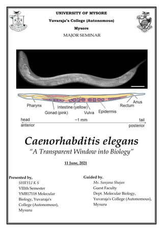
  • 1. Caenorhabditis elegans “A Transparent Window into Biology” Guided by, Ms. Sanjana Shajan Guest Faculty Dept. Molecular Biology, Yuvaraja's College (Autonomous), Mysuru Presented by, SHRYLI K S VIIIth Semester YMB17118 Molecular Biology, Yuvaraja's College (Autonomous), Mysuru 11 June, 2021
  • 2. Contents  Classification  History  Basic Introduction  Life Cycle  Genetics and Genomics  Why C. elegans?  Epidermis: a model for extracellular matrix production, wound healing, and cell fusion  Muscles—a model for controlling animal movement  The digestive system—a model for organogenesis and pathogenesis  The nervous system—a model for neurobiology  Reproductive tissue—a model for sex-specific anatomy  Key Discoveries  Conclusion  References
  • 3. Classification The binomial name fo C. elegans was first given in 1900, by Émile Maupas a French librarian, zoologist and botanist. Kingdom: Animalia Phylum: Nematoda Class: Chromadorea Order: Rhabditida Family: Rhabditidae Genus: Caenorhabditis Species: C. elegans History In the 1970s, Sydney Brenner and his students sought an organism wherein it might be possible to identify each gene involved in development as well as to trace the lineage of each and every cell. Nematode roundworms seemed like a good group to start with because embryologists such as Richard Goldschmidt and Theodor Boveri had already shown that several nematode species have a relatively small number of chromosomes and a small number of cells with invariant cell lineages. Brenner and his colleagues eventually settled on C. elegans, a small (1 mm long), free-living (i.e., nonparasitic) soil nematode with relatively few cell types. In Brenner's original vision, detailed elucidation of the development and anatomy of C. elegans would serve as the foundation for the subsequent analysis of mutants. Both of these efforts were completed primarily at the LMB (Laboratory of Molecular Biology) Cambridge. The transparency of the animal allowed John Sulston, Robert Horvitz, Judith Kimble, David Hirsh, and Einhard Schierenberg to describe every cell division starting with the single-celled zygote and ending with the adult male and hermaphrodite. These efforts produced the first and only entire cell lineage of any multicellular organism. During this same time, John White, Sydney Brenner, Donna Albertson, Eileen Southgate, Sam Ward, and Nichol Thomson described the anatomy and connectivity of all 302 neurons of the adult hermaphrodite. These projects set a standard for completeness in the understanding of the animal that has been a hallmark of C. elegans research. Sydney Brenner, Robert Horvitz, and John Sulston were awarded the 2002 Nobel Prize in Physiology or Medicine in part for the significance of the lineage project as a platform for discovery of genes that orchestrated developmental decisions. Introduction Caenorhabditis elegans is a tiny, free-living nematode found worldwide. Newly hatched larvae are 0.25 millimeters long and adults are 1 millimeter long. Their small size means that the animals are usually observed with either dissecting microscopes, which generally allow up to 100X magnification, or compound microscopes, which allow up to 1000X magnification. Because C. elegans is transparent, individual cells and subcellular details are easily visualized using Nomarski (differential interference contrast, DIC) optics. C. elegans has a rapid life cycle and exists primarily as a self-fertilizing hermaphrodite, although males arise at a frequency of <0.2%. These features have helped to make C. elegans a powerful model of choice for Fig 01: Émile Maupas
  • 4. eukaryotic genetic studies. In addition, because the animal has an invariant numbers of somatic cells, researchers have been able to track the fate of every cell between fertilization and adulthood in live animals and to generate a complete cell lineage. Researchers have also reconstructed the shape of all C. elegans cells from electron micrographs, including each of the 302 neurons of the adult hermaphrodite. Moreover, because of the invariant wild-type cell lineage and neuroanatomy of C. elegans, mutations that give rise to developmental and behavioral defects are readily identified in genetic screens. Finally, because C. elegans was the first multicellular organism with a complete genome sequence, forward and reverse genetics have led to the molecular identification of many key genes in developmental and cell biological processes. The experimental strengths and the similarities between the cellular and molecular processes present in C. elegans and other animals across evolutionary time (metabolism, organelle structure and function, gene regulation, protein biology, etc.) have made C. elegans an excellent organism with which to study general metazoan biology. At least 38% of the C. elegans protein-coding genes have predicted orthologs in the human genome, 60-80% of human genes have an ortholog in the C. elegans genome, and 40% of genes known to be associated with human diseases have clear orthologs in the C. elegans genome. Thus, many discoveries in C. elegans have relevance to the study of human health and disease. Anatomy Similar to other nematodes, C. elegans has an unsegmented, cylindrical body shape that is tapered at the ends. It shows the typical nematode body plan with an outer tube and an inner tube separated from each other by the pseudocoelomic space. The outer tube (body wall) consists of cuticle, hypodermis, excretory system, neurons and muscles, and the inner tube, the pharynx, intestine and, in the adult, gonad. All of these tissues are under an internal hydrostatic pressure, regulated by an osmoregulatory system Life cycle C. elegans embryogenesis takes approximately 16 hours at 20° (all of the subsequent times are also for development at 20°). A virtually impermeable eggshell is made after fertilization, allowing the embryo to develop completely independent of the mother. However, embryos are usually retained within the hermaphrodite until about the 24-cell stage at which time they are laid. The hermaphrodite embryo hatches with 558 nuclei (some nuclei are in multi-nuclear syncytia, so the cell count is lower) and becomes a first Fig 02: C. elegans anatomy. Major anatomical features of a hermaphrodite (A) and male (B) viewed laterally. (A) The dorsal nerve cord (DNC) and ventral nerve cord (VNC) run along the entire length of the animal from the nerve ring. Two of the four quadrants of body wall muscles are shown. (B) The nervous system and muscles are omitted in this view, more clearly revealing the pharynx and intestine. (C) Cross-section through the anterior region of the C. elegans hermaphrodite (location marked with a black line in A) showing the four muscle quadrants surrounded by the epidermis and cuticle with the intestine and gonad housed within the pseudocoelomic cavity.
  • 5. stage (L1) larva (PAR-3, PAR-6, PKC-3, PAR-1, PAR-2, SKN-1, PAL-1, PIE-1, MED-1, MED-2, POP-1, TCF, (pha-4) and muscles (hlh-1), MOM-2, MOM-5, GLP-1 and APX-1). The animals begin to eat and develop through four larval stages (L1-L4). The L1 stage is ~16 hr long; the other stages are ~12 hr long. Each stage ends with a sleep-like period of inactivity called lethargus in which a new cuticle (outer collagenous layer) is made. Lethargus ends with the molting of the old cuticle. Approximately 12 hr after the L4 molt, adult hermaphrodites begin producing progeny for a period of 2-3 days until they have utilized all of their self- produced sperm; additional progeny can be generated if the sperm-depleted hermaphrodite mates with a male. After the reproductive period, hermaphrodites can live several more weeks before dying of senescence. When bacteria are depleted and the animals are crowded, L2 larvae activate an alternative life cycle and molt into an alternative L3 larval stage called the “dauer” larva (“dauer” in German means “lasting"; the signal is actually processed by L1 animals, but its results are not seen until the so-called “L2d” stage). The dauer larva cuticle completely surrounds the animal and plugs the mouth preventing the animal from eating and thereby arresting development. The dauer cuticle has enhanced resistance to chemicals, so it provides the dauer with greater protection against environmental stresses and caustic agents. Dauer larvae can survive for many months and are the dispersal form most commonly encountered in the wild. When the dauer larvae are transferred onto plates with bacteria, they shed their mouth plugs, molt, and continue their development as slightly different L4 larvae. Life span regulating genes: DAF-2, AGE-1, DAF-16, math-33, JNK-1, CST-1, HSF-1, SKN-1 and PQM-1. Genetics and Genomics A major reason Sydney Brenner chose to study C. elegans was the ease of genetic manipulation. C. elegans is a well-established and powerful genetic system. Self-fertilization means that after hermaphrodites (P0s) are mutagenized, any mutant alleles (except dominant lethals) can be maintained through self-propagation in first-generation (F1) progeny, second-generation (F2) progeny, etc. without mating. This property makes obtaining these progeny easy. A second genetic advantage of C. elegans is that it grows quickly. Since animals take ~3 days at 25° (~3.5 days at 20°) to develop from fertilized eggs to adults producing their own fertilized eggs, mutant homozygotes can be detected two generations (~1 week) after mutagenesis. In addition, the ability to freeze and recover C. elegans makes it possible to preserve mutant strains without Fig 06: Life Cycle of C. elegans
  • 6. worrying that they have acquired unwanted suppressors, other modifiers, or additional background mutations or have lost important mutations, particularly if they are maintained as heterozygotes. Therefore, much less effort needs to be devoted to strain maintenance. The use of genetics in C. elegans is in forward genetics reverse genetics and Mutant-like phenotypes can also be obtained using RNA interference (RNAi), the use of double-stranded RNA (dsRNA) to reduce gene activity. C. elegans was the first multicellular eukaryotic organism to have its genome sequenced. The entire C. elegans genome is 100 Mb and has 20,444 protein-coding genes. Both C. elegans sexes contain five autosomal chromosomes named linkage group (LG ) I, II, III, IV, and V and the X chromosome. Compared to vertebrate genes, C. elegans genes are relatively small with the average gene size of 3 kb due primarily to the presence of very small introns. The chromosomes do not contain traditional centromeres; during mitosis the microtubule spindle attaches to more than one position along the chromosome. Why choose C. elegans? Sharing large amounts of genetic and cellular information has been central to the success of C. elegans research. In addition to being a powerful system for genetic studies, C. elegans has many inherent advantages as a model for eukaryotic biology. These features include its small size, large brood size, ease of cultivation, low maintenance expense, long-term cryopreservation, quick generation time, transparency, invariant cell number and development, and the ability to reduce gene activity using feeding RNAi. Although not usually mentioned, another favorable feature of C. elegans is the organisms are quite benign to humans. In fact, because they cannot grow at body temperatures, they cannot grow in humans. Some nematodes, e.g., Ascaris suum, induce a debilitating allergic reaction and must be studied in ventilated hoods. As far as we are aware, allergic reactions to C. elegans have not been documented. Studies of cell and developmental biology that use C. elegans are greatly aided by the transparency of the animal, which allows researchers to examine development and changes due to mutations or altered environments at the level of a single, identified cell within the context of the entire living organism. Thus, many biological problems can be studied “in miniature” at the single-cell level, instead of large numbers of cells in heterogeneous tissues. Transparency also enables a wealth of studies in living animals utilizing fluorescent protein reporters. By labelling cells and proteins in living cells, fluorescent proteins enable genetic screens to identify mutants defective in various cellular processes. In addition, fluorescent protein- based reporters, which fluoresce in response to calcium flux, provide neuron-specific detection of calcium flux under a fluorescent microscope, and therefore allow researchers to measure electrophysiological activity in vivo. Furthermore, mapping of cell-cell and synaptic contacts can be accomplished by expressing complementary fragments of GFP in different cells. Transparency also means that optogenetic tools, which alter the activity of individual neurons, are particularly effective in C. elegans. In all of these experiments, greater control of the animal's position and environment can be accomplished by microfluidic devices in which individual worms are mounted in custom-designed channels allowing the application of various compounds or other agents while simultaneously monitoring fluorescent readout of gene regulation or electrophysiological activity by microscopy. 1. Epidermis: a model for extracellular matrix production, wound healing, and cell fusion The outer epithelial layer, the epidermis, of the embryo undergoes a series of cell fusions to make large multinucleate, or syncytial, epidermal cells. These cells secrete the cuticle, a protective layer of specialized extracellular matrix (ECM) consisting primarily of collagen, lipids, and glycoproteins. The cuticle determines the shape of the body and, through connection from the epidermis to muscle, provides
  • 7. anchoring points for muscle contraction. The cuticle also serves as a model for ECM formation and function with molecules and pathways involved in cuticle biogenesis conserved in vertebrates. Mutations in several genes needed for cuticle formation produce visible phenotypes. Mutations in collagen genes can result in animals that move in a corkscrew fashion [the Roller (Rol) phenotype] or that have normal width but reduced length [the Dumpy (Dpy) phenotype]. Other mutations affect the struts formed between layers of the adult cuticle, resulting in fluid-filled blisters [the Blister (Bli) phenotype]. Still other mutations make the animals longer than normal [the Long (Lon) phenotype]. At the end of each larval stage, C. elegans sheds its cuticle and secretes a new one to accommodate the growing organism. Genes involved in cuticle formation are regulated so that the cuticle is re-established after each molt. Studying the epidermis has led to insights in early cell movements, wound healing, cell-cell fusions, and the establishment of epithelial layers in developing embryos. As the “skin” of C. elegans, the epidermis is a model for the innate immune response to pathogens and for repair after a physical wound such as a needle puncture. The wounded epidermis upregulates both Ca++ signalling to direct actin polymerization for repair and innate immune signalling pathways to help promote survival after injury. The cell-cell fusion events leading to the multinucleate epidermis and genes important for this process have been studied. This work has supported the idea that repression of fusion in some cells may be just as important for proper development as activation of cell fusion in other cells. 2. Muscles—a model for controlling animal movement Just interior and connected to the epidermis are four quadrants (95 cells) of body-wall muscles that run along the length of the body. The regular contraction and relaxation of the muscle cells leads to the “elegant” sinusoidal movement of the animal. These somatic muscles are striated (although, unusually, they are obliquely striated) and mononucleate (muscle cells do not fuse as they do in vertebrates) with multiple sarcomeres per cell. Genetic studies of muscle led to the first cloning and sequencing of a myosin gene (unc-54), and this finding provided major insights into the structure of all myosins. unc-54 and many other unc (uncoordinated) genes encoding proteins needed for muscle activity produce a “floppy paralytic” phenotype. The study of the assembly of sarcomeres into functional muscles and, in particular, the proteins mediating attachment to the plasma membrane has revealed many molecules in common with vertebrate focal adhesion complexes. Genetic screens designed to understand molecules involved in muscle contraction have also led to insights regarding muscle-wasting diseases such as Duchenne's Muscular Dystrophy and cardiomyopathies. In addition to the body-wall muscle, C. elegans has muscles that control eating (pharyngeal muscles), egg-laying (vulval and uterine muscles and the contractile gonad sheath), mating (male-specific tail muscles), and defecation (enteric muscles). 3. The digestive system—a model for organogenesis and pathogenesis Food (bacteria) enters the anterior of the animals and passes through the pharynx, a two-lobed neuromuscular pump that grinds the food before it is passed on to the intestine for digestion. The pumping behavior of the animals depends on the availability and the quality of the food; for example, animals pump more when hungry and less when full. Studying pharyngeal development has been a model for organogenesis, including how epithelial morphogenesis and cell-fate specification occur during development. For example, the transcription factor PHA-4 plays a major regulatory role in the organ identity of the pharynx. Animals defective in PHA-4 function do not contain a pharynx and embryos that overexpress PHA-4 have more pharyngeal cells. The vertebrate FoxA transcription factors are homologous
  • 8. to PHA-4 and are involved in gut development in many species. Intestinal development has been studied in detail. C. elegans has served as a model to study infection and response to infection by several different bacterial pathogens, microsporidia, and viruses that colonize the digestive system. 4. The nervous system—a model for neurobiology C. elegans has become an important model for the study of neurobiological questions. Researchers have identified genes and mechanisms needed for neuronal generation and specification, cell death, precursor migration, synapse formation, chemosensory and mechanosensory transduction, neuronal degeneration, neurite regeneration, and glial function. Researchers also have investigated a variety of behaviors, both simple and complex, including chemotaxis, thermotaxis, several responses to touch, male-specific mating rituals, social feeding, and both associative and non-associative learning. C. elegans also experiences periods of restful inactivity that are similar to aspects of mammalian sleep. The nervous system of the adult hermaphrodite has 302 neurons; that of the adult male has 383 neurons. The majority of neuronal cell bodies are arranged in a few ganglia in the head, in the ventral cord, and in the tail (the specialized male tail has the majority of the extra neurons). In addition to neurons, C. elegans has several glia-like support cells, which are primarily associated with sensory neurons, but are not as numerous as in vertebrates. C. elegans neurons make more than 7000 chemical synapses and gap junction connections. Chemical synapses are identified in electron micrographs by presynaptic darkening and synaptic vesicles; postsynaptic specializations are not obvious. C. elegans uses many of the most common neurotransmitters, including acetylcholine, glutamate, Îł-amino butyric acid (GABA), dopamine, and serotonin and has several receptors for their detection. In addition to the chemical synaptic and gap junction connections, C. elegans neurons are modulated by numerous neuroendocrine signals. From the perspective of cellular and molecular detail, most of the problems of neurobiology can be studied in the worm. In C. elegans, some individual neurons perform functions that would be performed by multiple neurons in vertebrates. For example, individual olfactory neurons express multiple G protein-coupled odorant receptors, rather than a single receptor as in vertebrates; each of the two mirror image bilateral ASE chemosensory cells respond to several different ions; and the connectivity of the touch receptor neurons suggests that their stimulation initiates several different activities. Thus, functions of multiple vertebrate sensory neurons are compressed into a single neuron in C. elegans. 5. Reproductive tissue—a model for sex-specific anatomy The C. elegans sexes display several obvious anatomical differences in the somatic gonad, secondary sexual structures, and body size. The somatic gonad is located in the center of the body alongside the intestine. In hermaphrodites the gonad consists of two mirror-image U-shaped tubes; in males the gonad consists of a single U-shaped lobe. Both gonads house the germline where the oocytes and sperm develop. The somatic gonad and the germline develop together during larval stages until animals reach maturity at the young adult stage. A powerful advantage for studies of the C. elegans germline is that one can observe all stages of meiosis at once as the germline is a visible gradient of development. Studying the germline has been a model for meiosis, gamete development, fertilization, stem cell biology, and even tumor formation. Secondary sexual mating structures are the vulva in hermaphrodites and the fan-shaped tail in males. The vulva develops in the center of the epidermis on the ventral side of the hermaphrodite and is the conduit for sperm entry from the male and egg laying from the uterus. Studying vulval morphogenesis has
  • 9. provided insights into signaling by the Notch, EGF, and Wnt pathways that coordinate the spatiotemporal development of the organ. Defects in these pathways can lead to animals with no vulva [the egg-laying defective (Egl) phenotype] or with many vulval-like protrusions [the multivulva (Muv) phenotype]. In many cases these animals cannot lay eggs (or mate), and their progeny develop internally, hatch within the hermaphrodite, and create the “bag-of-worms” phenotype whereby the larvae consume the mother (a process called endotokia matricidia). This phenotype, which also occurs when wild-type adult hermaphrodites are starved, has been used to identify numerous egg-laying defective mutants. For example, details of Ras GTPase/Map Kinase signaling have been elucidated through the identification of mutants in enhancer/suppressor screens of vulval phenotypes. Key Discoveries C. elegans has been used as a model organism in major research areas including but not limited to developmental biology, neurobiology, cell biology, and aging. The worm has contributed decisively to our understanding of fundamental cellular and developmental processes including cell signaling, programmed cell death, neuronal and synaptic properties and function, muscle biology, and sex determination. Similarly, C. elegans has contributed enormously to our understanding of human neurodegenerative disorders, cancer, and aging. Indeed, C. elegans has also emerged as a powerful experimental system to study the molecular and cellular aspects of human disease in vivo. It has been estimated that about 42% of the human disease genes have an ortholog in the genome of C. elegans including those genes associated with Alzheimer’s disease, juvenile Parkinson’s disease, spinal muscular atrophy, hereditary non-polyposis colon cancer, and many other age-related disorders. Modeling a human disease in a simple invertebrate, such as C. elegans, allows the dissection of complex molecular pathways into their component parts and thus provides meaningful insight into the pathogenesis of a complex disease phenotype. Notable examples are: Year Discovery References 1974 Identification of mutations that affect animal behavior Brenner 1974; Dusenberry et al. 1975; Hart 2006 1975 First description of mutations that affect thermotaxis and mechanotransduction Hedgecock and Russell 1975; Sulston et al. 1975; Chalfie and Sulston 1981; Mori and Ohshima 1995, 1977 First cloning and sequencing of a myosin gene Macleod et al. 1977 1977 Genetic pathways for sex determination and dosage compensation described Hodgkin and Brenner 1977; Meyer 2005; Zarkower 2006 1981 Identification of mutations affecting touch sensitivity Sulston et al. 1975; Chalfie and Sulston 1981 1981 First germline stem cell niche identified Kimble and White 1981; Kimble and Crittenden 2005 1983 Notch signaling, presenilins, ternary complex, and lateral inhibition roles in development described Greenwald et al. 1983; Levitan and Greenwald 1995; Petcherski and Kimble 2000; Greenwald and Kovall 2012 1983 First complete metazoan cell lineage Sulston and Horvitz 1977; Kimble and Hirsh 1979; Sulston et al. 1983 1983 Discovery of apoptosis (cell death) genes Hedgecock et al. 1983; Ellis and Horvitz 1986; Yuan and Horvitz 1992; Yuan et al. 1993; Conradt and Xue 2005 1984 Identification of heterochronic genes Ambros and Horvitz 1984; Slack and Ruvkun 1997 1986 First complete wiring diagram of a nervous system White et al. 1986; Jarrell et al. 2012; White 2013 1987 Discovery of the first axon guidance genes Hedgecock et al. 1987, 1990; Culotti 1994 1987 Identification of role of Notch signaling in embryonic blastomeres Priess et al. 1987; Priess 2005 1988 Discovery of par genes, whose products affect the asymmetric distribution of cellular components in embryos Kemphues et al. 1988; GĂśnczy and Rose 2005 1988 Identification of the first LIM and POU homeodomain transcription factors Way and Chalfie 1988; Finney et al. 1988; Hobert 2013 1990 First description of a role for RAS signaling function in Beitel et al. 1990; Han and Sternberg 1990; Sternberg
  • 10. metazoan development 2005; Sundaram 2013 1993 Demonstration of a role for insulin pathway genes in regulating lifespan Friedman and Johnson 1988; Kenyon et al. 1993; Kimura et al. 1997; Collins et al. 2007 1993 Identification of genes for conserved synaptic functions Gengyo-Ando et al. 1993; Richmond et al. 1999; Richmond 2007 1993 First microRNA (lin-4) and its mRNA target (lin- 14) described Lee et al. 1993; Wightman et al. 1993 Vella and Slack 2005 1993 Identification of nonsense-mediated decay genes Pulak and Anderson 1993; Hodgkin 2005b 1994 Introduction of GFP as a biological marker Chalfie et al. 1994; Boulin et al. 2006 1994 First demonstration of specific olfactory receptor/ligand pair Sengupta et al. 1994; Bargmann 2006 1998 First metazoan genome sequenced C. elegans Sequencing Consortium 1998; Schwarz 2005 1998 Discovery of RNA interference (RNAi) Fire et al. 1998 2000 Conservation and ubiquity of miRNAs Pasquinelli et al. 2000 2000 Development of genome-wide RNAi screening/first full genome-wide profiling of gene function Fraser et al. 2000; Kamath et al. 2001 2000 Transgenerational inheritance and its mediation by piRNA Grishok et al. 2000; Ashe et al. 2012 2002 First cytoplasmic polyA polymerase (gld-2) discovered Wang et al. 2002; Kimble and Crittenden 2005 2005 First full-genome RNAi profiling of early embryogenesis SĂśnnichsen et al. 2005 2005 First use of channelrhodopsin optogenetics in an intact animal Nagel et al. 2005 2011 Discovery of first nematode viruses FĂŠlix et al., 2011 Conclusion It all started when in 1963, Sydney Brenner sent a letter to Max Perutz, the chairman of the Medical Research Council's Laboratory of Molecular Biology (LMB), detailing his concerns that the “classical problems of molecular biology have either been solved or will be solved in the next decade” and proposing that the future of molecular biology lies in the extension to other fields, “notably development and the nervous system”. With the simplicity and power of prokaryotic genetics in mind, he proposed that a nematode (round worm), Caenorhabditis briggsae, would be an ideal system in which to tackle these problems. Later, he settled on the related nematode C. elegans as the focus of his efforts because the elegans strain grew better than the briggsae isolate in Brenner's laboratory. Since work on C. elegans genetics began in earnest during the 1970s, this animal has proven fruitful for making general discoveries about cell and developmental biology. These findings have helped us understand molecular genetic mechanisms in all animals. Evolution has maintained thousands of conserved genes that play similar, or in some cases nearly identical, functions in nematodes and other animals including humans. For example, the vertebrate apoptosis regulator Bcl-2 can functionally substitute for its C. elegans ortholog ced-9. Thus, discoveries with direct relevance to understanding all animals were made possible by what initially appeared to be an ambitious and esoteric undertaking to define in detail the structure and genetics of an apparently simple animal. Today, C. elegans is actively studied in over a thousand laboratories worldwide with over 1500 C. elegans research articles published each year for the last 5 years. References  Scott F. Gilbert and Michael J. F. Barres, Developmental Biology Eleventh Edition, Sinauer Associates, Inc., Publishers, Sunderland, Massachusetts U.S.A. 2016, 941pp  Ambros, V. and H. R. Horvitz, 1984 Heterochronic mutants of the nematode Caenorhabditis elegans. Science 226: 409-416.
  • 11.  Ardiel, E. L. and C. H. Rankin, 2010 An elegant mind: learning and memory in Caenorhabditis elegans. Learn. Mem. 17: 191-201.  Baldi, C., S. Cho, and R. E. Ellis, 2009 Mutations in two independent pathways are sufficient to create hermaphroditic nematodes. Science 326: 1002-1005.  Balla, K. M. and E. R. Troemel, 2013 Caenorhabditis elegans as a model for intracellular pathogen infection. Cell. Microbiol. 15: 1313-1322.  Bargmann, C. I., 1998 Neurobiology of the Caenorhabditis elegans genome. Science 282: 2028-2033.  Barr, M. M. and L. R. Garcia, 2006 Male mating behavior (June 19, 2006), WormBook, ed. The C. elegans Research Community, WormBook, doi/10.1895/wormbook.1.78.1.  Barrière A. and M. A. FĂŠlix, 2014 Isolation of C. elegans and related nematodes. WormBook ed. The C. elegans Research Community, WormBook, 1-19. doi: 10.1895/wormbook.1.115.2.  Benian, G. M. and H. F. Epstein, 2011 Caenorhabditis elegans muscle: a genetic and molecular model for protein interactions in the heart. Circ. Res. 109: 1082-1095.  Brenner, S., 1974 The genetics of Caenorhabditis elegans. Genetics 77: 71-94.  Brenner, S., 1988 Foreword. The Nematode Caenorhabditis elegans, edited by W. B. Wood, Cold Spring Harbor Laboratory Press, Cold Spring Harbor, NY.  C. elegans Sequencing Consortium, 1998 Genome sequence of the nematode C. elegans: a platform for investigating biology. Science 282: 2012-2018.  Carroll, S. B., J. K. Grenier, and S. D. Weatherbee, 2004 From DNA to Diversity: Molecular Genetics and the Evolution of Animal Design, 2nd edition, Wiley-Blackwell, Hoboken, NJ.  Corsi A.K., Wightman B., and Chalfie M. 2015 A Transparent window into biology: A primer on Caenorhabditis elegans. GENETICS 200: 387-407.  Diogo, J. and A. Bratanich, 2014 The nematode Caenorhabditis elegans as a model to study viruses. Arch. Virol. 159: 2843-2851.  Driscoll, M. and M. Chalfie, 1992 Developmental and abnormal cell death in C. elegans. Trends Neurosci. 15: 15-19. Dusenbery, D. B., R. E. Sheridan, and R. L. Russell, 1975 Chemotaxis-defective mutants of the nematode Caenorhabditis elegans. Genetics 80: 297-309.  Finney, M., G. Ruvkun, H. R. Horvitz, 1988 The C. elegans cell lineage and differentiation gene unc- 86 encodes a protein with a homeodomain and extended similarity to transcription factors. Cell 55: 757-769.  Friedman, D. B. and T. E. Johnson, 1988 A mutation in the age-1 gene in Caenorhabditis elegans lengthens life and reduces hermaphrodite fertility. Genetics 118: 75-86.  Golden, J. W. and D. L. Riddle 1984 The Caenorhabditis elegans dauer larva: developmental effects of pheromone, food, and temperature. Dev. Biol. 102: 368-378. Hedgecock, E. M., J. G. Culotti, and D. H. Hall, 1990 The unc-5, unc-6, and unc-40 genes guide circumferential migrations of pioneer axons and mesodermal cells on the epidermis in C. elegans. Neuron 4: 61-85.  Jorgensen, E. M. and S. E. Mango, 2002 The art and design of genetic screens: Caenorhabditis elegans. Nat. Rev. Genet. 3: 356-369.  Raizen, D. M., J. E. Zimmerman, M. H. Maycock, U. D. Ta, Y. J. You et al., 2008 Lethargus is a Caenorhabditis elegans sleep-like state. Nature 451: 569-572. (Erratum in: Nature, 2008, 453: 952)  Rao, A. U., L. K. Carta, E. Lesuisse, and I. Hamza, 2005 Lack of heme synthesis in a free-living eukaryote. Proc. Natl. Acad. Sci. USA, 102: 4270-4275.  Ruvkun, G. and O. Hobert, 1998 The taxonomy of developmental control in Caenorhabditis elegans. Science 282: 2033-2041.  San-Miguel A. and H. Lu, 2013 Microfluidics as a tool for C. elegans research. (September 24, 2013), WormBook, ed. The C. elegans Research Community, WormBook, doi/10.1895/wormbook.1.162.1.
  • 12.  Thompson, O., M. Edgley, P. Strasbourger, S. Flibotte, B. Ewing et al., 2013 The million mutation project: a new approach to genetics in Caenorhabditis elegans. Genome Res. 23: 1749-1762.  Yuan, J., S. Shaham, S. Ledoux, H. M. Ellis, and H. R. Horvitz, 1993 The C. elegans cell death gene ced-3 encodes a protein similar to mammalian interleukin-1 beta-converting enzyme. Cell 75: 641-652. Apendix • PAR- Partition defective protein • Unc: Uncoordinated • PKC: Protein Kinase C • SKN: Skinhead Protein. Controls the fate of the EMS blastomere, the cell that generates the posterior pharynx. • PAL: Phenylalanine ammonia lyase (PAL) is a key enzyme of the phenylpropanoid pathway that catalyzes the deamination of phenylalanine to trans-cinnamic acid, a precursor for the lignin and flavonoid biosynthetic pathways. • PIE: Pharynx and Intestine in Excess. Maternally provided PIE-1 is required for germline cell fate determination. Functions as a repressor of RNA polymerase II-dependent gene expression in the developing germline. Required for expression of nos-2 in P4 germline blastomere cells. Inhibits the histone deacetylase activity of hda-1. Represses transcriptional activation of cdk-9 and cit-1.1, which are members of the P-TEFb complex. • MED: Mesoderm and Endoderm Determination. Enables sequence-specific DNA binding activity. Is involved in endodermal cell fate specification and pharynx development. Located in nucleus. • POP: POsterior Pharynx. Enables several functions, including RNA polymerase II transcription factor binding activity; beta-catenin binding activity; and enzyme binding activity. Is involved in several processes, including mesodermal cell fate commitment; mitochondrial unfolded protein response; and regulation of transcription, DNA-templated. • TCF: T-cell factor/lymphoid enhancer factor (TCF/LEF). TCF/LEFs are multifunctional proteins that use their sequence-specific DNA-binding and context-dependent interactions to specify which genes will be regulated by Wnts. • HLH: Helix-loop-helix. Regulates numerous developmental events in eukaryotic cells. • MOM: Mom protein. Required in embryonic development for endoderm specification and the correct positioning and orientation of the mitotic spindles and division planes in blastomere cells. Involved in cleavage axis determination. • GLP: Germ Line Proliferation. Enables RNA polymerase II transcription factor binding activity and transcription coactivator activity. Is involved in several processes, including maintenance of dauer; pharyngeal muscle development; and positive regulation of cell population proliferation. Located in cytoplasm; endomembrane system; and lateral plasma membrane. Is spanning component of plasma membrane. Part of RNA polymerase II transcription regulator complex. Is expressed in several structures, including AB lineage cell; anus; germ line; neurons; and somatic nervous system. Is an ortholog of human NOTCH1 (notch receptor 1); NOTCH2 (notch receptor 2); and NOTCH3 (notch receptor 3). • APX: Anterior Pharynx in eXcess. Is involved in several processes, including embryonic pattern specification; lateral inhibition; and regulation of vulval development. Is expressed in several structures, including P6.paa; P6.pap; P6.ppa; non-striated muscle; and vulva. Is an ortholog of human DLK2 (delta like non-canonical Notch ligand 2) and DLL3 (delta like canonical Notch ligand 3). • DAF: abnormal DAuer Formation. Enables several functions, including 14-3-3 protein binding activity; DNA-binding transcription factor activity; and enzyme binding activity. Is involved in
  • 13. several processes, including defense response to other organism; regulation of dauer larval development; and regulation of transcription, DNA-templated. Is expressed in several structures, including gonad; hypodermis; neurons; somatic cell; and ventral nerve cord. Is an ortholog of human FOXO1 (forkhead box O1); FOXO3 (forkhead box O3); and FOXO4 (forkhead box O4). • AGE: Aging alteration. Is predicted to enable 1-phosphatidylinositol-3-kinase activity. Is involved in several processes, including dauer entry; insulin receptor signaling pathway involved in determination of adult lifespan; and regulation of synaptic growth at neuromuscular junction. Is expressed in amphid neurons; hypodermis; and pharyngeal-intestinal valve. Is an ortholog of human PIK3CA (phosphatidylinositol-4,5-bisphosphate 3-kinase catalytic subunit alpha) and PIK3CD (phosphatidylinositol-4,5-bisphosphate 3-kinase catalytic subunit delta) • MATH: Meprin-associated Traf homology. Encodes deubiquitylase, suppresses the extended lifespan. • JNK: c-Jun N-terminal kinases. JNKs (c-Jun N-terminal kinases) are a group of mitogen-activated protein kinases (MAPKs), originally called stress activated protein kinases (SAPKs), because they are activated by various environmental stresses. • CST: Caenorhabditis STE20-like kinase. Enables protein serine/threonine kinase activity. Is involved in determination of adult lifespan and muscle fiber development. Is expressed in hypodermis; sensory neurons; terminal bulb; and vulva. Is an ortholog of human STK3 (serine/threonine kinase 3). • HSF: Heat Shock Factor. Enables calmodulin binding activity; promoter-specific chromatin binding activity; and sequence-specific double-stranded DNA binding activity. Is involved in several processes, including dauer larval development; defense response to other organism; and determination of adult lifespan. Located in nuclear stress granule. Is expressed in several structures, including body wall musculature; hypodermis; intestine; neurons; and somatic cell. Is an ortholog of human HSF2 (heat shock transcription factor 2). • PQM: ParaQuat (Methylviologen) responsive. Is involved in defense response to Gram-negative bacterium; determination of adult lifespan; and innate immune response. Is expressed in head muscle; head neurons; intestinal cell; and pseudocoelom. • COVID: This C. elegans line expresses the human ACE2 (angiotensin-converting enzyme 2) receptor fused with GFP under a ubiquitous promoter. Upon treatment with Spike protein, the ACE2 receptor translocates from the cell surface to cytoplasmic vesicles. • Limitations: Simple body plan, and lack many defined organs/tissues including a brain, blood, a defined fat cell, internal organs, and is evolutionarily distant from humans. • C. elegans lack certain anatomical features of mammals, including a blood transport system, a blood- brain barrier, a first-pass metabolism process in the liver, and blood filtration in the kidney, which may be specific for certain signal pathways or epigenetic effects. • As a model system to predict human research outcomes, the lack of DNA methylation, an epigenetic tag that possibly has a greater function in mammals than in nematodes, is another limitation of C. elegans. • Forward genetics seeks to find the genetic basis of a phenotype or trait, reverse genetics seeks to find what phenotypes arise as a result of particular genetic sequences • DNA methylation is essential for silencing retroviral elements, regulating tissue-specific gene expression, genomic imprinting, and X chromosome inactivation. Importantly, DNA methylation in different genomic regions may exert different influences on gene activities based on the underlying genetic sequence. • C elegans relies solely on its innate immune system to defend itself against pathogens. Studies of the nematode response to infection with various fungal and bacterial pathogens have revealed that the innate immune system of C. elegans employs evolutionary conserved signalling pathways.
  • 14. • ATP8: The P-type adenosinetriphosphatases (P-type ATPases) are a family of proteins which use the free energy of ATP hydrolysis to drive uphill transport of ions across membranes. • A homologous gene is a gene inherited in two species by a common ancestor. Orthologous genes are homologous gene, where the gene diverges after a speciation event. Main function is conserved.