

Recommended
More Related Content
What's hot
What's hot (20)
Similar to Fertilization 1
Similar to Fertilization 1 (20)
More from Shoeb Ahmad
More from Shoeb Ahmad (11)
Recently uploaded
Recently uploaded (20)
Fertilization 1
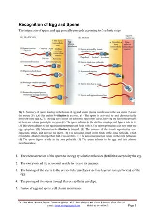
- 1. Dr. Shoeb Ahmad, Assistant Professor, Department of Zoology, AKI’s Poona College of Arts, Science & Commerce, Camp, Pune -01 e-mail: shoeb.zoology@gmail.com Mobile no 9919509673 Page 1 Recognition of Egg and Sperm The interaction of sperm and egg generally proceeds according to five basic steps Fig 1. Summary of events leading to the fusion of egg and sperm plasma membranes in the sea urchin (A) and the mouse (B). (A) Sea urchin fertilization is external. (1) The sperm is activated by and chemotactically attracted to the egg. (2, 3) The egg jelly causes the acrosomal reaction to occur, allowing the acrosomal process to form and release proteolytic enzymes. (4) The sperm adheres to the vitelline envelope and lyses a hole in it. (5) The sperm adheres to the egg plasma membrane and fuses with it. The sperm pronucleus can now enter the egg cytoplasm. (B) Mammalian fertilization is internal. (1) The contents of the female reproductive tract capacitate, attract, and activate the sperm. (2) The acrosome-intact sperm binds to the zona pellucida, which constitutes a thicker envelope than that of sea urchins. (3) The acrosomal reaction occurs on the zona pellucida. (4) The sperm digests a hole in the zona pellucida. (5) The sperm adheres to the egg, and their plasma membranes fuse. 1. The chemoattraction of the sperm to the egg by soluble molecules (fertilizin) secreted by the egg. 2. The exocytosis of the acrosomal vesicle to release its enzymes. 3. The binding of the sperm to the extracellular envelope (vitelline layer or zona pellucida) sof the egg. 4. The passing of the sperm through this extracellular envelope. 5. Fusion of egg and sperm cell plasma membranes
- 2. Dr. Shoeb Ahmad, Assistant Professor, Department of Zoology, AKI’s Poona College of Arts, Science & Commerce, Camp, Pune -01 e-mail: shoeb.zoology@gmail.com Mobile no 9919509673 Page 2 Sometimes steps 2 and 3 are reversed (as in mammalian fertilization) and the sperm binds to the egg before releasing the contents of the acrosome. After these five steps are accomplished, the haploid sperm and egg nuclei can meet, and the reactions that initiate development can begin. In many species, the meeting of sperm and egg is not a simple matter. Many marine organisms release their gametes into the environment. That environment may be as small as a tide pool or as large as an ocean. Moreover, it is shared with other species that may shed their sex cells at the same time. These organisms are faced with two problems: How can sperm and eggs meet in such a dilute concentration, and how can sperm be prevented from trying to fertilize eggs of another species? Two major mechanisms have evolved to solve these problems: species-specific attraction of sperm and species-specific sperm activation. Sperm attraction: Action at a distance Species-specific sperm attraction has been documented in numerous species, including cnidarians, molluscs, echinoderms, and urochordates. In many species, sperm are attracted toward eggs of their species by chemotaxis, that is, by following a gradient of a chemical secreted by the egg. The mechanisms of chemotaxis differ among species. One chemotactic molecule, a 14-amino acid peptide called resact, has been isolated from the egg jelly of the sea urchin (Arbacia punctulata). Resact diffuses readily in seawater and has a profound effect at very low concentrations when added to a suspension of Arbacia sperm. When a drop of seawater containing Arbacia sperm is placed on a microscope slide, the sperm generally swim in circles about 50 μm in diameter. Within seconds after a minute amount of resact is injected into the drop, sperm migrate into the region of the injection and congregate there. As resact continues to diffuse from the area of injection, more sperm are recruited into the growing cluster. Resact is specific for A. punctulata and does not attract sperm of other species. A. punctulata sperm have receptors in their plasma membranes that bind resact and can swim up a concentration gradient of this compound until they reach the egg. Resact also acts as a sperm-activating peptide. Sperm-activating peptides cause dramatic and immediate increases in mitochondrial respiration and sperm motility. The sperm receptor for resact is a transmembrane protein, and when it binds resact on the extracellular side, a conformational change on the cytoplasmic side activates the receptor’s enzymatic activity. This activates the mitochondrial ATP-generating apparatus as well as the dynein ATPase that stimulates flagellar movement in the sperm The acrosomal reaction in sea urchins A second interaction between sperm and egg is the acrosomal reaction. In most marine invertebrates, the acrosomal reaction has two components: the fusion of the acrosomal vesicle with the sperm plasma membrane (an exocytosis that results in the release of the contents of the acrosomal vesicle) and the extension of the acrosomal process. The acrosomal reaction in sea urchins is initiated by contact of the sperm with the egg jelly. Contact with egg jelly
- 3. Dr. Shoeb Ahmad, Assistant Professor, Department of Zoology, AKI’s Poona College of Arts, Science & Commerce, Camp, Pune -01 e-mail: shoeb.zoology@gmail.com Mobile no 9919509673 Page 3 causes the exocytosis of the sperm’s acrosomal vesicle and the release of proteolytic enzymes that can digest a path through the jelly coat to the egg surface. The sequence of these events is outlined in Fig. 2 Fig. 2 Acrosomal reaction in sea urchin sperm. (A–C) The portion of the acrosomal membrane lying directly beneath the sperm plasma membrane fuses with the plasma membrane to release the contents of the acrosomal vesicle. (D) The actin molecules assemble to produce microfilaments, extending the acrosomal process outward. Actual photographs of the acrosomal reaction in sea urchin sperm are shown below the diagrams. The exocytosis of the acrosomal vesicle is caused by the calcium-mediated fusion of the acrosomal membrane with the adjacent sperm plasma membrane. The egg jelly factors that initiate the acrosomal reaction in sea urchins are often highly specific to each species. The second part of the acrosomal reaction involves the extension of the acrosomal process (see Fig. 2). This protrusion arises through the polymerization of globular actin molecules into actin filaments. Species-specific recognition in sea urchins Once the sea urchin sperm has penetrated the egg jelly, the acrosomal process of the sperm contacts the surface of the egg (Fig. 3). A major species-specific recognition step occurs at this point. The acrosomal protein mediating this recognition is called bindin. Further, its interaction with eggs is relatively species-specific. Bindin isolated from the acrosomes of a species will bind to eggs of same species.
- 4. Dr. Shoeb Ahmad, Assistant Professor, Department of Zoology, AKI’s Poona College of Arts, Science & Commerce, Camp, Pune -01 e-mail: shoeb.zoology@gmail.com Mobile no 9919509673 Page 4 Fig. 4 Species-specific binding of acrosomal process to egg cell surface in sea urchins. (A) Actual contact of a sea urchin sperm acrosomal process with an egg microvillus. (B) In vitro model of species-specific binding. The agglutination of dejellied eggs by bindin was measured by adding bindin aggregates to a plastic well containing a suspension of eggs. After 2–5 minutes of gentle shaking, the wells were photographed. Each bindin bound to and agglutinated only eggs from its own species. Biochemical studies have shown that the bindins of closely related sea urchin species are indeed different. This finding implies the existence of species-specific bindin receptors on the egg, vitelline envelope, or plasma membrane. The bindin receptors are thought to be aggregated into complexes on the egg cell surface, and hundreds of these complexes may be needed to tether the sperm to the egg. Thus, species-specific recognition of sea urchin gametes occurs at the levels of sperm attraction, sperm activation, and sperm adhesion to the egg surface. Gamete binding and recognition in mammals ZP3: the sperm-binding protein of the mouse zona pellucida The zona pellucida in mammals plays a role analogous to that of the vitelline envelope in invertebrates. This glycoprotein matrix, which is synthesized and secreted by the growing oocyte, plays two major roles during fertilization: it binds the sperm, and it initiates the acrosomal reaction after the sperm is bound. The binding of sperm to the zona is relatively, but not absolutely, species-specific. (Species-specific gamete recognition is not a major problem when fertilization occurs internally.)
- 5. Dr. Shoeb Ahmad, Assistant Professor, Department of Zoology, AKI’s Poona College of Arts, Science & Commerce, Camp, Pune -01 e-mail: shoeb.zoology@gmail.com Mobile no 9919509673 Page 5 The binding of mouse sperm to the mouse zona pellucida can be inhibited by first incubating the sperm with zona glycoproteins. Bleil and Wassarman (1980, 1986 1988) isolated an 83- kDa glycoprotein, ZP3, from the mouse zona that was the active competitor for binding in this inhibition assay. The other two zona glycoproteins they found, ZP1 and ZP2, failed to compete for sperm binding (Fig.5). Moreover, they found that radiolabeled ZP3 bound to the heads of mouse sperm with intact acrosomes. Thus, ZP3 is the specific glycoprotein in the mouse zona pellucida to which the sperm bind. ZP3 also initiates the acrosomal reaction after sperm have bound to it. The mouse sperm can thereby concentrate its proteolytic enzymes directly at the point of attachment at the zona pellucida. Fig. 5. Mouse ZP3 as the zona protein that binds sperm. (A) Diagram of the fibrillar structure of the mouse zona pellucida. The major strands of the zona are composed of repeating dimers of proteins ZP2 and ZP3. These strands are occasionally crosslinked together by ZP1, forming a meshlike network. (B) Inhibition assay showing the specific decrease of mouse sperm binding to zonae pellucidae when sperm and zonae were first incubated with increasingly large amounts of the glycoprotein ZP3. The importance of the carbohydrate portion of ZP3 is also indicated by this graph