disease management in livestock and its prevention
•Download as PPTX, PDF•
11 likes•10,565 views

The document discusses disease management and prevention in livestock. It provides information on signs of good health in livestock and general symptoms of disease. It then discusses specific infectious diseases like anthrax, mastitis, foot and mouth disease, rabies, PPR in goats, and non-infectious diseases caused by faulty nutrition, metabolic disorders, and trauma. The document also provides information on prevention measures, vaccination schedules, and discusses specific poultry diseases like avian influenza, infectious bronchitis, chronic respiratory disease, and E. coli infections.

Recommended
More Related Content
What's hot
What's hot (20)
Similar to disease management in livestock and its prevention
Similar to disease management in livestock and its prevention (20)
More from ShekhAlisha
More from ShekhAlisha (13)
Recently uploaded
Recently uploaded (20)
disease management in livestock and its prevention
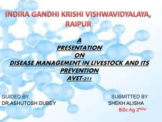
- 1. A PRESENTATION ON DISEASE MANAGEMENT IN LIVESTOCK AND ITS PREVENTION AVET-211 GUIDED BY SUBMITTED BY DR.ASHUTOSH DUBEY SHEKH ALISHA BSc Ag 2NDyr
- 3. A. Signs of good health include: 1. Contented animals look free from all anxiety. 2.Some animals have specific signs to look for and others will take a trained eye to recognize. Sheep will stay quiet and cattle will chew their cud. 3. Body temperature, respiration, and pulse rate should be monitored because unusual highs and lows can be symptoms. 4. Alertness can be judged by checking if an animal perks its ears when you draw near. 5. The skin and coat of most animals should be oily and elastic like. 6. One of the easiest things to notice is when an animal goes off feed. All healthy animals should eat aggressively when fed and ruminants should be seen chewing their cud.
- 4. General symptoms specific to disease: 1. Animal losing fetus 2. Shaking 3. Coughing 4. Poor growth or decrease in production 5. Rough coat 6. Loss of appetite
- 5. NORMAL CLINICAL VALUES IN ANIMAL Species Temp ture1 Pulse rate, per minute Respiration rate per minute Cattle& buffalo Sheep & Goat . Poultry 101.6 102.6 107.0 42 – 60 70 – 80 130 – 160 16 – 24 18 – 30 15 – 30 Table: Normal clinical values in animals:
- 7. TYPE OF DISEASE : INFECTIONOUS DISEASE NON INFECTIONOUS DISEASE DISEASE OF LIVESTOCKS INFECTIONOUS VIRAL DISEASE BACTERIAL DISEASE NON INFECTIONOUS Metabolic disorder Faulty nutrition Trauma
- 10. Anthrax • Causative agent: Bacillus anthracis • Mode of Transmission: By ingestion, from soil when grazing or in contaminated food/wound infection • Host: All Species (Cattle, sheep and goats are most susceptible) • Symptoms: In peracute sepeticemia death occurs within 2 hours after animal collapsing with convulsions, sudden death in animals that appeared normal is common. In acute septicemia death occurs within 48 to 96 hours clinical signs include fever, anorexia, ruminal stasis, hematuria and blood tinged diarrhea. Pregnant animals may abort and milk production often abruptly decreases. Terminal signs include severe depression, respiratory distress and convulsions. Oozing of tarry coloured unclotted blood from all the orifices of the animals body • Prevention and Control: By active immunization. The organism is susceptible to penicillin-G, tetracyclines, erythromycin and chloramphenicol.
- 13. Mastitis • Introduction: Mastitis is an inflammation of the mammary gland. In which the milk undergo physical, chemical and microbiological changes where as mammary glandular tissue under go physical and pathological changes. In which infected milk colour, consistency change and contains more amount of leucocytes. • Clinical Signs: Per acute form: Pyrexia, anorexia, respiratory distress, swollen, hot and painful udder. Cessation of milk production. Exudate are often blood stained. Acute form: Swollen udder, changes in quality of milk. Milk become curd like, yellow, brown fluid with flakes and clots. Subacute form: No changes in the udder tissue. Chronic form: Udder is haemorrhagic, and fibrotic. Swollen and palpable supra mammary lymphnode,. Udder is thick, firm, nodular and atrophic, yellowish or white fluid with clots and flakes. Treatment: Stripping out the milk from the infected quarters. Cleaning of infected quarters with normal saline and distilled water. Infusion of antibiotic therapies immediately after the infection. Continuous use antibiotics as per the antibiogram. Control: Hygenic measures are important. Animals diagnosed positive should be milked at last. Milkers should wash their hands before milking and should use well washed white overalls. A separate clean cloth for each cow is used for washing the udder with a disinfectant. The first stream of milk from each quarter should not be allowed to drop on floor but collected in a separate container. Milkers should not wet their hands with first stream of milk.
- 20. Foot and Mouth Disease (FMD) • Causative agent: Picorna Virus • Mode of Transmission: By direct contact i.e. Through water, manure, pastureBy direct contact i.e. Through water, manure, pasture and cattle attendant • Host: In cloven-hoofed animals (cattle, pigs, sheep, goats, and water buffalo) • Symptoms: The formation of vesicles (fluid-filled blisters) and erosions in the mouth, nose, teats and feet. Initial signs are pyrexia (39.4-40.6ºC), dullness, anorexia, and fall in milk production. These signs are followed by excessive salivation; drooling, serous nasal discharge; shaking, kicking of the feet or lameness; and vesicle (blister) formation in the tongue, dental pad, gums, soft palate, nostrils, muzzle, interdigital space, coronary band, and teats. Pregnant cows may abort, and young calves may die without developing any vesicle The course of an FMD infection is 2 to 3 weeks. Secondary infection may delay recovery. Presence of ropy saliva • Prevention & Control: Thorough disinfection of shed, utensils, clothes of attendants.Vaccination – polyvalent – once – 4 months or varies with type of vaccine
- 21. Rabies • Causative agent:- Rhabdovirus • Initial Signs:- pain at the bite site, a general feeling of illness fever, headache, muscle aches poor appetite, nausea, and vomitin, sore throat depression • Later Symptoms:- anxiety, confusion, excessive saliva production hallucinations, high level of excitement insomnia, restlessness paralysis of lower legs, problems swallowing due to painful throat and voice box spasms hydrophobia (fear at the sight of water despite an intense thirst) Prevention & Treatment: By vaccination (Pre or Post bite)
- 26. PPR in Goats • Causative agent: Morbilli virus of family Paramyxovirus • Transmission: By direct contact i.e. Through water, manure, pasture • Symptoms: Fever Occular and nasal mucous discharge, Mouth lesion Respiratory distress Prevention: Yearly vaccination. Separation of infected one from healthy animals
- 27. NON INFECTION DISEASE Faulty nutrition . >Ration is not balanced Metabolic disorder. >Not adequate digested. Trauma >Wound and injuries
- 28. FAULTY NUTRITION :EXAMPLE Bloat Rapid fermentation (breakdown of carbohydrate by enzyme to much gas ). Bovine pulmonary emphysema : Feedlot problem ,panting,coughing,difficult breathing . Fescue foot Pasture problem,cattle grazing in a fescue pasture ,the animal shifting from one hind to other,sometime with one in the air. Enterotoxemia (overeating disease) Affect cattle /sheep on high concentration rations.animal may die in 1 or 24 hours . Founder: Footlot problem :swelling of tissue thar=ts attaches to hoof of the feet
- 29. METABOLIC DISORDER ;EXAMPLE Grass tetany most often affects cows that are lactating ,it is caused by a lack of magnesium . Hardware disease: Animal swallows metal objects that ruminant stomach. Nitrate poisoning : Caused by the animal eating or drinking a product that has too much nitrogen in it . Poisonous plant animal eating poisonous plant in a graze land setting ..
- 30. METABOLIC DISORDER : Rumentitis (liver abcess complex) Cattle on a high conc rations .the soil and manure content levels play a factor in the prevalence of thus disease. white muscle (selenium difficiency) Common where the soil is lacking in selection in selenum . The muscle turns a white colour. Pic: white muscle
- 33. PREVENTION MEASURES General Measures for Prevention of Contagious Diseases •Identification of isolation of infected,-rd in contact animal. •Treatment of' affected animals •Slaughter of animals suffering from incurable diseases. •Disposal of Deal animals either burning or deep burial. •Destroy contaminant folder by burning. •Proper disposal of contaminated water. •Regular cleaning and disinfection of cattle shed and its premises. •Don’t allow animals from affected to clean area. •Restrict the movement of animals from effected to clean area. •Don’t allow animals to drink water from ponds, rivers etc. during out break of disease.
- 34. •Close animal markets, cattle shows etc. during outbreak of disease. •Regular spraying of insecticide to control external parasites. •Regular de-worming to control internal parasites. •Avoid stress associated with along distance transportation, inclement weather and under nutrition •Provide adequate ventilation and sufficient space.
- 35. VACCINATION SCHEDULE Disease Age Interval Month FMD 3rd month Every six month Jan-Feb, June-July BG 6th Month Every year Aug-Sep HS 6th Month Every Year Sep-Oct Anthrax 6th Month Every Year ( Affected area only) April - May Brucellosis 4-8th month of Heifer -- Mar - April Vaccination schedule
- 36. Avian influenza •Avian Influenza (AI) is a viral infection affecting wild and domestic birds. •Many species of birds are susceptible to AI including chickens, turkeys, guinea fowl, and other domestic birds, as well as a some wild avian species. AI is caused by viruses that are members of the family Orthomyxoviridae, and the genus Influenza virus, Type A.
- 37. Clinical Signs •The severity of clinical signs depends on factors such as: •age •species •concurrent infections •environment •the pathogenicity of the virus Symptoms: •In domestic poultry, clinical signs reflect abnormalities in the respiratory, digestive, urinary, and reproductive organs. •Generalized signs include decreased activity, ruffled feathers, decreased feed and water consumed occasionally greenish diarrhea. •Respiratory manifestations include mild to severe coughing, sneezing, excessive lacrimation, accumulation of liquids in eyelids, and nasal discharge. •decrease in egg production are observed in laying hens and breeders.
- 38. Prevention and Control The recommended strategy for controlling AI is eradication. •This requires different components including: 1) biosecurity practices •controlling human traffic, quarantining birds before introduction, proper cleaning and disinfection of facilities, keeping healthy birds away from contact with sick birds and wild birds, and incubating eggs only from clean flocks 2) increasing host resistance through vaccination •Inactivated vaccines have been shown to be effective but are fairly expensive
- 39. Infectious bronchitis(IB) •Cause •Corona-virus is the causal agent. Several different genotypes of IB virus are known to exist. •Transmission •The virus is transmitted from bird to bird through the airborne route. The virus can also be transmitted via the air between chicken houses and even from farm to farm. •Species affected •Only chickens are susceptible to IB virus. •Clinical signs •In young chicks IB virus infection causes a cheesy exudate in the bifurcation of the bronchi •In older birds IB does not cause mortality. •Egg productionwill decrease dramatically, deformed eggs with wrinkled shells will often be laid
- 40. •Treatment and control •There is no treatment for infectious bronchitis. •Secondary bacterial infections may be prevented by, or treated with antibiotics. •Prevention by vaccinationis the best method to control IB
- 41. Chronic respiratory Disease(CRD) •Cause •The underlying cause of CRD is Mycoplasma gallisepticum (Mg). •TheconditionisfrequentlytriggeredbyrespiratoryvirusessuchasNDandI Bandsubsequentlycomplicatedbybacterialinvasion. •The main agents involved in the infection are Mycoplasma gallisepticum and E. coli. •Stresscausedbymovingthebirds,bydebeakingorotheroperationsorothe runfavorableconditionse.g.coldorbadventilation,makethebirdsmoresus ceptible. •Transmission •ThemainproblemisthatparentbirdsinfectedwithMycoplasmagalliseptic umcantransmittheorganismthroughtheeggtotheiroffspring. •Inaddition,infectioncanoccurbycontactorbyairbornedustordroplets
- 42. •Clinical signs •Young chickens (broiler chicks or layer pullets) will show respiratory distress •The birds frequently show a lack of appetite, decreased weight gain and increased feed conversion ratios. •In adult birds the most common symptoms are sneezing, coughingand general signs of respiratory congestion. •In laying birds a drop of egg productionbetween 20-30 % can occur. •CRD does not normally cause an alarming number of deaths.. •Treatment •Treatment of Mg-infected chickens or turkeys with suitable antibiotics or chemotherapeutics has been found to be of economic value. •control by medicationor vaccinationand eradication of Mg infections •Fertile eggs from infected birds can be treated with antibiotics such as tylosin to eliminate the Mycoplasma gallisepticum organisms. •Methods used are the injection of fertile eggs or egg dipping. Blood serum testing of breeder chickens for Mg antibodies has become a routine to test flocks for a Mg infection
- 43. ESCHERICHIA COLI INFECTIONS •Escherichia coli infections are widely distributed among poultry of all ages and categories. •They are primarily related to poor hygienic conditions neglected technological requirements or to respiratory and immuno suppressive diseases. •A common sequel of navel infections is local or diffuse peritonitis . •Omphalitis (navel infection) •It is characterized with reddening and tissue oedema in the umbilical region . The delayed absorption of the yolk sac is a prerequisite for E. coli infections and peritonitis
- 44. •Salpingitis (inflammation of the oviduct). Salpingites due to E. coli infections could be also observed in growing birds. •The oviduct is dilated, with thinned wall and filled with caseous exudate all along its length . •Salpingites are among the commonest causes for death in layer hens. E. coli penetrates from the cloa via an ascendant route. •Egg yolk peritonitis in a layer hen consequently to E. coli salpingitis. The chickens could be hatched with a latent infection, when E. coli is present in ovaries and the oviduct. In these instances, the infection could turn into an overt infection under the influence of some stress factors or lesions •Panophthalmitis (inflammation of all tissues of the eyeball). Generally, it develops secondary to E. coli septicaemia and is usually unilateral
- 45. •E. coli septicaemia of a respiratory origin. In such cases, the respiratory mucosa damaged by infectious and non-infections agents (ND viruses including vaccinal strains, IB, mycoplasmae, high ammonia levels) is the entrance door of the E. coli infection. The lesions are principally observed in the respiratory tract (trachea, lungs and air sacs), but some adjacent serous coats (pericardium, peritoneum) are also affected and thus, the picture of a typical serofibrinous polyserositis is produced