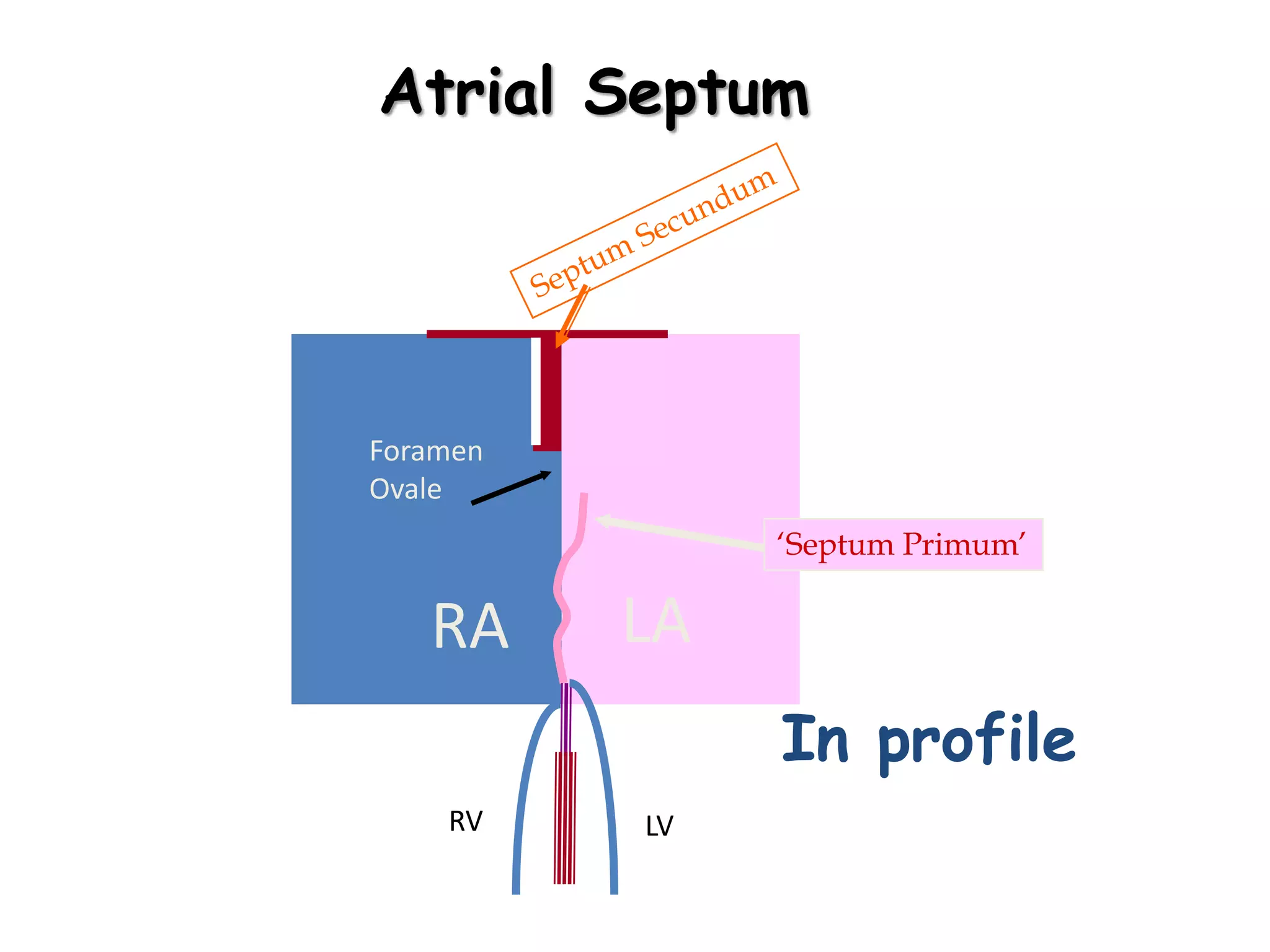
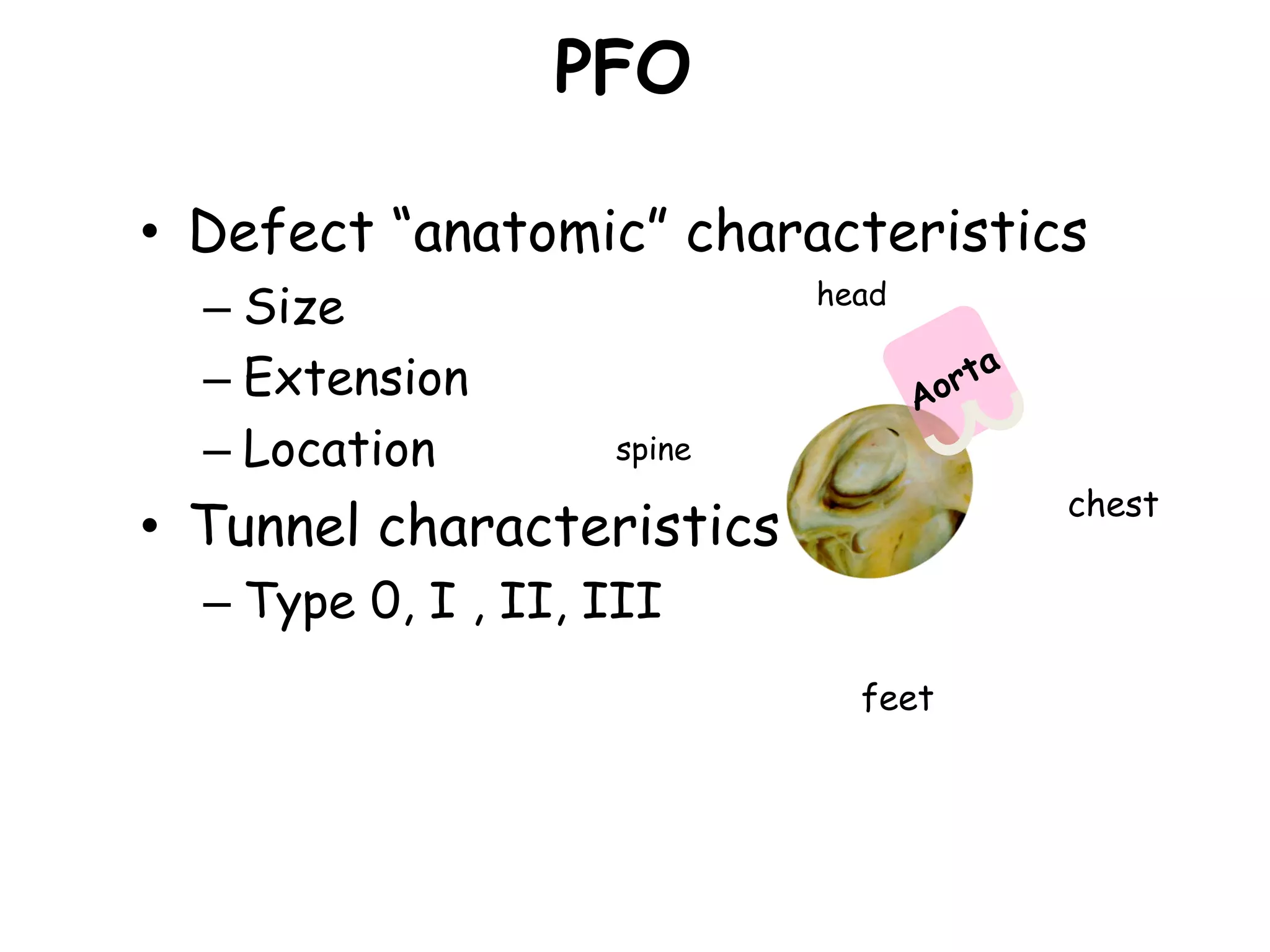
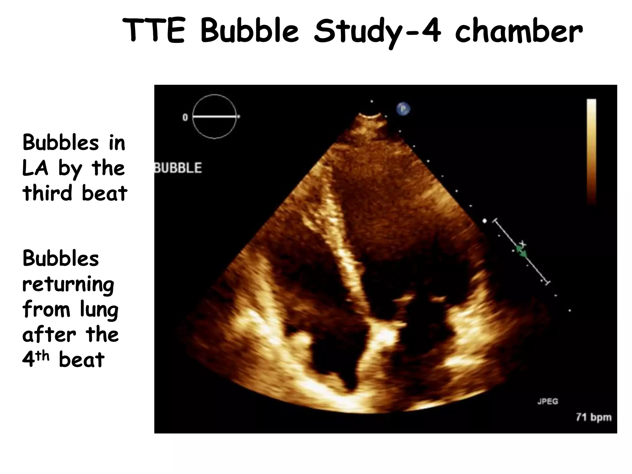
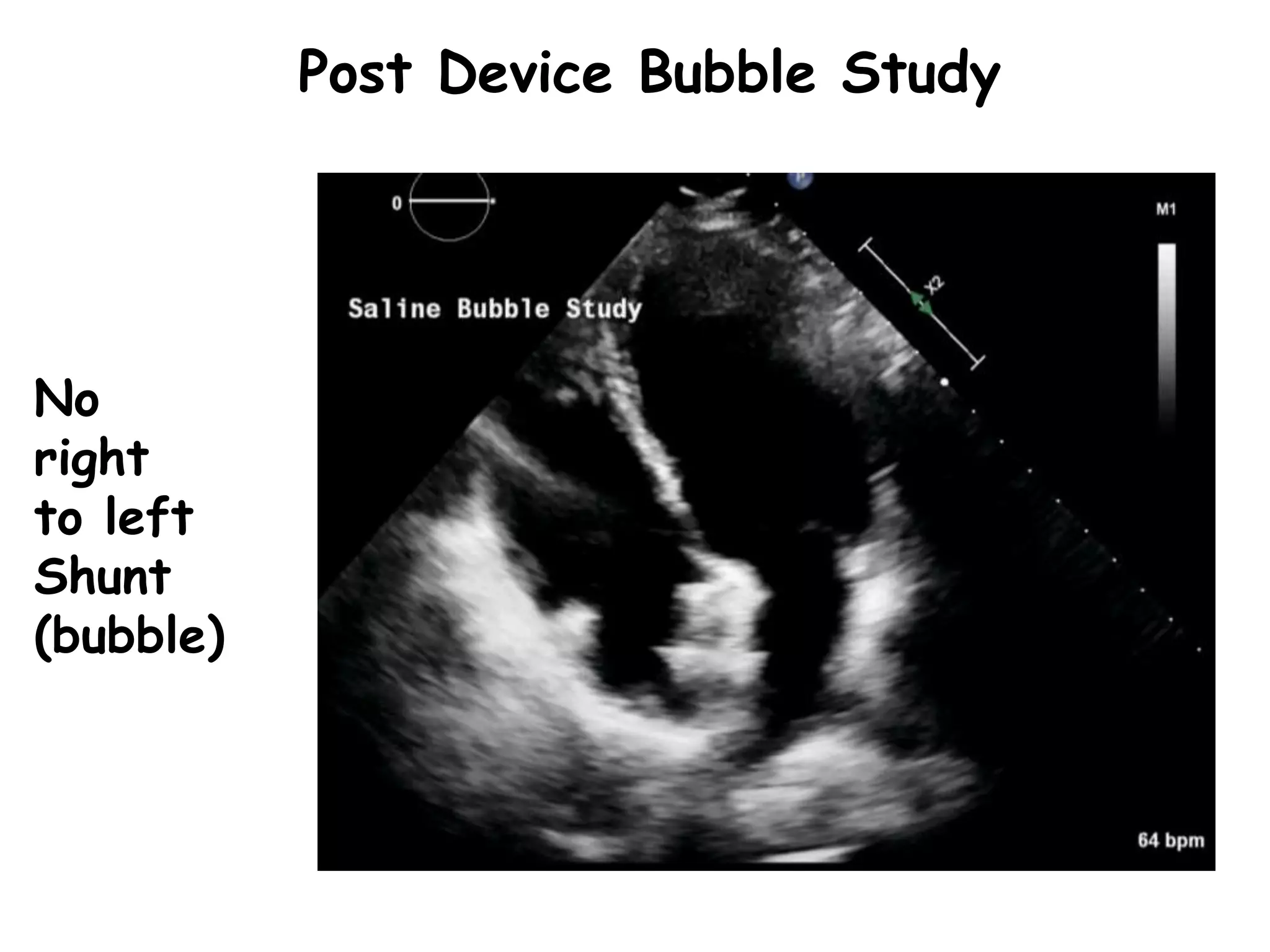
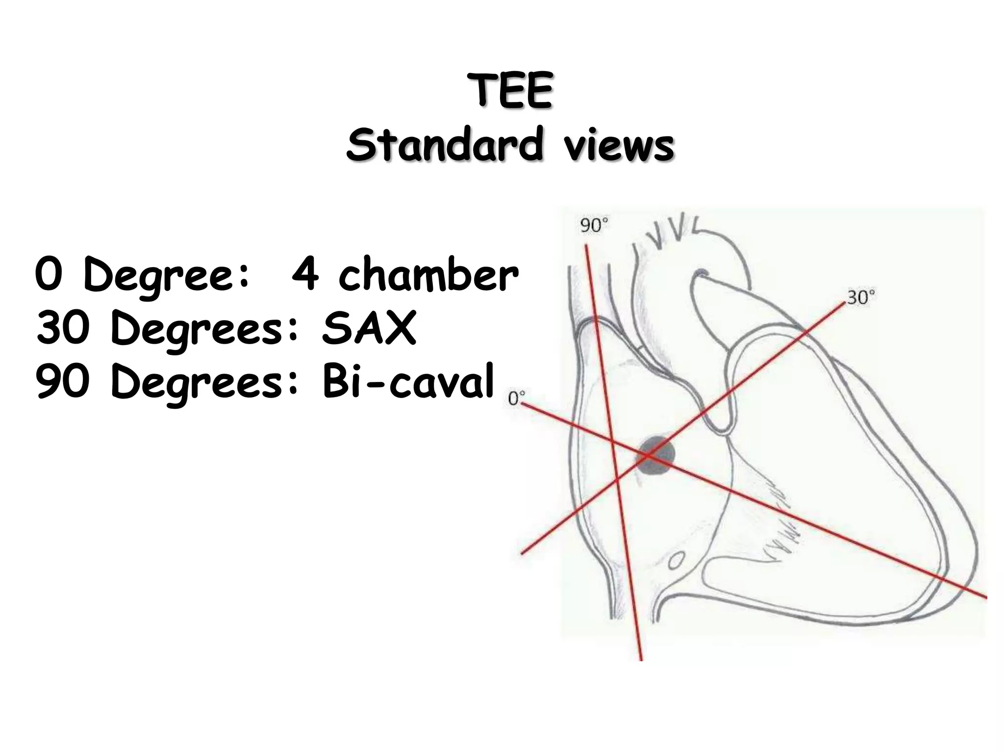
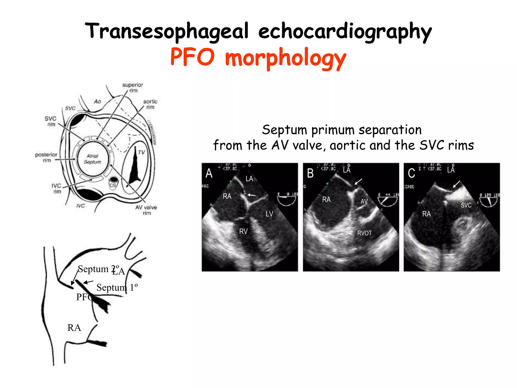
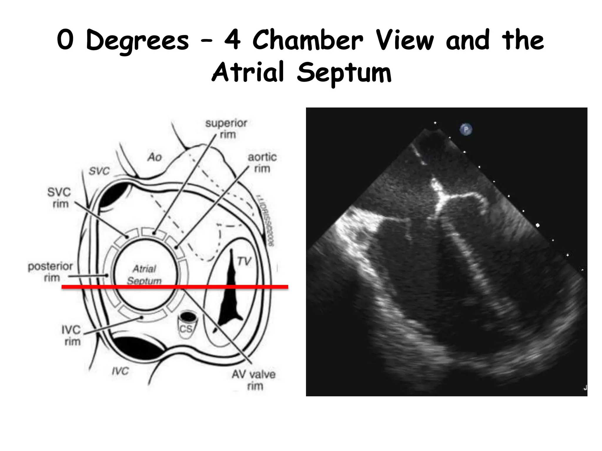
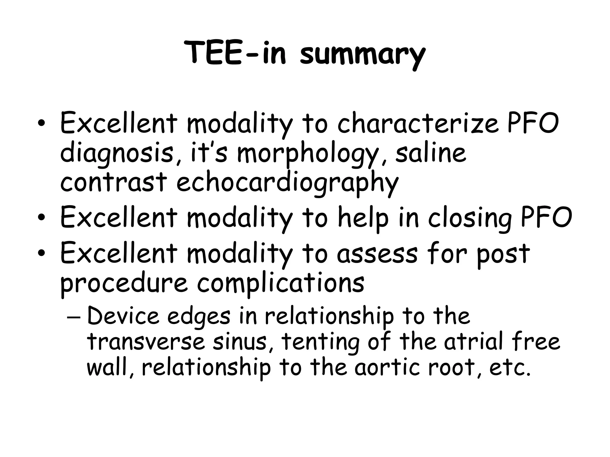
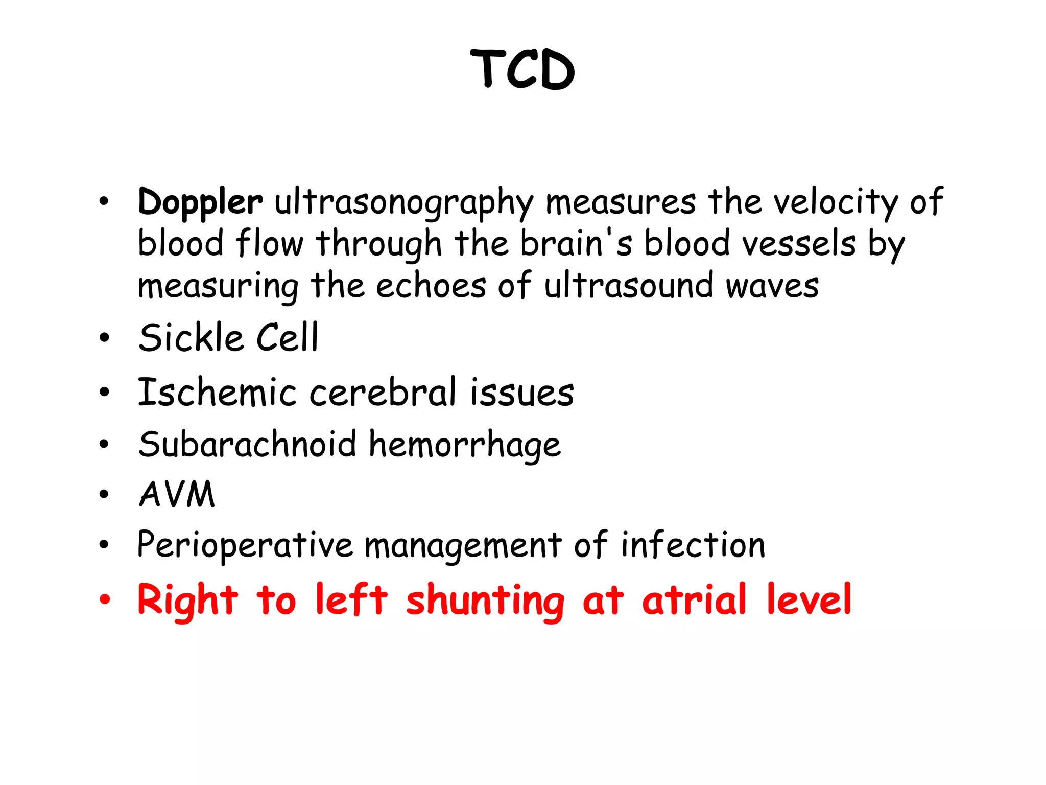
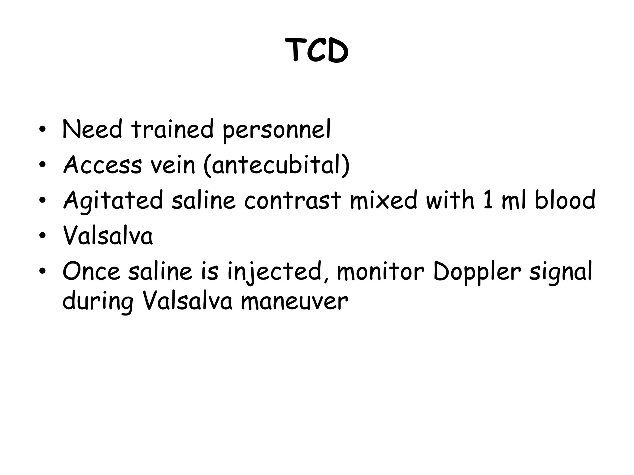
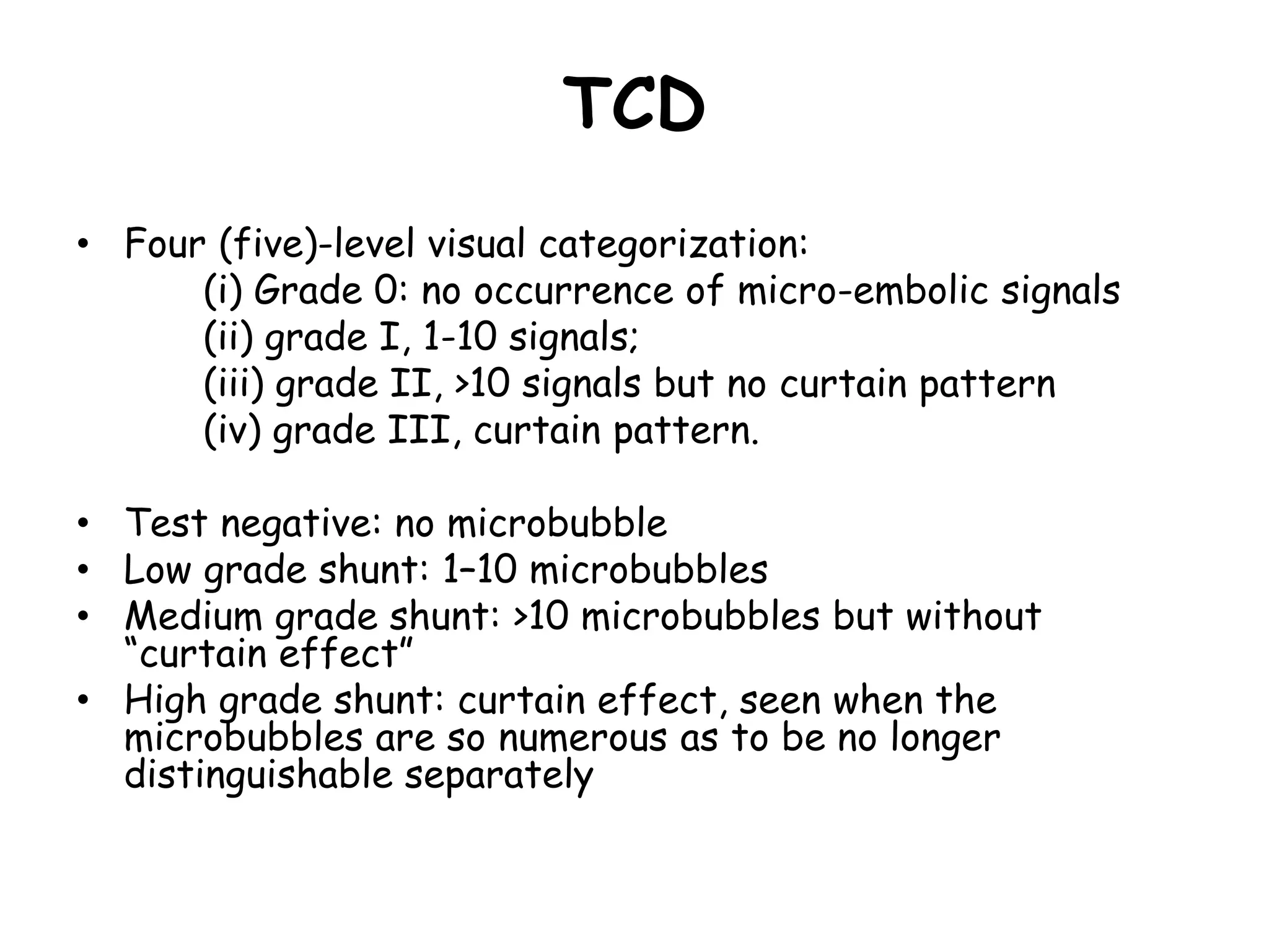
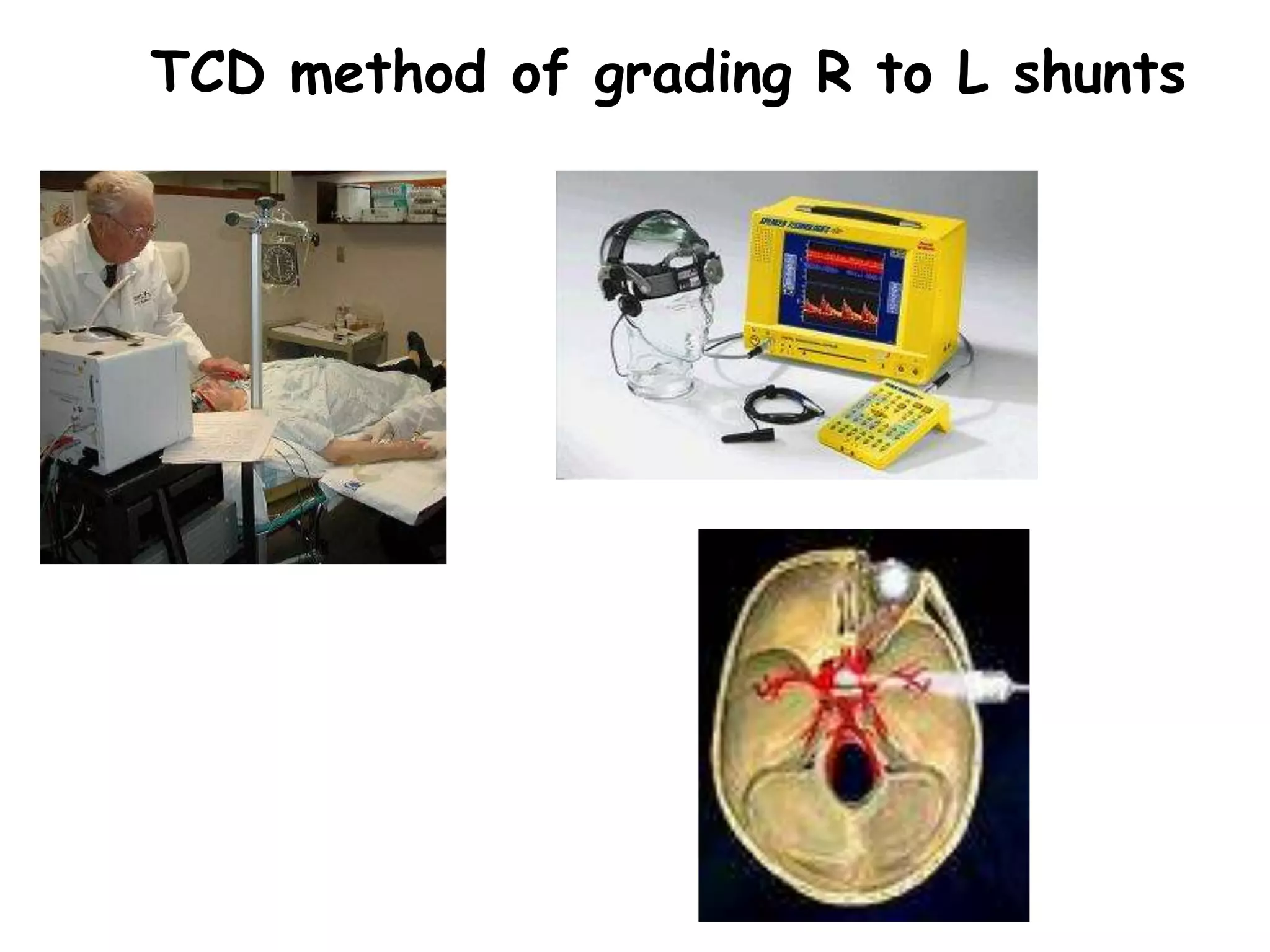
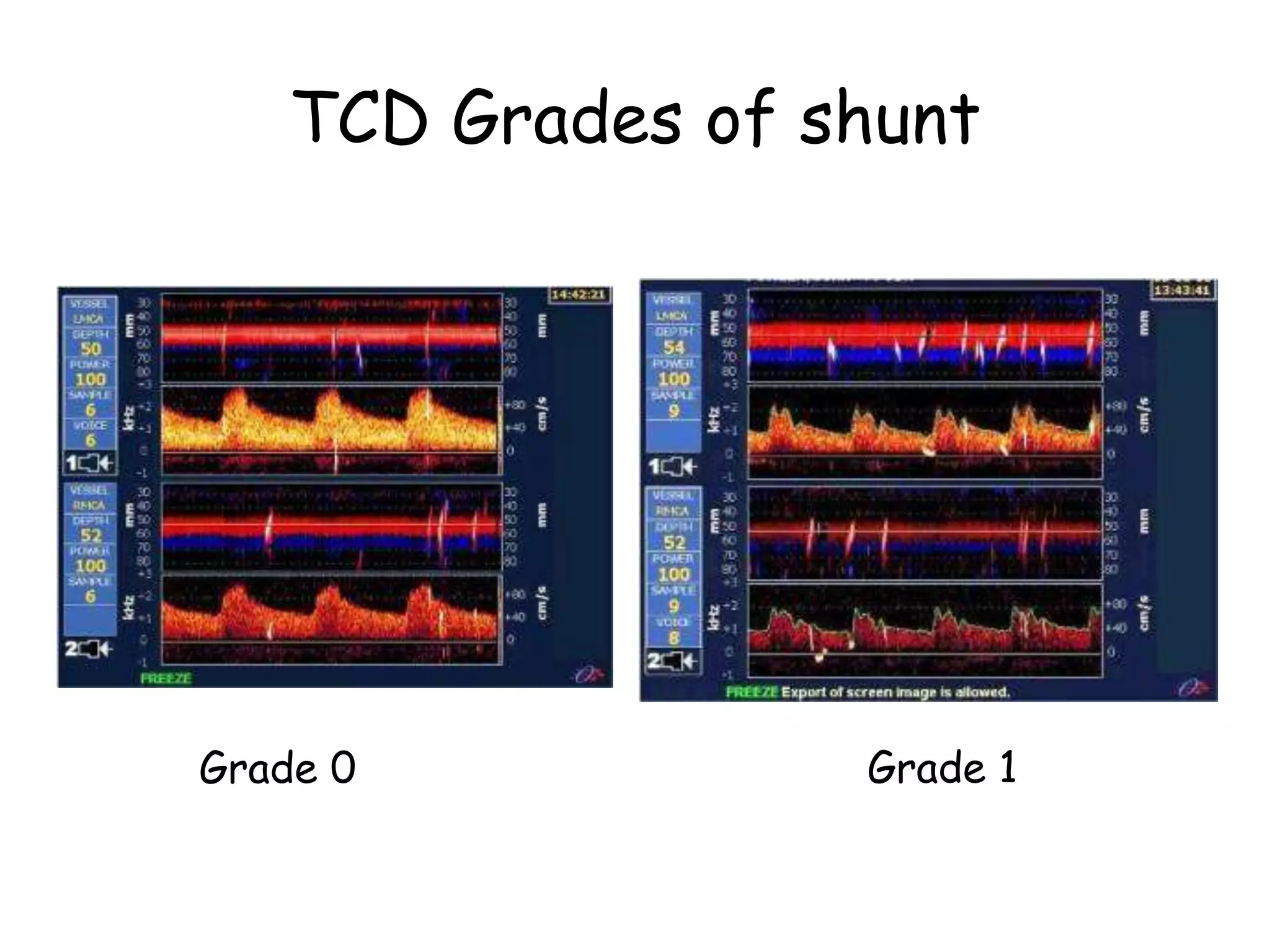
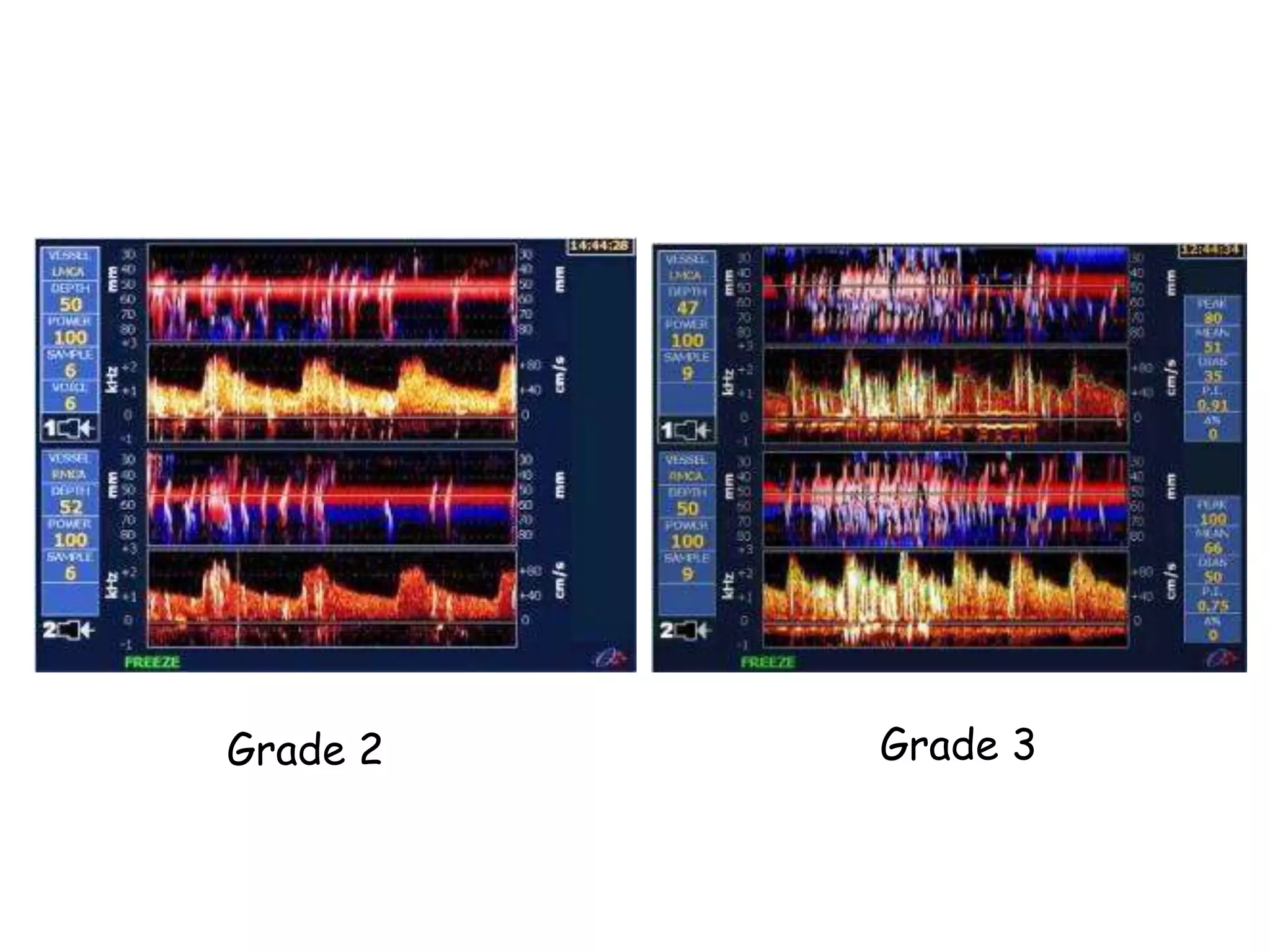
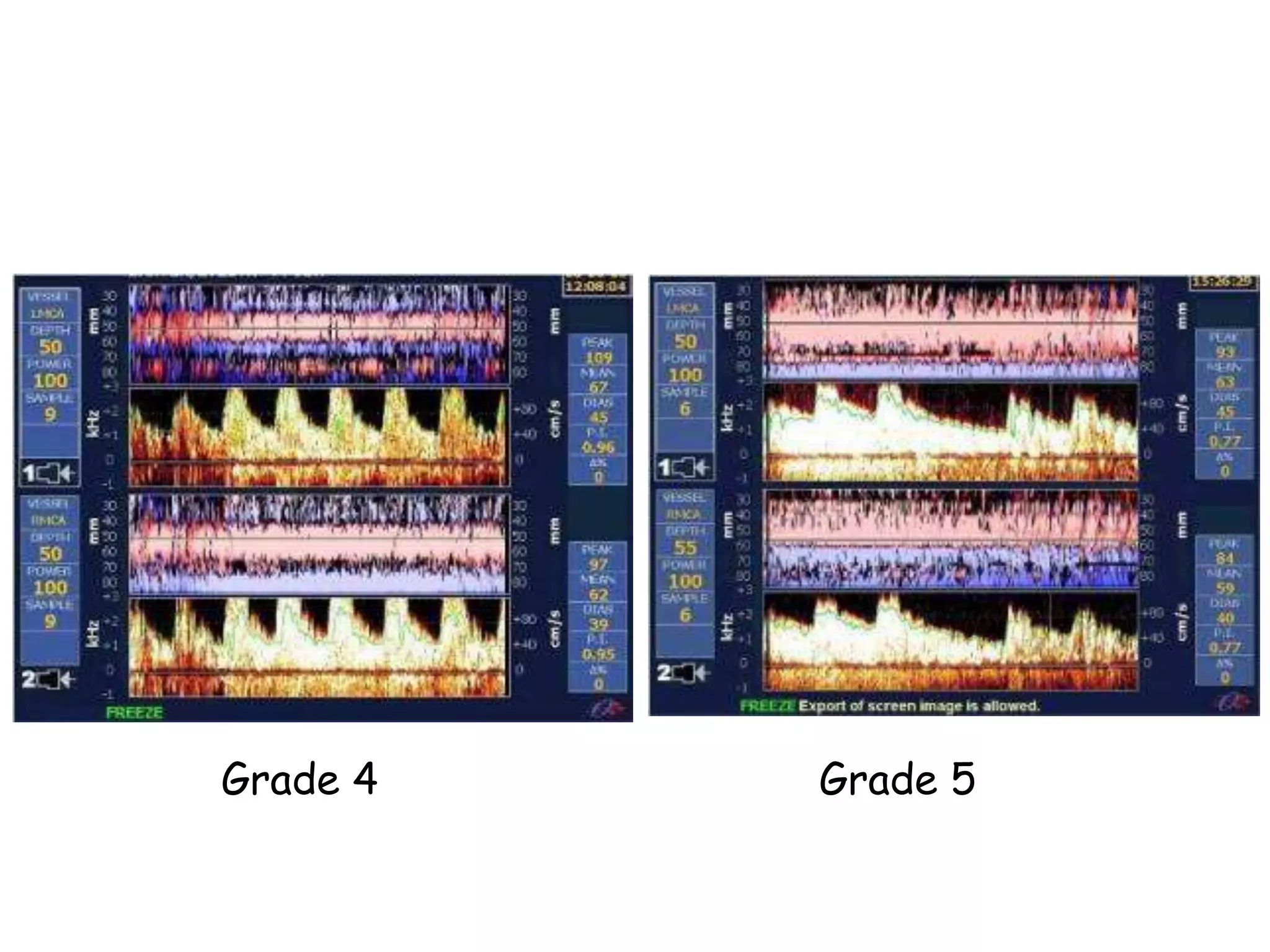
Pre-Procedural Imaging to
Characterize the PFO: TTE,
TEE, TCD
Zahid Amin, MD
William B. Strong Endowed Chair
Augusta University; Children’s
Hospital of Georgia, Augusta, GA
Disclosure Statement of Financial
Interest
Within the past 12 months, I or my
spouse/partner have had a financial
interest/arrangement or affiliation with the
organization(s) listed below.
Affiliation/Financial Relationship Company
Consulting Fees/Honoraria Abbott Vascular; W.L. Gore &
Associates
Characterize the PFO
• Characterize means: to describe the
distinctive nature and features of..
• By using TTE, we can diagnose PFO but
we may not be able to elaborate on it
– Extension
– Tunnel length
– Size
– Aneurysmal nature (we can a little, but it
may not be crisp)
PFO
Potential For Opening
• Lack of apposition between septum
secundum and primum that results in PFO
– May be small and slit like
– May be long and lunar in shape
• Potential For Overlap
– Significant overlap = Long tunnel
– No overlap means no tunnel
• Potential For Other issues
– Aneurysmal (redundant septum primum)
– Large defect
– Difficult to cross occasionally
RA LA
Atrial Septum
Foramen
Ovale
In profile
RV LV
‘Septum Primum’
Atrial Septum
AV valve
Foramen
Ovale
Right Atrial
View
Atrial Septum
Left Atrial view
Foram
en
Ovale
‘Flap valve’
FRONT BACK
Atrial Septum
4- Chamber View
Patent
Foramen
Ovale
RA LA
RV LV
narrow
oblique
channel
PFO
• Defect “anatomic” characteristics
– Size
– Extension
– Location
• Tunnel characteristics
– Type 0, I , II, III
chest
spine
head
feet
TTE
• Usually used for initial evaluation of
PFO
– For detection and diagnostic purposes
– Sufficient in pediatric patients
• Images may not be “good enough” to
characterize the atrial septum and
hence the anatomy of PFO
TTE
• Good for Detecting PFO:
– Adequate to appreciate lack of apposition
between septum primum and secundum
– Color flow at the atrial level
• Diagnosis
– Demonstration of right to left shunt by
• Color flow
• Saline bubble contrast study that shows right
to left shunt by the first three heart beats
TTE Bubble Study-4 chamber
Bubbles in
LA by the
third beat
Bubbles
returning
from lung
after the
4th beat
Aneurysmal Atrial Septum Parasyernal
SAX
Aneurysmal
septum >
10 mm
excursion
Aneurysmal Septum Seen in Parasternal
LAX view
Post Device Bubble Study
No
right
to left
Shunt
(bubble)
TEE
Standard views
0 Degree: 4 chamber
30 Degrees: SAX
90 Degrees: Bi-caval
Transesophageal echocardiography
PFO morphology
A LA
RA
LV
RV
B LA
RA AV
RVOT
C LA
RA
SVC
RA
LA
PFO
Septum 1º
Septum 2º
Septum primum separation
from the AV valve, aortic and the SVC rims
0 Degrees – 4 Chamber View and the
Atrial Septum
TEE 0º Inferior
0 Degrees – 4 Chamber View
(Caudal)
0 Degrees – 4 Chamber View
(Cranial)
T0º
45 Degrees - Short Axis View
90-120 Degrees – Bicaval View
TEE-in summary
• Excellent modality to characterize PFO
diagnosis, it’s morphology, saline
contrast echocardiography
• Excellent modality to help in closing PFO
• Excellent modality to assess for post
procedure complications
– Device edges in relationship to the
transverse sinus, tenting of the atrial free
wall, relationship to the aortic root, etc.
TCD
• Doppler ultrasonography measures the velocity of
blood flow through the brain's blood vessels by
measuring the echoes of ultrasound waves
• Sickle Cell
• Ischemic cerebral issues
• Subarachnoid hemorrhage
• AVM
• Perioperative management of infection
• Right to left shunting at atrial level
TCD
• Need trained personnel
• Access vein (antecubital)
• Agitated saline contrast mixed with 1 ml blood
• Valsalva
• Once saline is injected, monitor Doppler signal
during Valsalva maneuver
TCD
• Four (five)-level visual categorization:
(i) Grade 0: no occurrence of micro-embolic signals
(ii) grade I, 1-10 signals;
(iii) grade II, >10 signals but no curtain pattern
(iv) grade III, curtain pattern.
• Test negative: no microbubble
• Low grade shunt: 1–10 microbubbles
• Medium grade shunt: >10 microbubbles but without
“curtain effect”
• High grade shunt: curtain effect, seen when the
microbubbles are so numerous as to be no longer
distinguishable separately
TCD method of grading R to L shunts
TCD Grades of shunt
Grade 0 Grade 1
Grade 2 Grade 3
Grade 5
Grade 4
To Summarize
• TCD is not used commonly
• Physicians who use it are strong supporters
• Requires trained personnel
• It does not have higher specificity
• Grade 3 or higher shunts have high
specificity
• Not an absolute requirement to have TCD
but can be useful
Thank you

Pre-Procedural Imaging to Characterize the PFO- TTE, TEE, TCD.pptx

  • 1.
    Pre-Procedural Imaging to Characterizethe PFO: TTE, TEE, TCD Zahid Amin, MD William B. Strong Endowed Chair Augusta University; Children’s Hospital of Georgia, Augusta, GA
  • 2.
    Disclosure Statement ofFinancial Interest Within the past 12 months, I or my spouse/partner have had a financial interest/arrangement or affiliation with the organization(s) listed below. Affiliation/Financial Relationship Company Consulting Fees/Honoraria Abbott Vascular; W.L. Gore & Associates
  • 3.
    Characterize the PFO •Characterize means: to describe the distinctive nature and features of.. • By using TTE, we can diagnose PFO but we may not be able to elaborate on it – Extension – Tunnel length – Size – Aneurysmal nature (we can a little, but it may not be crisp)
  • 4.
    PFO Potential For Opening •Lack of apposition between septum secundum and primum that results in PFO – May be small and slit like – May be long and lunar in shape • Potential For Overlap – Significant overlap = Long tunnel – No overlap means no tunnel • Potential For Other issues – Aneurysmal (redundant septum primum) – Large defect – Difficult to cross occasionally
  • 5.
    RA LA Atrial Septum Foramen Ovale Inprofile RV LV ‘Septum Primum’
  • 6.
  • 7.
    Atrial Septum Left Atrialview Foram en Ovale ‘Flap valve’ FRONT BACK
  • 8.
    Atrial Septum 4- ChamberView Patent Foramen Ovale RA LA RV LV narrow oblique channel
  • 9.
    PFO • Defect “anatomic”characteristics – Size – Extension – Location • Tunnel characteristics – Type 0, I , II, III chest spine head feet
  • 10.
    TTE • Usually usedfor initial evaluation of PFO – For detection and diagnostic purposes – Sufficient in pediatric patients • Images may not be “good enough” to characterize the atrial septum and hence the anatomy of PFO
  • 11.
    TTE • Good forDetecting PFO: – Adequate to appreciate lack of apposition between septum primum and secundum – Color flow at the atrial level • Diagnosis – Demonstration of right to left shunt by • Color flow • Saline bubble contrast study that shows right to left shunt by the first three heart beats
  • 12.
    TTE Bubble Study-4chamber Bubbles in LA by the third beat Bubbles returning from lung after the 4th beat
  • 13.
    Aneurysmal Atrial SeptumParasyernal SAX Aneurysmal septum > 10 mm excursion
  • 14.
    Aneurysmal Septum Seenin Parasternal LAX view
  • 15.
    Post Device BubbleStudy No right to left Shunt (bubble)
  • 16.
    TEE Standard views 0 Degree:4 chamber 30 Degrees: SAX 90 Degrees: Bi-caval
  • 17.
    Transesophageal echocardiography PFO morphology ALA RA LV RV B LA RA AV RVOT C LA RA SVC RA LA PFO Septum 1º Septum 2º Septum primum separation from the AV valve, aortic and the SVC rims
  • 18.
    0 Degrees –4 Chamber View and the Atrial Septum
  • 19.
    TEE 0º Inferior 0Degrees – 4 Chamber View (Caudal)
  • 20.
    0 Degrees –4 Chamber View (Cranial)
  • 21.
    T0º 45 Degrees -Short Axis View
  • 22.
    90-120 Degrees –Bicaval View
  • 23.
    TEE-in summary • Excellentmodality to characterize PFO diagnosis, it’s morphology, saline contrast echocardiography • Excellent modality to help in closing PFO • Excellent modality to assess for post procedure complications – Device edges in relationship to the transverse sinus, tenting of the atrial free wall, relationship to the aortic root, etc.
  • 24.
    TCD • Doppler ultrasonographymeasures the velocity of blood flow through the brain's blood vessels by measuring the echoes of ultrasound waves • Sickle Cell • Ischemic cerebral issues • Subarachnoid hemorrhage • AVM • Perioperative management of infection • Right to left shunting at atrial level
  • 25.
    TCD • Need trainedpersonnel • Access vein (antecubital) • Agitated saline contrast mixed with 1 ml blood • Valsalva • Once saline is injected, monitor Doppler signal during Valsalva maneuver
  • 26.
    TCD • Four (five)-levelvisual categorization: (i) Grade 0: no occurrence of micro-embolic signals (ii) grade I, 1-10 signals; (iii) grade II, >10 signals but no curtain pattern (iv) grade III, curtain pattern. • Test negative: no microbubble • Low grade shunt: 1–10 microbubbles • Medium grade shunt: >10 microbubbles but without “curtain effect” • High grade shunt: curtain effect, seen when the microbubbles are so numerous as to be no longer distinguishable separately
  • 27.
    TCD method ofgrading R to L shunts
  • 28.
    TCD Grades ofshunt Grade 0 Grade 1
  • 29.
  • 30.
  • 31.
    To Summarize • TCDis not used commonly • Physicians who use it are strong supporters • Requires trained personnel • It does not have higher specificity • Grade 3 or higher shunts have high specificity • Not an absolute requirement to have TCD but can be useful
  • 32.