Download to read offline
![jets as opposed to eccentric jets. However, a 20% to 30% underesti-
mation of TR severity can occur using the proximal isovelocity surface
area or jet area method.38
In practice, the proximal isovelocity surface
area method is seldom used for TR severity. In addition to evaluating
the TR jet by color Doppler and measuring the vena contracta width,
the size of the inferior vena cava (IVC) and right atrium and hepatic
venous flow reversal are used to assess TR severity. However, these
indirect signs are also influenced by other factors, such as RV compli-
ance, RV preload, and atrial tachyarrhythmias.
Right Ventricle. i. Size.–Challenges in determining RV size by
echocardiography include its retrosternal position, highly variable ge-
ometry that does not conform to standard geometric models, and dif-
ficulties in imaging the entire chamber by 3D echocardiography in a
significant number of patients. RV size is determined by 2D echocar-
diography from multiple acoustic windows.23
The normally
crescentic shape of the right ventricle is best appreciated in the
short-axis view; if the right ventricle’s short-axis anteroposterior
(AP) diameter is larger than that of the left ventricle at the level of
the papillary muscles, it is considered severely enlarged. The right
ventricle is measured from an RV-focused apical four-chamber view
with both the crux and the apex visible to avoid foreshortening. RV
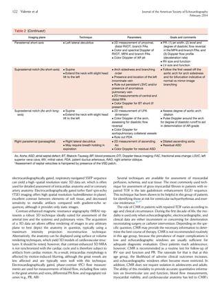
end-diastolic cross-sectional area 20 cm2
/m2
body surface area
(BSA) has been associated with CMR-measured RVend-diastolic vol-
ume index 170 mL/m2
.39
A diameter 42 mm at the base and
35 mm at the midventricular level indicate RV dilatation.23
Note that
the latter cutoff values are not adjusted to body size.
ii. Function.–Although guidelines for assessment of RV size and
function in adults are available,23
only limited information exists on
the accuracy, reproducibility, and prognostic value of these
echocardiography-derived data in patients with repaired TOF.
Nevertheless, a quantitative approach to assessment of RV size and
function is preferred to qualitative assessment (the ‘‘eyeball method’’)
because the latter has been shown to be inadequate.40
In general, 2D-based measurements correlate only modestly with
CMR-derived RV volumes and ejection fraction (EF), and the degree
of discrepancy increases as the right ventricle dilates.41
Evidence sug-
gests that compared with 2D-based measurements, 3D echocardiog-
raphy provides more accurate and reproducible quantification of RV
volumes.23,42-46
However, a meta-analysis suggested that 3D echo-
cardiography consistently underestimates RV volumes and EF,47
a
discrepancy that might increase as the right ventricle becomes
severely enlarged.44
However, only limited normative data are avail-
able to allow routine use of 3D echocardiography in the quantifica-
tion of RV volumes and EF. In a study of 70 patients with a variety
of CHDs undergoing transthoracic 3D echocardiography, Renella
et al.48
reported that RV volume and EF could not be measured in
42% because of technical limitations, mostly because of restricted
acoustic windows and an inability to include the entire chamber
within the 3D volume.
RV dilatation and dysfunction in patients with repaired TOF
adversely affect LV geometry and function.49
RV volume and pressure
overload are associated with flattening or leftward displacement of the
intraventricular septum, which results in a D-shaped left ventricle,
which can interfere with diastolic filling. Ventricular-ventricular interac-
tion is a term that has been used to describe the association between
worsening RV dilatation and dysfunction and declining LV systolic
function.50,51
Factors contributing to this phenomenon include the
above-mentioned geometric remodeling, shared myofibers, and a
shared pericardial space.11
Furthermore, the pathophysiology of the
right ventricle after TOF repair is associated with impaired electrome-
chanical synchrony, which affects global biventricular function.52,53
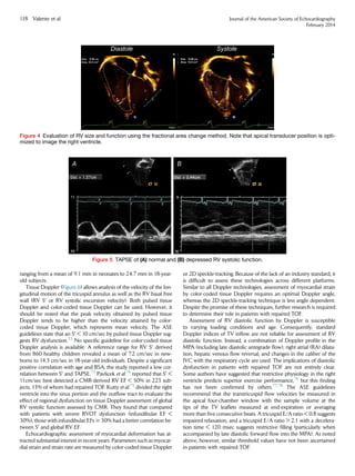
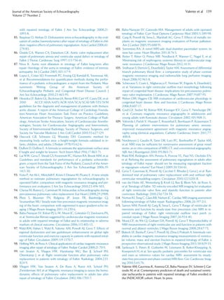
The percentage RV fractional area change is a measure of RV sys-
tolic function and is defined as ([end-diastolic area À end-systolic
area]/end-diastolic area) Â 100. The RVendocardium is traced in sys-
tole (minimal area) and end-diastole (maximal area), as shown in
Figure 4. Care must be taken to trace the endocardium beneath the
trabeculations along the free wall. The lower reference value for
normal RV systolic function using this method is 35%. This value cor-
relates modestly with CMR measurements in patients with
CHD.54,55
In patients with repaired TOF, studies have shown low
to modest correlations between RV fractional area change and
CMR-derived RV EF.41,56
Nongeometric methods used to evaluate RV function include the
rate of pressure rise in the right ventricle (dP/dt),57
myocardial accel-
eration during isovolumic contraction,58-60
and the Tei index61
(also
known as the myocardial performance index). However, the clinical
utility of these parameters in patients with repaired TOF is un-
clear.62-65
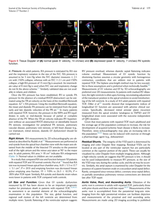
Another method of measuring longitudinal RV function is the
tricuspid annular motion or the tricuspid annular plane systolic excur-
sion (TAPSE). This parameter measures the excursion distance of the
lateral TV annulus during systole from the apical four-chamber win-
dow (Figure 5). The measurement can be obtained by M-mode or
2D imaging. The assumption that underlies this parameter is that it re-
flects global RV function, which may not be the case in patients with
repaired TOF. Studies in non-CHD patients have reported a modest
correlation between TAPSE and CMR-derived RV EF.66-68
Importantly, in patients with repaired TOF, the correlations
between TAPSE and CMR-derived RV EF and RV end-diastolic vol-
ume index are weak.56,69-71
American Society of Echocardiography
(ASE) guidelines indicate TAPSE of 16 mm as the lower limit of
normal RV systolic function in adult patients. Koestenberger et al.72
reported TAPSE values in 640 healthy children and demonstrated
that TAPSE had a positive nonlinear relationship with age and BSA,
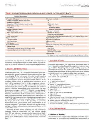
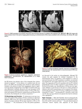
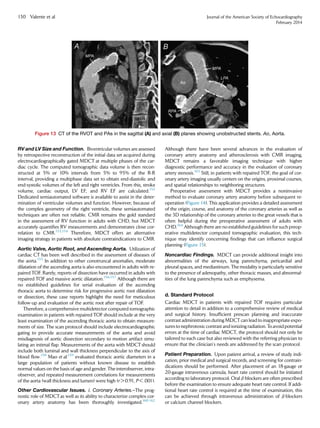
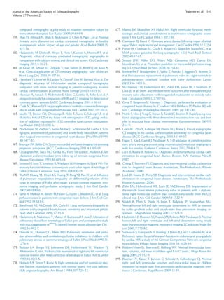
Figure 3 Apical four-chamber view (systolic frame) showing
measurement of TV tethering height.
Journal of the American Society of Echocardiography
Volume 27 Number 2
Valente et al 117](https://image.slidesharecdn.com/repairedtoffeb2014-160606145940/85/Repaired-tof-feb2014-7-320.jpg)
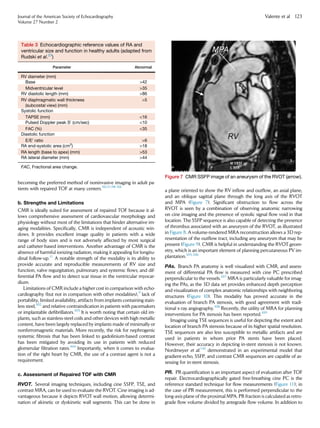
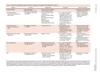
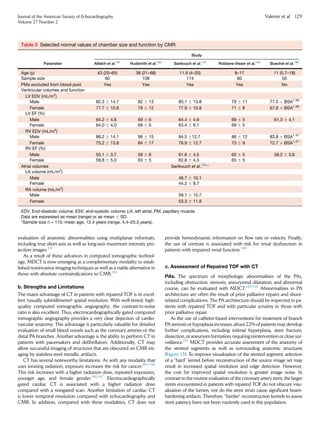
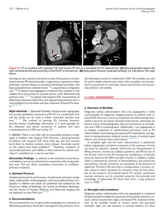

This document provides guidelines for using multimodality imaging to evaluate patients with repaired tetralogy of Fallot (TOF). It describes the role of echocardiography, cardiovascular magnetic resonance (CMR), computed tomography (CT), nuclear scintigraphy, and angiography. Echocardiography and CMR are well-suited for longitudinal follow-up due to lack of radiation. CMR is considered the reference standard for assessing right ventricular size and function and pulmonary regurgitation. A multimodality approach is recommended to comprehensively evaluate the complex anatomy and physiology while considering each patient's needs and institutional resources.


