Exam 2 structure function Protiens Biochem f2016 Syed A Jamal
•
1 like•125 views

Exam, on structure and function of protiens
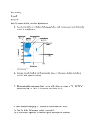
![[Hormone] nm θ θ θ
Protein A Protein B Protein C
0.1 0.024 0.15 .08
0.2 0.04 0.28 0.17
0.5 0.10 0.50 0.34
1 0.2 0.67 0.5
2 0.3 0.75 0.65
4 0.5 0.91 0.79
10 0.8 0.98 0.94
.
5. (a) Using the graph below, show that Log(Y/1-Y) =0 corresponds to the half-
saturation point.
(b)Why is the slope not constant for Hb?
(c)For Hb, why does the slope tend towards 1.00 at both high and low pressures of
oxygen?](data:image/gif;base64,R0lGODlhAQABAIAAAAAAAP///yH5BAEAAAAALAAAAAABAAEAAAIBRAA7)
Recommended
Recommended
More Related Content
What's hot
What's hot (20)
Similar to Exam 2 structure function Protiens Biochem f2016 Syed A Jamal
Similar to Exam 2 structure function Protiens Biochem f2016 Syed A Jamal (20)
More from Sayed Jamal
Recently uploaded
Recently uploaded (20)
Exam 2 structure function Protiens Biochem f2016 Syed A Jamal
- 1. Biochemistry Exam 2 Points 80 Best 20 answers will be graded for 4 points each. 1. Based on the tables provided on the last page below, pick 3 amino acids least likely to be present in an alpha-helix. . 2. Drawing peptide bond(s), briefly explain the kinds of information that the plot above provides with regard to proteins. 3. The protein alpha-glucosidase binds glucose with a dissociation rate of 7.6 * 103 M_1 s_1 and an overall Kd of 10nM. Calculate the association rate, ka. 4. Three proteins bind tightly to a hormone as shown in the data below. (a) Find the Kd for the hormone binding by protein C. (b) Which of these 3 proteins exhibits the tightest binding to the hormone?
- 2. [Hormone] nm θ θ θ Protein A Protein B Protein C 0.1 0.024 0.15 .08 0.2 0.04 0.28 0.17 0.5 0.10 0.50 0.34 1 0.2 0.67 0.5 2 0.3 0.75 0.65 4 0.5 0.91 0.79 10 0.8 0.98 0.94 . 5. (a) Using the graph below, show that Log(Y/1-Y) =0 corresponds to the half- saturation point. (b)Why is the slope not constant for Hb? (c)For Hb, why does the slope tend towards 1.00 at both high and low pressures of oxygen?
- 3. 8. 6. (a) What is the difference between sickle cell trait and sickle cell disease?
- 4. (b) Why do HbS heterozygotes have an advantage against the malarial parasite? Please focus on the biochemical and molecular basis. (c) Explain how the effects of sickle cell disease demonstrate that hemoglobin undergoes a conformational change upon releasing oxygen 7. (a) Prions (pronounced pree-ahns) have been implicated in Bovine spongiform encephalopathy (BSE or mad cow disease), sheep scrapie and Creutzfeldt- Jakob disease (CJD) of humans. Upon entering cells, prions convert normal proteins into prions just like themselves. Normal cell proteins have all the same "parts" as the prions--specifically the same amino acid building blocks--but they fold differently. Do you expect both the normal and the infectious versions of a prion protein to have the same primary structure? Why or why not? (b) Using the diagram above as a guide, explain the biochemical basis of Alzheimer’s disease. 8. Read the passage below and compare the oxygen binding curves for adult and fetal hemoglobins?
- 5. (a) Why does fetal hemoglobin have a greater affinity for oxygen? (b) What is this effect caused by? (c) How does Hemoglobin act as a blood buffer? Fetal hemoglobin's affinity for oxygen is substantially greater than that of adult hemoglobin. Notably, the P50 value for fetal hemoglobin is lower than adult hemoglobin (i.e., the partial pressure of oxygen at which the protein is 50% saturated; lower values indicate greater affinity). The P50 of fetal hemoglobin is roughly 19 mmHg, whereas adult hemoglobin is approximately 26.8 mmHg. As a result, the "oxygen saturation curve", which plots percent saturation vs. pO2, is left-shifted for fetal hemoglobin as compared to adult hemoglobin.
- 6. This greater affinity for oxygen is explained by the lack of fetal hemoglobin's interaction with 2,3-bisphosphoglycerate (2,3-BPG or 2,3-DPG). In adult red blood cells, this substance decreases the affinity of hemoglobin for oxygen. 2,3-BPG is also present in fetal red blood cells, but interacts less efficiently with fetal hemoglobin than adult hemoglobin. This is due to a change in a single amino acid (residue 143) found in the 2,3-BPG 'binding pocket': from histidine to serine, which gives rise to the greater oxygen affinity. Whereas histidine is positively charged and interacts well with the negative charges found on the surface of 2,3-BPG, Serine has a neutrally charged side chain at physiological pH, and interacts less well. This change results in less binding of 2,3-BPG to fetal Hb, and as a result oxygen will bind to it with higher affinity than adult hemoglobin.[2] For mothers to deliver oxygen to a fetus, it is necessary for the fetal hemoglobin to extract oxygen from the maternal oxygenated hemoglobin across the placenta. The higher oxygen affinity required for fetal hemoglobin is achieved by the protein subunit γ (gamma), instead of the β (beta) subunit. Because the γ subunit has fewer positive charges than the (adult) β subunit, 2,3-BPG is less electrostatically bound to fetal hemoglobin compared to adult hemoglobin. This lowered affinity of HbF for 2,3-BPG allows for adult hemoglobin (maternal hemoglobin) to readily transfer its oxygen to the fetal bloodstream. (9) (a) What is one known treatment of sickle cell disease and how does it work? (b) Suggest a technique that you would use in your laboratory to confirm that this treatment is effective at a molecular level. (10) (a) How is the tertiary structure of a protein generated?
- 7. (b) In the above diagram, name the process shown and also name the mediator(s) of the process. Is this a random process? Yes or no. Why? .
- 8. 11. (a) Label the carbons on the R group of Arginine. 12. What is desmosine? What is its clinical utility? 13. Which protein (one from lecture) is broken down in chronic obstructive pulmonary disease, with tobacco use, and in cystic fibrosis? 14. (a) Calculate the pI for glutamate using the titration curve below.
- 9. (b) Why is histidine frequent in the catalytic sites of enzymes and as a ligand in metalloproteins? 15. What is the basis of protein separation in ion-exchange and column chromatography? 16. You have a hypothesis that a novel protein is involved in a signaling pathway that triggers adipocyte differentiation. Signal transduction processes do have roles for phosphorylated proteins. Suggest the techniques you would use to show that this protein is indeed expressed at high levels in nuclear fractions of adipocytes. 17. What is the purpose of isoelectric focusing? Where and why is IE focusing used? 18. .What is molecular motion or breathing in the protein structure? (b)Why do glycine and proline act as helix breakers? 19. Fill in the blanks: Beta-2-microglobulins are components of ____________.
- 10. Proline is favored to take the second position in a _____ turn because the cis isomer, the conversion to which is catalyzed by _______________, has a __________degree amide bond or omega angle that allows a turn 20. Nitration of tyrosine is a free radical reaction that can affect proteins such as tyrosine. Draw the chemical structure of the product. 21. (a) Define a motif. (b) Draw a beta-alpha-beta motif. 22. Helix-turn-helix motif is common in DNA-binding proteins. (d) When the helix turn helix motif binds the major groove of DNA, what kind of interactions play a role? 23. The EF hand shown below is a helix-loop-helix structural domain or motif in a large family of calcium-binding proteins.
- 11. (a) In the diagram above, name an amino acid that you would expect to find in the region where divalent calcium binds. (b) Name one technique each that we use to study the secondary and tertiary structure of proteins. (c)What are some roles of intrinsically disordered proteins? Name the forces that are involved in ligand-protein binding. 24. (a) Define Kd as shown in the diagram below.
- 13. (b) In the graph above, why is the P50 of HbF different from that of HbA? 25. (a) In the graph above, determine the P50 for the oxygen-binding protein at different concentrations of BPG. What is the significance of elevated BPG levels in RBCs at high altitudes?
- 14. (b) In the diagram above, BPG interact with several positively charged groups (shown in blue) in a pocket between the β subunits on the surface of deoxyhemoglobin in the T state. A mutation has placed several amino acids with nonpolar aliphatic R groups in the areas shown in blue. Will BPG binding weaken or strengthen? Why or why not? (c) What are the interactions below that stabilize the T state of Hb? What happens to these interactions in the R state? What is the significance of these changes?
- 15. 26. (a) In the graph above, determine the P50 of Hb at each pH?
- 16. (b) What happens to the secondary structure of T state, above, upon oxygen binding? (c) What is allosteric regulation? (d) Why does carbon monoxide not bind to Hb as easily as it would bind free heme groups?