Functional group interconversions(oxidation reduction)
Main Exam Applied biochemistry final year
1. 1
SEMESTER ONE EXAMINATION – January 2024
MAIN
DEPARTMENT: Biosciences and Chemistry
MODULE TITLE: Applied Biochemistry
MODULE LEADER: Prof David Smith
EXAM DATE: 17th January at 09:30 UK time
DURATION: 4 hours
__________________________________________________________________
EXAMINATION CONDUCT:
1. The University Academic Conduct Regulation outlines the behavioural expectations of
candidates completing any examination.
2. Students are responsible for ensuring that they know how to submit their exam script,
when the deadline is and that they submit the script in enough time before the deadline
expires. It is anticipated that Blackboard will be slower around submission times.
3. It is a fundamental principle that students are assessed fairly and equitably. The
University Academic Conduct Regulation defines unfair behaviour relating to an
examination to be 'cheating'. The University will investigate and may sanction any acts
or behaviours which breach the Code of Academic Conduct.
4. Students are reminded that this is an individual task and that students who contact or
collude with other students to complete their exam may be subject to sanction later.
INSTRUCTIONS TO CANDIDATES:
1. This is a time limited examination; you are responsible for managing your time
appropriately. The duration is shown at the top of this page.
2. Answer all of Section A and TWO from FOUR questions in Section B
3. Academic support will be available for the 30 minutes of reading time from 09:30 via
https://shu.zoom.us/my/hwbds1
4. It is possible that you may encounter technical issues during the exam; if you have any
difficulty with IT you should consult the below student guidance document on My
Hallam which contains useful information on hints and tips, contact numbers and links
to support: https://www.shu.ac.uk/~/media/home/myhallam/Guides/student-exam-
guidance.docx
5. Any changes or clarification to the exam paper will be communicated via the module
Blackboard site announcements. It is recommended that students monitor Blackboard
announcements prior to submission of their final script but particularly in the first hour
after release of the exam paper.
2. 2
Section A - data analysis (50 marks)
The following sections require you to interpret data sets and apply information you that you have
encountered during the module. Answer ALL questions in this section.
Protein Expression (10 marks)
You are studying Protein Z, a crucial enzyme involved in human cellular signalling. Given the need for
a large amount of this protein for in vitro studies, you are using a bacterial expression system,
specifically E. coli.
A1 Describe the steps to express Protein Z in E. coli, assuming you already have the coding sequence
for Protein Z in a cloning vector. (4 marks)
You can use heat shock to express your protein in bacteria by first, taking the Protein Z cloning vector
and introducing it into the E. coli cells, then performing heat shock at 39℃-41℃, this will cause the
plasma membrane to open and allow the plasmid in. These cells should then be incubated on ice,
then at approximately 37℃, this should start the expression of Protein Z.
A2 Upon expression, you find that Protein Z is forming inclusion bodies in E. coli. What are inclusion
bodies, and why might they form during the overexpression of eukaryotic proteins in bacteria? (3
marks)
Inclusion bodies are aggregates, clumps of proteins that have formed due to the overexpression of E.
coli. They form during overexpression as it can overwhelm the folding process and lead to misfolding
which results in in inclusion bodies. Also, there may be a lack of or no molecular chaperones that
accompany the protein and allow it to unfold properly which could lead to inclusion bodies as well.
A3 Propose a series of strategies to optimise the expression conditions in E. coli to reduce the
formation of inclusion bodies and increase the yield of soluble Protein Z. (3 marks)
Use molecular chaperones, co-expression of the target protein with a molecular chaperone will
increase the recovery of the proteins, these chaperones can also be overexpressed. Secreting the
proteins into the periplasmic space or outside the cell will help proteins to unfold properly and cause
fewer inclusion bodies however, yield may be low but this may happen anyway if many inclusion
bodies form, suggesting it is a better method. Disulphide Isomerase is an enzyme that can help
protein unfolding, and co-expression with the protein this enzyme will help create functional
proteins that will result in a higher yield.
3. 3
Protein Structure (10 marks)
The figure below shows the thermal unfolding of recombinant human Apo A-1 as seen by Far-UV
Circular Dichroism (CD)spectra (buffer-subtracted) between 5°C (black line) and 90°C (olive-green line)
recorded in 5°C steps. The Apo A-1 concentration was 101 μM in PBS buffer adjusted to pH 7·4.
A4 State with justification the secondary structural changes that have occurred to the protein when
heated. (6 marks)
When starting the protein was a ⍺-helical shown by the peaks at 193nm, 208nm and 222nm. The
structure has changed into a less clear ⍺-helical but remains this structure suggested by the peaks at
193nm, 208nm and 222nm.
Given the CD spectra above, you observe the secondary structural changes in recombinant human
ApoA-1 upon heating. Now, consider the HSQC (Heteronuclear Single Quantum Coherence) spectra
below, representing the same protein at 5°C (Folded) and 90°C (Unfolded).
A5 Describe the differences observed in the HSQC spectra between the folded and unfolded protein
states. What do these changes suggest about the tertiary structure of the recombinant protein (Apo-
A-1) upon heating? (4 marks)
4. 4
The Folded spectra have dispersed peaks that are clearly separated and strong. The unfolded spectra
has peaks that are narrow, they are overlapping and close together, and they are also weaker. These
changes suggest that heating has lost the structural integrity of the protein as it is less defined, also
the fact that the spectra is less spread out suggests the protein may be more coil-like or have a
random structure rather than its previous defined structure.
Protein localisation (10 marks)
Eukaryotic Initiation Factor 2B (eIF2B) is a heterotrimer eukaryotic initiation factor. It is required for
most forms of eukaryotic translation initiation. During stress it associates with larges cellular
condensates known as Stress granules (SG). The image below shows the co-localisation analysis of a
mark for SGs (α-G3BP) and the alpha subunit of eIF2B (α-eIF2Ba) following 1h treatment with
500 M Sodium Arsenate (SA).
A6 Describe a possible method to allow the co-localisation of the two proteins to be imaged in
mammalian cells. (4 marks)
Immunofluorescence and fluorescence resonance energy transfer (FRET) can be used for co-
localisation of two proteins to be imaged in mammalian cells. Using the mammalian cells that
express the proteins of interest, they can be transfected with fluorescently tagged proteins such as
green fluorescent protein (GFP), and fixed for later staining. They need to be permeabilised to get
inside the cell as antibodies will not get through the cell membrane without this. The cells need to be
incubated with first the primary antibody then the second, then put through FRET, which is a process
where energy is transferred non-radioactively from an excited donor fluorochrome to another
molecule or acceptor. The donor fluorochrome will be excited and signals will be taken from the
donor and acceptor. You will be able to see the the proteins of interest.
A7 For your selected method, give one advantage and one disadvantage for its use in this
experimental context. (2 marks)
5. 5
The disadvantage of this method is that FRET will not work if the donor and acceptor molecules are
not within 10nm of each other as it depends on proximity. The advantage of using fluorophores is
that if the experiment is successful, you will be able to clearly see the interactions of two proteins in
different colours and enhance the experiment to gain better results.
A8 In the lower row, α-eIF2Ba has been mutated. What has been the effect on the complex's ability
to form? Describe how you have come to your conclusion. (4 marks)
The mutated α-eIF2Ba shows less co-localisation in comparison to the wild-type suggesting that the
mutation has affected the interaction between the SGs and eIf2B. The wild-type results show clear
images with different colours whereas the mutated results show weaker images that are not very
clear demonstrating that co-localisation has been affected and the complex has not fully formed. We
know that some fluorescent proteins such as GFP can affect the expression of proteins as some may
form aggregates or cause loss of function which may explain the mutant results.
6. 6
Gene Editing (10 marks)
The image below shows the Western blotting results from a CRISPR gene knockout experiment using
two different Cas9:gRNA complexes, to two different regions of a target protein. The control lane
shows the full-length protein of 50 kDa, lanes 1 and 2 shows the protein expression from the
resulting cell lines.
A9 Discuss the potential genetic alterations and molecular mechanisms that could lead to the
observed protein expression seen lane 1. (4 marks)
The observed protein expression in lane 1 may be due to incomplete or partial gene knockout which
has not led to complete loss of the gene. Also, the gene knockout may have been successful, but
another molecule may be expressing that protein as well. The experiment for the gene knockout may
have led to mutations that affected other mRNA leading to the protein still being coded for.
A10 Explain the implications of the absence of a band in lane 2. (2 marks)
The gene knockout was successful leading to the protein not being expressed and therefore not
having a band represent it, on the other hand, It could also be due to problems with the experiment
leading to inaccurate results.
A11 Comment on the variability of outcomes when using the CRISPR/Cas9 system for gene editing
and how this can be avoided in future experiments. (2 marks)
Unintended mutations or incomplete gene knockout may occur when using CRISPR/Cas9, but these
can be avoided with precautions and research, properly monitoring every step taken and its results
can help avoid mutations and ensure gene knockout. Performing the experiments as they should be
done will also help this.
7. 7
A12 How can the effectiveness of CRISPR knockout be further verified. (2 marks)
You can perform quantitative PCR to check the effectiveness of CRISPR knockouts, by
measuring the expression of the target gene, if there is no expression your experiment was
successful.
8. 8
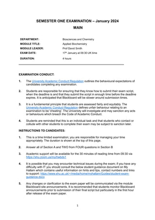
RNAi Manipulation (10 marks)
Figure. Main stages of the RNAi process. The image above shows the main stages of gene knockdown that could be used to
silence a gene within cell culture.
A12 Write a short (~200 word) figure legend for this image (above) that would be understandable by
an A-level student studying Chemistry and/or Biology. Directly reference the four steps in the answer.
(10 marks)
Step 1 consists of dicer, an endoribonuclease protein that cuts the RNA into smaller pieces and,
siRNAs are small interfering RNAs taken from long double-stranded RNAs. In step 2, a small RNA
complex called argonaute binds to the short RNA and a RNA-induced silencing complex (RISC)
effector complex will formed. In step 3, the siRNA has unwounded and bind to mRNA which directs
RISC to bind to specific mRNAs that are needed. In step 4, the argonaute will catalyse the cleavage of
the mRNA and it will be degraded as siRNAs can stop the production of certain proteins the mRNAs
are targeted.
(We will be looking at the accurate descriptions of the stages rather than the spelling and structure
of the answer)
10. 10
Section B Problem Solving (50 marks)
TWO questions from FOUR each are worth 25 marks.
Each question should take ~40 min to complete.
In this section you are required to address two biochemical scenarios from a choice of four. In each
case you are required to:
- Design an experimental plan to address the scenarios. Your approach should be feasible with
current technology and expected outcomes given.
- Describe the theoretical background to the main method(s) you have chosen.
The total answer should not exceed 600 words (approximately a page of typed text)
B1 You are a researcher investigating the effects of the chemotactic hormone vbr-1 on neutrophils.
The hormone vbr-1, is known to have diverse effects on the immune system. Previous data has
shown that:
• Acting on macrophages vbr-1 binds to a receptor called VR-1, leading these cells to migrate
towards the source of the hormone.
• Acting on T-cells it binds to a different receptor, VR-2, and is chemosuppressive, inhibiting
migration to the source of the hormone.
• You have made the novel observation that neutrophils express both receptor VR-1 and VR-2, and
are interested in understanding the effect of vbr-1 on these cells.
Given that neutrophils express both VR-1 and VR-2 (both potential receptors for vbr-1), design a
series of experiments to;
(1) Demonstrate that the chemotactic hormone vbr-1 binds to, and is internalised by, neutrophils
cultured in the laboratory.
(2) Show definitively which of the two receptors VR-1 or VR-2 is targeted by vbr-1 on neutrophils. To
do this you:
• have access to a drug that can irreversibly block binding to the VR-1 receptor but has no effect on
the VR-2 receptor.
• also know the genetic sequences of both VR-1 and VR-2.
Please record your answer here
11. 11
B2 You want to determine the structure of a soluble protein complex in two co-existing
conformational states. A proposed model with open and closed conformations is shown below.
(1) Given that each of the domains (shown in orange and red) can be purified independently,
design a series of experiments to gain the structure of the complex in its two states.
(2) Describe in your answer the limitations of each method and how together they can be used
to gain the complete structure.
Please record your answer here
B3: Severe combined immunodeficiency (SCID) is a group of rare disorders caused by mutations in
different genes involved in the development and function of infection-fighting immune cells. The
target gene for this study has been identified and coded as SHU21.
(1) Develop a plan for the ex vivo gene therapy of SCID.
First, you would isolate haematopoietic stem cells (HSCs) from bone marrow through a bone marrow
aspiration. Then you culture the HSCs to stimulate proliferation to have enough cells for gene editing
with growth factors. The cells should be exposed to the retroviral vector that has been replaced with
a healthy replica of SHU21 containing the cDNA copy of the defective gene. A lentiviral vector should
be used as can transduce dividing cells and can integrate into the host genome, it is the best choice
for immunological conditions. Incubate these cells to ensure successful reimplantation of the cells
and proliferation of the health SHU21 in the body. Also, any endogenous bone marrow progenitor
cells must be eliminated as well as avoid genotoxicity as some gene therapies have resulted in
leukaemia. The cells can then be infused back into the patient, where they will pass on the healthy
gene to all cells resulting in a possible cure for SCID. The cells should be tested to confirm that they
have been modified and the healthy SHU21 is being expressed, as well as to make sure there have
not been any negative effects or mutations. The patients will require regular checks and monitoring
to ensure the immune system of the patient is functioning as it should.
B4 The eukaryotic translation initiation factor complex eIF2B is known to form eIF2B bodies in
mammalian cells. Recently, in yeast, the protein SAC-1, a PI3K phosphatase, has been found to
interact with eIF2B. However, this interaction has only been observed in vitro, and SAC-1 is known to
interact with microtubules. It has not yet been confirmed whether this interaction occurs in
12. 12
mammalian cells. To investigate this further, you have access to a stable cell line that expresses
eIF2B-GFP.
Provide an outline of the experimental approach you would take to determine if this interaction is
conserved in mammalian cells. Specifically:
(1) Show that SAC-1 interacts with eIF2B in mammalian cells.
Co-localisation can be done using green fluorescent protein tagging and FRET. You can transfect the
stable cell line with a plasmid that will express the protein required and tag it with fluorescently
tagged antibodies to later view your cells. Controls should also be used to ensure accuracy in your
results for example, you may have one experiment that just expresses eIF2B, with a different
fluorescent which would also help compare the accuracy of different fluorophores. The expression of
the proteins can be confirmed with a qPCR or a western blot to maintain accuracy withing the
experiment. The cells must be fixed with reagents like paraformaldehyde to maintain structure
carefully to avoid false negative or positive results. Also, the cells have to be permeabilised will a
substance like detergent to allow the antibodies to probe inside the cell as they would not get
through the cell membrane.
FRET can be used to see the interactions between the proteins, when the donor fluorophore is
excited energy will be transferred non-radioactively to the acceptor as long as the two proteins are
withing 10nm of each other, this will confirm whether or not the two proteins are interacting. If the
experiment does not show any viable results, its suggests that the two proteins are not interacting. If
there are viable results you should be able to view the interaction of the two proteins from the
images.
(2) Design an experimental approach to test whether there is any interaction with
microtubules and test (assess/determine) whether microtubules are important for this
interaction.
A similar method can be applied to test the microtubules, the eIF2B protein can have its microtubule
structures distorted with an agent and both proteins can be tagged with a fluorescent protein. The
cells will be fixed and permeabilised to allow the antibodies to go into the cells. Using just
immunofluorescent microscopes, you should be able to view the changes that disrupting the
microtubules made on the structure of the protein. Also, using FRET again should allow you to see
the differences between the interactions of the proteins with their normal microtubule structure and
with it disrupted. If the results are very similar it would suggest the microtubules had no or very little
effect and if there have been a big change, its could prove that the structures has a big effect.