Atypical Metastatic Presentations in Colorectal Cancer A Case Series.pdf
•
0 likes•4 views

This case report describes 6 cases of unusual metastatic presentations in colorectal cancer patients. Case 1 involved bone and soft tissue metastases to the foot and calf. Case 2 involved a thyroid gland metastasis. Cases 3 and 4 involved scalp metastases. Case 5 involved pancreatic body and mandibular metastases over many years. Case 6 involved a young patient with mandibular and bone metastases. All patients received standard chemotherapy and biologic therapy. Localized therapies were used for symptom management or metastatic eradication in most cases. Atypical metastatic presentations can substantially impact quality of life and treatment decisions remain difficult due to limited information.

Recommended
Recommended
More Related Content
Similar to Atypical Metastatic Presentations in Colorectal Cancer A Case Series.pdf
Similar to Atypical Metastatic Presentations in Colorectal Cancer A Case Series.pdf (15)
More from Sarah Adams
More from Sarah Adams (20)
Recently uploaded
Recently uploaded (20)
Atypical Metastatic Presentations in Colorectal Cancer A Case Series.pdf
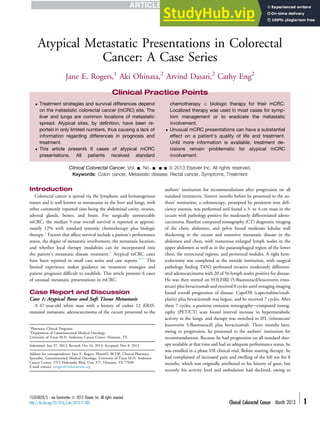
- 1. Case Report Atypical Metastatic Presentations in Colorectal Cancer: A Case Series Jane E. Rogers,1 Aki Ohinata,2 Arvind Dasari,2 Cathy Eng2 Clinical Colorectal Cancer, Vol. -, No. -, --- ª 2013 Elsevier Inc. All rights reserved. Keywords: Colon cancer, Metastatic disease, Rectal cancer, Symptoms, Treatment Introduction Colorectal cancer is spread via the lymphatic and hematogenous routes and is well known to metastasize to the liver and lungs, with other commonly reported sites being the abdominal cavity, ovaries, adrenal glands, bones, and brain. For surgically unresectable mCRC, the median 5-year overall survival is reported at approxi- mately 12% with standard systemic chemotherapy plus biologic therapy.1 Factors that affect survival include a patient’s performance status, the degree of metastatic involvement, the metastatic location, and whether local therapy modalities can be incorporated into the patient’s metastatic disease treatment.2 Atypical mCRC cases have been reported in small case series and case reports.3-11 This limited experience makes guidance on treatment strategies and patient prognosis difficult to establish. This article presents 6 cases of unusual metastatic presentations in mCRC. Case Report and Discussion Case 1: Atypical Bone and Soft Tissue Metastasis A 67-year-old white man with a history of codon 12 KRAS- mutated metastatic adenocarcinoma of the cecum presented to the authors’ institution for recommendations after progression on all standard treatments. Sixteen months before he presented to the au- thors’ institution, a colonoscopy, prompted by persistent iron defi- ciency anemia, was performed and found a 3- to 4-cm mass in the cecum with pathology positive for moderately differentiated adeno- carcinoma. Baseline computed tomography (CT) diagnostic imaging of the chest, abdomen, and pelvis found moderate lobular wall thickening in the cecum and extensive metastatic disease in the abdomen and chest, with numerous enlarged lymph nodes in the upper abdomen as well as in the paraesophageal region of the lower chest, the retrocrural regions, and peritoneal nodules. A right hem- icolectomy was completed at the outside institution, with surgical pathology finding T4N2 perforated invasive moderately differenti- ated adenocarcinoma with 20 of 56 lymph nodes positive for disease. He was then started on FOLFIRI (5-fluorouracil/leucovorin irino- tecan) plus bevacizumab and received 8 cycles until restaging imaging found overall progression of disease. CapeOX (capecitabine/oxali- platin) plus bevacizumab was begun, and he received 7 cycles. After these 7 cycles, a positron emission tomographyecomputed tomog- raphy (PET/CT) scan found interval increase in hypermetabolic activity in the lungs, and therapy was switched to IFL (irinotecan/ leucovorin 5-fluorouracil) plus bevacizumab. Three months later, owing to progression, he presented to the authors’ institution for recommendations. Because he had progression on all standard ther- apy available at that time and had an adequate performance status, he was enrolled in a phase I/II clinical trial. Before starting therapy, he had complained of increased pain and swelling of the left toe for 8 months, which was originally attributed to his history of gout, but recently his activity level and ambulation had declined, owing to Clinical Practice Points Treatment strategies and survival differences depend on the metastatic colorectal cancer (mCRC) site. The liver and lungs are common locations of metastatic spread. Atypical sites, by definition, have been re- ported in only limited numbers, thus causing a lack of information regarding differences in prognosis and treatment. This article presents 6 cases of atypical mCRC presentations. All patients received standard chemotherapy biologic therapy for their mCRC. Localized therapy was used in most cases for symp- tom management or to eradicate the metastatic involvement. Unusual mCRC presentations can have a substantial effect on a patient’s quality of life and treatment. Until more information is available, treatment de- cisions remain problematic for atypical mCRC involvement. 1 Pharmacy Clinical Programs 2 Department of Gastrointestinal Medical Oncology University of Texas M.D. Anderson Cancer Center, Houston, TX Submitted: Jun 27, 2013; Revised: Oct 16, 2013; Accepted: Nov 8, 2013 Address for correspondence: Jane E. Rogers, PharmD, BCOP, Clinical Pharmacy Specialist, Gastrointestinal Medical Oncology, University of Texas M.D. Anderson Cancer Center, 1515 Holcombe Blvd, Unit 377, Houston, TX 77030 E-mail contact: jerogers@mdanderson.org 1533-0028/$ - see frontmatter ª 2013 Elsevier Inc. All rights reserved. http://dx.doi.org/10.1016/j.clcc.2013.11.005 Clinical Colorectal Cancer Month 2013 - 1
- 2. persistent pain. Diagnostic workup and biopsy of the proximal phalanx of the hallux was consistent with adenocarcinoma compat- ible with a colon primary. Although his disease was stable, his pain in the left known metastatic toe became unmanageable at this time with supportive medication measures. The decision was made to hold systemic therapy for 4 weeks and proceed with palliative radiation therapy delivering 30 Gy in 10 fractions. Unfortunately, palliative radiation provided minimal relief of the patient’s symptoms. To complicate matters, before cycle 5 of therapy, the patient developed a soft tissue mass in the mid-left calf confirmed by ul- trasound that found a soft tissue intramuscular mass consistent with metastatic disease. At this time, the patient had a magnetic reso- nance imaging (MRI) scan of the foot, which found large expansile lytic metastases involving bones of the first ray of the left foot and nearby soft tissue; lytic metastases in the left calcaneus; probable small lytic metastases at the bases of the second, third, and fourth metatarsals; and small lytic intracortical metastasis in the left mid- tibial shaft, all consistent with metastatic progressive disease. The patient continued to have increasing pain and discomfort, resulting in lack of sleep and jerking movements throughout the night due to pain, increased fatigue, and emotional distress. He was referred to orthopedic consultation for potential options, including amputa- tion, for quality of life. He underwent radical resection of the left calf tumor and left foot partial amputation including amputations of the first and second toes. Unfortunately, the patient’s pain was ongoing, and he died shortly after the operation. Case 2: Thyroid Metastasis During the course of metastatic colon cancer treatment, a 65-year-old white woman was found to have a thyroid metastasis. She was originally diagnosed to have a T3N0M1 sigmoid colon carcinoma status post sigmoid resection, of pelvic peritoneum, and left ureteronephrectomy. She proceeded to receive adjuvant CapeOX plus bevacizumab for 3 months at an outside institution. She was lost to follow-up but then presented again 4 years later with recurrent disease in the lung. PET/CT scan reported a 5.5 5.1-cm left upper lobe mass with a standardized uptake value of 11.3 and evidence of mediastinal lymph node enlargement in addition to uptake in the left thyroid. A soft tissue neck ultrasound found an inferior nodule in the left lobe of the thyroid measuring 1.6 1.4 0.9 cm with a superior colloid nodule. Lung biopsy and fine-needle aspiration biopsy of the left thyroid were both posi- tive for metastatic adenocarcinoma from a colorectal primary. She began treatment with FOLFOX (5-fluorouracil/leucovorin oxali- platin) and bevacizumab. After 12 cycles, response was seen, and the patient underwent lung resection with segmentectomy of the chest wall, rib resection, and left upper lobe apical and posterior segmen- tectomy with en bloc resection of ribs 1, 2, and 3. Surgical pathology found metastatic adenocarcinoma consistent with a colorectal pri- mary with treatment response. Tumor mutation testing was consis- tent with KRAS and BRAF wild-type tumor. The patient was given 3 months of adjuvant therapy with capecitabine and bevacizumab. After adjuvant therapy, a CT scan of the chest, abdomen, and pelvis found no evidence of metastatic disease. Thyroid ultrasound found stable colloid nodules. The 1.6-cm mass identified previously was not seen on this ultrasound. On examination, her thyroid moved easily without deglutition; no nodularity was appreciated in the thyroid; no voice changes or hoarseness were identified; she had stable weight; and she had a relatively good energy level. She was not thyroid hormone dependent. She underwent a left thyroid lobectomy and isthmusectomy for curative isolated metastatic treat- ment. Pathology was negative for malignancy. She was placed on surveillance. One year after her thyroid surgery, a PET/CT scan found recurrent lung metastasis. She has since received treatment with capecitabine and bevacizumab and is currently on this regimen. Cases 3 and 4: Scalp Metastasis A 50-year-old white man with an 8-year history of moderately differentiated rectal adenocarcinoma was most recently found to have a small nodule on his scalp. He was treated at an outside institution with stage III rectal adenocarcinoma after neoadjuvant chemo- radiotherapy with capecitabine and oxaliplatin, with surgical pa- thology resulting in a T3N1M0 moderately differentiated rectal adenocarcinoma. He received 3 months of adjuvant FOLFIRI. Five years after adjuvant therapy, he was found to have biopsy-confirmed recurrent metastatic disease to a left upper lobe nodule measuring 1.5 cm, with a left perihilar adenopathy. He began systemic chemo- therapy with FOLFIRI plus bevacizumab for 8 cycles. He subse- quently underwent a left upper lobectomy and mediastinal lymph node dissection. He received chemoradiotherapy with capecitabine to a residual paratracheal node. He was then followed up on surveil- lance. Approximately 1 year after completion of chemoradiotherapy, the patient noted a small nodule on his scalp believed initially to be a cyst. The patient did not report any symptoms. Pathology from a wide local excision of the frontal scalp involving the dermis and subcutaneous adipose tissue with negative margins was consistent with mCRC. No other scalp lesions have developed. A 45-year-old white woman with unresectable metastatic colon cancer recently presented to the authors’ institution for treatment recommendations. Approximately 1 year earlier, she had increased abdominal cramping and dysmenorrhea. She was found to have a stage I, grade 1 endometrial cancer and underwent a total abdominal hysterectomy with bilateral salpingo-oophorectomy and extensive pelvic lymphadenectomy. Postoperatively, she recovered well but continued to have right upper quadrant abdominal discomfort that prompted CT imaging. Imaging found multiple large heterogeneous liver lesions throughout the liver, with the largest lesion measuring 9 8.6 cm. A biopsy found moderately differentiated adenocarcinoma with a CK20-positive and likely gastrointestinal primary. CT chest imaging found multiple bilateral 2- to 3-mm pulmonary nodules. A colonoscopy was performed and identified a near obstructing mass located at the splenic flexure and consistent with moderately differentiated adenocarcinoma. She was advised to begin FOLFIRI therapy with the addition of bevacizumab once her current rectal bleeding symptoms are resolved. During her surgical port placement, she was found to have a new nodule on the left side of her scalp. She did not report any symptoms associated with this nodule. The lesion was surgically removed and was positive for mCRC with involved margins. To date, no other scalp lesions have developed. Case 5: Pancreatic Body and Mandibular Metastases A 58-year-old white woman with an extensive colon cancer his- tory dating back 17 years since initial diagnosis was found to have Unusual Metastatic Colorectal Cancer Sites 2 - Clinical Colorectal Cancer Month 2013
- 3. metastatic disease to the pancreas. Seven years after adjuvant 5-fluorouracil therapy for a stage III colon cancer, the patient was found to have bilateral lung nodules. She underwent resection of these lung nodules but deferred any adjuvant therapy. Three years later, she developed another lung recurrence in the right lower lobe of the lung. Surgery again was performed to remove the metastatic disease. She received adjuvant capecitabine for 6 months. A right submandibular lesion and a lesion in the right lower lobe of the lung were seen on imaging 2 years after her adjuvant therapy. Surgeries for both were performed, and pathology was consistent with metastatic colon carcinoma. She completed 6 months of adjuvant capecitabine. One year after completion of her most recent adjuvant capecitabine therapy, a PET/CT scan found a hypermetabolic lesion in the pancreatic body. Symptoms consisted of mild abdominal pain and heartburn. No other symptoms were reported. This was followed up for 4 months, when a PET/CT again found persistent hypermetabolic focus in the pancreatic body with distal pancreatic ductal dilatation. CT imaging found a mass measuring 2.8 1.8 cm in the pancreatic body. The patient had an endo- scopic ultrasound and fine-needle aspiration biopsy. Pathology was positive for metastatic adenocarcinoma consistent with a colonic primary. She underwent a distal pancreatosplenectomy, and pa- thology found moderately differentiated metastatic colorectal adenocarcinoma replacing 80% to 90% of the pancreas, with 1 of 29 lymph nodes positive. The spleen showed no pathologic change. She presented to the authors’ institution after this surgery. She was advised to begin FOLFOX adjuvant therapy for 6 months. Nine months after adjuvant therapy, she developed unresectable liver metastases and was given systemic chemotherapy with FOLFOX plus bevacizumab. Tumor mutational analysis was consistent with KRAS and BRAF wild-type tumor. Case 6: Mandibular Metastasis A recently diagnosed 26-year-old man with a KRAS and BRAF wild-type metastatic rectal cancer presented to the authors’ insti- tution. Three months earlier, a colonoscopy, prompted by rectal bleeding, had been performed and had found a poorly differentiated invasive adenocarcinoma of the rectum. His symptoms consisted of hematochezia, poor appetite, a 100-pound weight loss over 6 months, and difficulty eating with a loose mandibular molar. He also complained of left lower extremity swelling and knee pain that was difficult to bear weight on and impaired his work duties. A dental examination found ill-defined radiolucency of 1.5- to 2-cm diameter in the left posterior mandible, apical to tooth 17 (3rd molar) and extending to the base of the root tips and to tooth 18 (2nd molar). Radiographic evidence of cortical bone loss on the lingual aspect of the left mandible was present. A PET/CT scan found the known rectal mass, multiple pelvic nodal metastases, multiple osseous metastases, bilateral cervical lymph node involve- ment, and a left mandibular metastasis. An MRI scan of the lower extremity showed findings consistent with metastatic disease within the distal femur and the proximal tibia, with a left tibia core-biopsy- proven metastatic poorly differentiated adenocarcinoma. The pa- tient began FOLFOX and bevacizumab plus zoledronic acid. Restaging PET/CT found excellent reduction in metabolic ac- tivity in the rectal/perirectal mass and reduction in metabolic ac- tivity associated with known adenopathy and bony metastases. Symptom improvement was also evident. He reported no pain or swelling in his left knee, ambulating without assistance, appetite improvement, and a stable pain management regimen with no new pains. The patient continued his current chemotherapy regimen plus zoledronic acid. Restaging PET/CT scan 2 months later found progressive disease in the primary rectal tumor. All other areas of disease were stable. He was continued on his current treatment regimen. Approximately 1 month after this PET/CT scan, he reported 10 days of constant, diffuse headaches that progressed from moderate to severe, double vision for 1 week, and difficulty swallowing and taking medications. Neurologic examination found multiple cranial nerve palsies including III, V, VI, VII, IX, X, and XII, with diplopia, dysarthria, and difficulty swallowing. MRI found leptomeningeal disease. Lumbar puncture found malignant cells consistent with mCRC. The patient’s functional status rapidly declined, and hospice care was recommended. Conclusion An improvement in systemic chemotherapy and biotherapy op- tions and an increased role of localized therapy over the past decade has significantly increased the survival for patients with mCRC. With longer survival and better diagnostic techniques, atypical metastatic presentations in colorectal cancer have been identified. Unusual metastatic sites reported as cases have included thyroid, breast, urethral, penile, testicular, cardiac, soft tissue, and subcu- taneous involvement.3-11 The limited number of reports makes it difficult to identify the best treatment approach and to determine chemotherapy/biotherapy sensitivity for these atypical sites. This article reports 6 cases of unusual metastatic presentations consisting of thyroid, pancreatic, and cutaneous scalp lesions, as well as bone and muscle metastasis to the toe, mandible, tibia, mid-calf, and femur. Five of the 6 cases incorporated localized therapy to the atypical metastatic site. Local therapies were incorporated to alle- viate symptoms, to eradicate the metastatic site, or to pursue curative intent. As evidenced by 1 case, quality of life and contin- uation of systemic therapy can be severely affected by the presence of atypical metastatic site development. The occurrence of atypical metastatic sites underscores the importance of conducting a comprehensive history, physical examination, diagnostic imaging, and follow-up of all reported symptoms for patients being treated for mCRC. Disclosure The authors have stated that they have no conflicts of interest. References 1. American Cancer Society. Colorectal Cancer Facts Figures 2011-2013. Atlanta, GA: American Cancer Society; 2011. 2. Mahmoud N, Bullard Dunn K. Metastasectomy for stage IV colorectal cancer. Dis Colon Rectum 2010; 53:1080-92. 3. Cozzolino I, Malapelle U, Carlomagno C, et al. Metastasis of colon cancer to the thyroid gland: a case diagnosed on fine-needle aspirate by a combined cytological, immunocytochemical, and molecular approach. Diagn Cytopathol 2010; 38:932-5. 4. Wakeham NR, Satchithananda K, Svensson WE, et al. Colorectal breast metastases presenting with atypical imaging features. Br J Radiol 2008; 81:e149-53. 5. Noorani S, Rao AR, Callaghan PS, et al. Urethral metastasis: an uncommon presentation of a colonic adenocarcinoma. Int Urol Nephrol 2007; 39:837-9, E-pub 2007 Feb 22. 6. Lo Russo G, Accarpio F, Spinelli GP, et al. Subcutaneous metastases from colon cancer: a case report. J Med Case Rep 2012; 6:212. 7. Rampa M, Battaglia L, Caprotti A, et al. Metastasis of sigmoid colon cancer in cryptorchid testis: report of a case. Tumori 2012; 98:63e-6e. Jane E. Rogers et al Clinical Colorectal Cancer Month 2013 - 3
- 4. 8. Lee JI, Kang WK, Kim HJ, et al. Unusual metastasis from a rectal adenocarcinoma: penile metastasis. ANZ J Surg 2011; 81:102. 9. Ishikawa N, Tanaka N, Yokoi K, et al. A case of rectal metastatic tumor in the soft tissue of the hand. J Nippon Med Sch 2007; 74:309-13. 10. Choi PW, Kim CN, Chang SH, et al. Cardiac metastasis from colorectal cancer: a case report. World J Gastroenterol 2009; 15:2675-8. 11. Moonda A, Fatteh S. Metastatic colorectal carcinoma: an unusual presentation. J Cutan Pathol 2009; 36:64-6. Unusual Metastatic Colorectal Cancer Sites 4 - Clinical Colorectal Cancer Month 2013