Acute Leukemia Cytogenetics
•
4 likes•1,300 views

General overview of WHO classification of Acute Leukemia, Genetic abnormalities Cytogenetics, Immunophenotyping

Recommended
Recommended
More Related Content
What's hot
What's hot (20)
Similar to Acute Leukemia Cytogenetics
Similar to Acute Leukemia Cytogenetics (20)
More from Sansar Babu Tiwari
More from Sansar Babu Tiwari (15)
Recently uploaded
Recently uploaded (20)
Acute Leukemia Cytogenetics
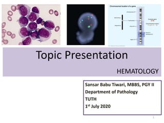
- 1. Topic Presentation HEMATOLOGY Sansar Babu Tiwari, MBBS, PGY II Department of Pathology TUTH 1st July 2020 1
- 2. Case A 52-year-old male Features of lethargy Complete blood count (CBC) showed pancytopenia • WBC: 1,620/µl – Neutrophils - 11% – Lymphocytes - 61% – Monocytes - 27% – Eosinophils - <1% – Basophils - <1% – Leukemic cells - 8% • Hemoglobin-8.8 g/dL • Platelets-98,000/µL Fibrinogen and d-dimer is increased. 2
- 3. Case AML M3 Suspected on clinical grounds. Bone Marrow biopsy with cytogenetics and flow cytometry ordered. BM findings: Bone marrow biopsy revealed about 100% cellularity, and that 80% of the nucleated elements were leukemic cells. The leukemic cells showed medium to large size, irregular shape, finely chromatinized nuclei with distinct nucleoli, and moderate amount of blue cytoplasm with azurophilic granules. The leukemic cells showed multiple Auer rods and frequently showed faggot cells 3
- 4. Case 4 MPO +
- 5. Case 5 Patient is started on Isotretinoin therapy. Did not show any improvement. So anthracycline derivative (daunorubicin) was added. Flow cytometry showed CD13, CD33, CD117, HLA-DR, and cytoplasmic-MPO, Negative CD34 ZBTB16-RARA translocation was detected on the leukemia gene screening test by reverse transcription-nested polymerase chain reaction. Cytogenetic study revealed t(11;17)(q23;q21)
- 6. Case 6
- 7. Case 7
- 8. Case Diagnosis reviewed: t(11;17); ZBTB16-RARA ATRA is now stopped and patient is doing well on Standard induction therapy. 7+3 therapy: • Cytarabine for 7 days • Daunorubicin on Day1, 2 and 3 • If FLT3 mutation: Add Midostaurin (Tyrosine kinase inhibitor) to the regimen. 8
- 9. Case Summary The present case emphasizes the importance of combining morphologic, immuno-phenotypic, cytogenetic, and molecular studies to distinguish variant APL cases from classical APL cases before initiating chemotherapy, regardless of whether the morphological study reveals findings consistent with those of classical APL. 9
- 12. Morphological difference between AML and ALL 12
- 13. Prognosis of AML and ALL 13
- 20. 20 Genetics: Cytogenetics VS Paracentric inversion
- 22. Acute Myeloid Leukemia 22 Blast count <20% = AML
- 23. Acute Myeloid Leukemia • The Cancer Genome Atlas Research Network – 200 cases of AML – 13 mutations per case – Overall: at least 23 recurrent mutations 23
- 24. AML with Recurrent genetic abnormalities. 24
- 26. • CD34+: Immature cells – t(8;21): 8 looks like B, so B cell markers seen (CD19, PAX5, CD79a) • RUNX: Run away from Killers (NK: CD56), to be safe. When CD56 present, bad prognosis. Auer Rods. – inv(16): CD2 + (e) – inv(3 = Mega): CD41, CD61. Aberrant expression of CD7 – BCR-ABL1: Aberrant expression of CD7 and CD19 – CEBPA: Absent Monocytic markers (CD14, CD64) – RUNX1: Present Monocytic markers • CD34-: Differentiated cells – t(15;17): Promyelocyte – t(9;11): Monocyte – t(6;9): Monocyte (CD15+) – t(1;22): Megakaryocyte (1+2=3=Mega) – NPM1 - Myelomonocytic 26 Immuno-phenotype in AML
- 27. 27 Chromosomal abnormalities in AML
- 28. AML with Recurrent genetic abnormalities: Balanced translocations or inversions. 28
- 29. AML: t(8;21);RUNX1-RUNX1T1 - 1-5% of all AML - Most frequent abnormality in children - D/D: FAB M2 29Dr. Kiril Lyapichev, MD Anderson
- 30. AML: t(8;21);RUNX1-RUNX1T1 - Myeloblast - Indented nuclei - Basophilic cytoplasm with a prominent perinuclear hof that may contain azurophilic granules. - Some may show large granules (Pseudo Chediak Higashi granules) - Promyelocyte, myelocyte and metamyelocyte - Often large and their cytoplasm has a waxy orange appearance and lacks a granular texture in Romanowsky stain - Pseudo Pelger Huet anomaly - Neutrophils: Homogenous pink cytoplasm (Salmon pink) - Auer rods, with tapering ends and present in blasts and maturing neutrophils. 30
- 31. AML: t(8;21);RUNX1-RUNX1T1 - Bone marrow eosinophilia is common but not dysplastic as in inv(16) - Erythroblastic and megakaryocytic lineage show normal morphology - Myeloid sarcoma may be present at diagnosis, in which case the blast percentage may be low. - Sometimes the manifestations of concurrent systemic mastocytosis is obscured by the acute leukemic infiltration of the bone marrow. - Good prognosis except in CD56 or CKIT. - More than 70% cases will show additional cytogenetic abnormality including loss of sex chromosome or del(9q) 31
- 32. AML: t(8;21);RUNX1-RUNX1T1 - Blasts: - Usually have myeloid markers like strong CD13, CD34, HLA-DR, MPO; and weak CD33. - Neutrophils: - CD15, CD65 - Maturation asynchrony: - CD15+ CD34+ - Lymphoid markers: - 8 = B: CD19, CD79a, PAX-5 - NK cell: - CD56 32
- 34. AML: t(8;21);RUNX1-RUNX1T1 34 Myeloblasts with abundant cytoplasm Indented nuclei and perinuclear hof Auer rods
- 35. 35
- 36. AML: inv(16) or t(16;16); CBFB-MYH11 - 5-8% of AML cases - Previously classified as AMML with abnormal eosinophils (FAB M4E0) - Younger patient - Extramedullary myeloid sarcoma (CNS predilection) - WBC count is significantly higher than in t(8;21) - No peripheral blood eosinophilia. 36
- 37. AML: inv(16) or t(16;16); CBFB-MYH11 37 Dr. Kiril Lyapichev, MD Anderson
- 38. AML: inv(16) or t(16;16); CBFB-MYH11 - Myeloblasts - Monocytic component (may be NSE negative) - Abnormal immature eosinophils without maturation arrest. - Atypical large purple-violet granules in addition to eosinophilic granules in their cytoplasm - Immature eosinophilic granule in late promyelocyte and myelocyte stages of development - Mature eosinophils often show nuclear hyposegmentation. 38
- 39. AML: inv(16) or t(16;16); CBFB-MYH11 - Occasional cases with this abnormality lack eosinophils, show only granulocytic maturation without a monocytic component, or show only monocytic differentiation. - The naphthol AS-D chloroacetate esterase (CAE) reaction which is normally negative in eosinophils can be faintly positive in abnormal eosinophils. (Significant finding) 39
- 40. AML: inv(16) or t(16;16); CBFB-MYH11 - Immature blasts - CD34 and CD117 (KIT) - Granulocyte lineage - CD13, CD33, CD15, CD65 - Monocyte lineage - CD4, CD14, CD64 - Aberrant expression of CD2 - Used for monitoring MRD 40
- 41. AML: inv(16) or t(16;16); CBFB-MYH11 - 40% cases have secondary genetic abnormality: - Gain of chromosome 22 and 8 - Trisomy 22 is fairly specific for inv(16) good outcome - Gain of chromosome 8 can be seen with patients with other primary aberrations - Del(7q) - Gain of chromosome 21 - KIT mutation has higher risk of relapse and worse survival (less significant than with t 8;21) 41
- 42. AML: inv(16) or t(16;16); CBFB-MYH11 42
- 43. AML: inv(16) or t(16;16); CBFB-MYH11 43
- 44. 44
- 45. AML: t(15;17); PML-RARA - AML with similar morphology resembling APL (FAB M3) may be seen. - Patients are relatively young and present with hemorrhagic manifestations. - Clinically: - Anemia and thrombocytopenia (due to platelet consumption in DIC) - Clinical or laboratory evidence of DIC and fibrinolysis, which may worsen with initial cytolytic response to chemotherapy. 45
- 46. AML: t(15;17); PML-RARA - Morphology: - Promyelocytes with coarse azurophilic granules and multiple Auer rods. - Characteristic folded, reniform or bilobed nucleus. - Hypergranular/typical variant: More common: Leukopenia. Auer rod is typically larger and ultrastructurally, they have hexagonal arrangement. If present, few promyelocytes in PBS. - Microgranular variant: Granules not visible on light microscopy. Leukocytosis. Promyelocytes can be seen on PBS. - Abnormal promyelocytes with deeply basophilic cytoplasm in relapse patients treated with isotretinoin. 46
- 47. AML: t(15;17); PML-RARA - Morphology: - MPO reaction is always strongly positive in all leukemic promyelocytes. Entire cytoplasm and nucleus. SBB and CAE also positive. - NSE is weakly positive in aprrox. 25% cases. - When in doubt between microgranular APL and monocytic AML, MPO often helps to break the tie. - Treatment: - Tretinoin (Maturation and macrophages contain Auer rods) and Arsenic trioxide (Increased osteoblasts surrounding trabeculae) 47
- 49. 49
- 51. AML: t(15;17); PML-RARA - Low or absent expression of: - HLA-DR, CD34 - Positive for: - CD 13 (heterogenous) and CD 33 homogenous) - Weak positive: - CD117 - Negative: - CD15, CD65 - Aberrant expression: - CD56 (worst outcome) - CD2 (17-15=2) 51
- 52. AML: t(V;17); v-RARA - Variant RARA translocations: - ZBTB16 { t(11;17); ZBTB16-RARA) - Regular nuclei - Many granules - Absence of Auer rods - Pelgeroid neutrophils - Strong MPO activity - NUMA1 t(11;17) (Respond to tretinoin) - NPM1 t(5;17) (Respond to tretinoin) - STAT5B t(17;17) 52
- 53. AML: t(9;11); KTM2A-MLLT3 - Common in children, but can occur at any age - D/D: M5a - Clinical features: - DIC - Extramedullary myeloid sarcoma and/or tissue infiltration (gingiva, skin) - Monoblasts ± promonocytes usually show strong positive NSE. - Strong expression of HLA-DR, CD4, CD33, CD65 - Few monocytic differentiation: CD14, CD64 - Few immature markers: CD34 (usually negative) 53
- 54. AML: t(9;11); KTM2A-MLLT3 54 Dr. Kiril Lyapichev, MD Anderson
- 56. AML: t(6;9); DEK-NUP214 - Occurs in both children and adults - D/D: FAB M2 or M4 - Basophilia and multilineage dysplasia (b;d) - Clinical features: - Pancytopenia - WBC usually lower than in other AML. (12000/MicroL) - Morphologically similar to AML with maturation and AMML. - Bone marrow or PB basophils ≥2%. - Erythrocytic and granulocytic dysplasia - Ring sideroblasts (69) are also seen. 56
- 57. AML: t(6;9); DEK-NUP214 - MPO ± NSE - Blasts: Non-specific myeloid immuno- phenotype: HLA-DR, CD13, CD33 - Few immature markers: - CD34, KIT - tDT in 50% - Monocyte marker: CD64 - Basophils: CD123, CD33 and CD38 but negative HLA-DR. - Poor prognosis 57
- 59. AML: inv(3) or t(3;3); GATA2, MECOM - Common in adults - D/D: Any FAB except M3 - Normal or elevated platelet count - Increased dysplastic megakaryocyte with unilobed or bilobed nuclei and tri(3)lineage dysplasia in the bone marrow. - PBS: - Hypogranular pelgeroid neutrophils ± Blasts - Bare megakaryocyte nuclei may be present - BM: - Hypocellular BM in some cases - Morphologically similar to M1, M4, M7 59
- 60. AML: inv(3) or t(3;3); GATA2, MECOM - HLA-DR, KIT, CD13, CD33, CD34 - High CD34 expression with inv(3) than t(3;3) - Megakaryocytic markers in few: - CD41, CD61 - Aberrant expression of CD7 (lymphoid marker) - Second abnormality: - Monosomy 7 in half of the cases - 5q deletions - RAS mutations (NRAS in 27%, KRAS in 11%, NF1 in 9%) 60
- 61. AML: inv(3) or t(3;3); GATA2, MECOM - Patients with BCR-ABL1 positive CML may acquire inv(3) or t(3;3) and such a findings indicates accelerated or blast phase of the disease. - Patients having t(9;22) and inv(3) or t(3;3) are considered an aggressive phase of CML rather than AML. - Poor prognosis. Even poorer if Monosomy 7 is present. 61
- 62. 62
- 63. AML: t(1;22); RBM15-MKL1 - Maturation in the megakaryocytic lineage (1+2=3=Mega) - Associated with FAB M7 - Most commonly occurs in infants without Down syndrome, with a female predominance. - This is a de novo AML restricted to infants and young children aged ≤3 years. - HSM, anemia, thrombocytopenia and a moderately elevated WBC. 63
- 64. AML: t(1;22); RBM15-MKL1 - Morphology: - Similar to M7 - The megakaryoblasts are usually medium-sized to large blasts with a round, slightly irregular or indented nucleus with fine reticular chromatin and 1-3 nucleoli. - Distinct blebs or pseudopod formation - Micromegas common, but dysgenesis in erythroid and granulocytes are usually not seen. - Dense marrow fibrosis, may mimic that of metastatic tumor. - SBB and MPO negative 64
- 65. AML: t(1;22); RBM15-MKL1 - Megakaryoblasts: - CD 41 (Gp IIb/IIIa) - CD 61 (Gp IIIa) - CD 42b (Gp Ib) - Myeloid: - CD 13 - CD 33 - Cytoplasmic staining of CD41 and CD61 is more specific and sensitive than membrane staining. - FLT3-internal tandem duplication: Poor prognosis 65
- 66. 66
- 67. AML: BCR-ABL1 - < 1% of all AML - < 1% of BCR-ABL1 positive acute and chronic leukemias - D/D: FAB M0-M2 - It occurs primarily in adults with a possible male predominance - Leukocytosis - In contrast to myeloid blast transformation in CML, patients with AML have less frequent splenomegaly (but if present it is massive) and lower peripheral blood basophilia (<2%). 67
- 68. AML: BCR-ABL1 - Morphology: - Less cellular marrow (80%) versus 95-100% in blast transformation of CML. - Dwarf megakaryocytes are less common than in CML. - MPO and SBB: Positive - CD34+ - Myeloid antigens: - CD13 and CD33 - Aberrant expression of CD19, CD7 and TdT - Second abnormality - Loss of chr 7 - Gain of chr 8 - Poor prognosis 68
- 69. 69 AML: BCR-ABL1
- 70. AML with Recurrent genetic abnormalities: Gene mutations. 70
- 71. AML: Mutated NPM1 - This AML type has myelomonocytic or monocytic features. - D/D: FAB M4 or M5 - 2-8% of childhood cases - 27-35% of adult cases - Female predominance - Anemia and thrombocytopenia - Higher WBC and platelet count - Extramedullary involvement: Gingiva, lymph nodes and skin. 71
- 72. AML: Mutated NPM1 - Immunostaining with anti-NPM1 antibodies (nuclear + cytoplasmic) reveals involvement of two or more bone marrow lineages (myeloid, monocytic, erythroid, megakaryocytic) in the vast majority of cases. - CD33 and CD13 expression - KIT, CD123, and CD110 positive - HLA-DR and CD34 negative - If CD34 +, worst prognosis 72
- 74. AML: Biallelic mutation of CEBPA - Provisional entity in 2008, definitive entity in 2016 - Children and young adults - Morphological resemblance to AML with or without maturation (FAB M1 or M2) - Higher hemoglobin levels - Lower platelet counts - Lower LDH levels - Multilineage dysplasia in 26% of cases of denovo AML with mutated CEBPA 74
- 75. AML: Biallelic mutation of CEBPA - Myeloid markers: - CD15, CD65, CD13, CD33 - Blasts: - HLA-DR, CD34 - Monocytic markers like CD14 and CD64 are usually absent. - Aberrant CD7 expression. - Favourable prognosis 75
- 76. 76 AML: Biallelic mutation of CEBPA
- 77. AML: Mutated RUNX1 - Provisional entity in WHO 2016 - 4-16 % of cases of AML - Common in older adults - Prior radiation exposure or alkylating agents - Lower hemoglobin and LDH - Low WBC count - Morphological resemblance to AML with monocytic or myelo-monocytic features (FAB M4 or M5) - HLA-DR, CD34, CD13 positive - Variable expression of CD33, monocytic markers and MPO - Poor prognosis 77
- 79. Lymphoblastic leukemia: Recurrent cytogenetic abnormalities. 79
- 80. Distinctive features and Considerations in ALL • Morphology is not helpful in differentiating types of ALL as for AML. • Immuno-phenotyping and cytogenetics are important. • t(5;14); IGH-IL3 has increased eosinophil as a reactive population. – Is the only morphological clue among ALL 80
- 81. 81 Distinctive features and Considerations in ALL
- 82. 82 Role of lumbar puncture in ALL Significance of PBS during CSF analysis for blasts.
- 83. 83 Role of lumbar puncture in ALL
- 87. 87 Genetic abnormalities in ALL
- 88. B-ALL: t(9;22) BCR-ABL1 - More common in adults - Positive for CD10, CD19 and TdT - Occasional positive for CD13, CD33 - CD25 is highly associated with B-ALL with BCR- ABL1. - Worst prognosis 88
- 89. B-ALL: t(9;22) BCR-ABL1 89 Black is a normal ABL Green is p210 Red is p190
- 90. B-ALL: t(v;11) KTM2A-rearranged - More common in infants < 1 year - High WBC count - High frequency of CNS involvement - t(4;11): CD19+, CD10- proB phenotype and also CD15+ - Others: t(19;11), t(9;11) - t(4;11) has a poorer prognosis 90
- 91. B-ALL: t(12;21) ETV6-RUNX1 - Not in infants but common in children - CD19+, CD10+, CD34+, CD20- - Very good prognosis, with cure seen in >90% - Even in with poor prognostic factors like high WBC and age > 10 years, these patients do far better 91
- 92. B-ALL: Hyperdiploidy - > 50 chromosomes without translocations or other structural aberrations - Common in children (not in infants) - CD19+, CD10+, CD34+ - Chromosome 21, X, 14 and 4 are usually involved, 1,2 and 3 least common - Trisomies 4 and 10 have good prognosis - Overall 90% patients are cured 92
- 94. B-ALL: Hypodiploidy - < 46 chromosomes - Child and adults - CD10+, CD19+ - Subtypes: - Near-haploid ALL (23-29): Childhood: RAS or receptor tyrosine kinase mutation - Low hypodiploid (33-39): Loss of function mutation in TP53 and/or RB1 - High hypodiploid (40-43) - Poor prognosis: Near haploid the worst one. 94
- 96. 96
- 97. B-ALL: t(5;14)- IGH/IL3 - Children and adults - Eosinophilia may be the presenting feature - This is a reactive population and not part of the leukemic clone - CD19+, CD10+ blasts in a presence of eosinophilia suggest this diagnosis - Unfavourable prognosis 97
- 99. B-ALL: t(1;19) – TCF3-PBX1 - Common in children - CD19+, CD10+, Cytoplasmic mu heavy chain + - Strong expression of CD9 and lack CD34 - Intermediate prognosis 99
- 100. B-ALL: BCR-ABL1 like - Common in high risk ALL children, adolescents and adults - Down syndrome children have high frequency of CRLF2 translocations - High WBC count - CD19+, CD10+ - Poor prognosis 100
- 101. B-ALL: iAMP21 - Amplification of a portion of chromosome 21 - Detected by FISH with a probe for RUNX1 - ≥5 copies of the gene - Or ≥3 extra copies on a single abnormal chromosome 21 - Common in older children - Poor prognosis 101
- 102. • Thankyou!!! 102
Editor's Notes
- There is overexpression of Annexin II on promyelocyte , which is a receptor for plasminogen and PAF. This leads to overproduction of plasmin and fibrinolysis.