Slide Presentation HISTOLOGY (Banff Classification
•
3 likes•356 views

Acute Graft Rejection of Kidney Transplantation

Recommended
More Related Content
What's hot
What's hot (20)
Similar to Slide Presentation HISTOLOGY (Banff Classification
Similar to Slide Presentation HISTOLOGY (Banff Classification (20)
More from Sansar Babu Tiwari
More from Sansar Babu Tiwari (14)
Recently uploaded
Recently uploaded (20)
Slide Presentation HISTOLOGY (Banff Classification
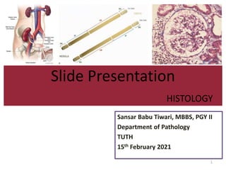
- 1. Slide Presentation HISTOLOGY Sansar Babu Tiwari, MBBS, PGY II Department of Pathology TUTH 15th February 2021 1
- 2. 1. History of transplant 2. Basic overview of immune responses in graft rejection 3. Normal Anatomy of kidney 4. Banff Classification of Renal Allograft Rejection 5. Case details 2
- 3. – Modern era of transplantation began in the 1950s – First successful kidney transplant was done in 1954 in Brigham Hospital in Boston between identical twins survived for 8 years and recurrence (major risk factor in twin recipients) – 1945 (Cadaver, septicemia), 1952 (Mother to son, single kidney, accident), 1954 (twin without immunosuppression) – Medawar found that allograft rejection is due to the immune response rather than non-specific inflammatory response. 3
- 4. – There is no need to take account of rhesus antigen compatibility in organ transplantation. – Permissible transplants are from: • Group O donor to O, A, B or AB recipient • Group A donor to group A or AB recipient • Group B donor to group B or AB recipient • Group AB donor to group AB recipient 4
- 5. 5
- 6. 6
- 7. 7
- 8. 8
- 9. 9
- 10. 10
- 11. 11
- 12. 12
- 13. 13
- 14. 14
- 15. 15
- 16. 16
- 17. 17
- 18. 18
- 19. 19 Rubin Zhang CJASN 2018;13:182-192
- 20. 20
- 21. 21
- 22. 22
- 23. 23
- 24. 24
- 25. 25
- 26. 26
- 27. 27
- 28. 28
- 29. 29
- 30. 30
- 31. 31
- 32. 32
- 33. Perplexing challenge to the pathologists. Why? • There is little time for consultation • Several disease can occur simultaneously • Wide range of potent therapy is possible whose selection depends on the accuracy of the diagnosis 33
- 34. 34
- 35. 35
- 36. 36
- 37. Calcineurin inhibitors are the mainstay of most standard protocols. (Cyclosporin, Tacrolimus) + Corticosteroids + Mycophenolate mofetil or Azathiprine 37
- 38. Short course of high dose steroids (oral) for 2 – 3 days If necessary rescue by ATG (anti- thymocyte globulin) 38
- 39. Renal biopsy: Gold standard to determine the cause of graft dysfunction. – 30% of recipients early after transplant. – 2 to 4 % per year after the first year. Biopsy findings change the clinical diagnosis in an average of 36% of patients and therapy in 59 %. 39
- 40. The sensitivity of single core is 90%. The predicted sensitivity of 2 cores is 99%. Ultrasound guided biopsy. No death among 5026 patients. (safe) 16-gauge better than 18-gauge (No complications) 40
- 41. 41
- 42. 5 sections at 3 levels (3 slides) 5 sections for Trichome (1 slide) 5 sections for PAS (1 slide) Total (5 slides) 42
- 43. Each section is carefully examined for a. Nature and degree of interstitial cellular infiltrate b. Arterial and arteriolar lesions c. Tubular injury, inflammation (tubulitis), viral inclusion bodies d. Glomerular lesions 43
- 44. At least 10 non-sclerotic glomeruli 2 arteries A normal medulla does not rule out rejection (low sensitivity) C4d staining can be done in tubules if sparse tissue 44
- 45. 45
- 46. Introduced in Banff, Canada in 1993 Had undergone number of significant modifications The most important of these were the incorporation of NIH Cooperative Clinical Trials in Transplant in 1999 which separated the category of endarteritis and addition of AMR in 2003. 46
- 47. 47
- 48. Areas that must not be considered for Banff “I” • Fibrotic areas • Immediate subcapsular cortex • Adventitia around large veins and lymphatics Asterisk (*) shall be placed if there are more than 5% to 10% eosinophils, neutrophils or plasma cells. 48
- 49. 49
- 50. 50
- 51. The presence of mononuclear infiltrate in the basolateral aspect of the renal tubule epithelium. (Reversed from 2015 leukocyte to 1997 mononuclear cells) The most severely affected tubule determines the score. Severely atrophic tubules (<25% of unaffected kidney) excluded. 51
- 52. 52
- 53. 53
- 54. 54
- 55. Arteries are defined as having 2 layers of smooth muscles in the media. Intimal arteritis (endothelialitis or endarteritis) is defined by the presence of inflammatory cells, mainly lymphocytes and monocytes in the subendothelial space of one or more arteries. Seen in both Acute TCMR and Active AMR. 55
- 56. Similar lesions in arterioles are only coded as an asterisk behind the Banff lesion score “ah” and are disregarded for “v”. Infiltrates buried deep into the intima are not considered for “v” but have been recognized as Chronic Active TCMR since 2005 and graded in the 2017 update as Grade II. 56
- 57. Asterisk (*) in “v” is attached when there is presence of tubulointerstitial hemorrhage and/or infarct. 57
- 58. 58
- 59. Glomerulitis is a form of microvascular inflammation (MVI) and is a feature of activity and antibody interaction with tissues in AMR. Defined as “complete or partial occlusion of 1 or more glomerular capillary by leukocyte infiltration and endothelial cell enlargement”. 59
- 60. 60
- 61. Determines the degree of inflammation within the peritubular capillaries. Together with glomerulitis, ptc constitutes MVI as a feature of Active AMR or Chronic active AMR. Peritubular capillaritis can be observed with pure acute TCMR or Borderline as well. 61
- 62. PTC are by definition found in the cortex. The number of luminal inflammatory cells includes PMN and mononuclear leukocytes. Asterisk (*) is used to indicate only mononuclear cells in the absence of neutrophils. 62
- 63. The extent of ptc inflammation should be documented either as Focal (10-50% of cortical area) Diffuse (>50% of cortical area) But this does not contribute to the score. Inflammatory cells within veins and medullary capillaries should not be scored. 63
- 64. 64
- 65. 65
- 66. 66
- 67. 67
- 68. 68
- 69. 69
- 70. 70
- 71. 71
- 72. 72
- 73. Not used to reach a diagnostic category and is purely descriptive 73
- 74. PAS positive arteriolar hyaline thickening. Asterisk (*) is added when arteriolitis is present. Not used to reach a diagnostic category and is purely descriptive 74
- 75. 75
- 76. Not used to reach a diagnostic category and is purely descriptive 76
- 77. All of the cortical parenchyma including areas of interstitial fibrosis and tubular atrophy (IFTA), subcapsular cortex and perivascular cortex including nodular infiltrates are considered for “ti” scoring. 77
- 78. 78
- 79. 79
- 80. 80
- 81. 81
- 82. 82
- 83. 83
- 84. 84 Category 3: Suspicious (Borderline) for Acute TCMR Foci of Banff t > 0 AND Banff i ≤ 1 OR Foci of Banff t1 AND Banff i ≥ 2
- 85. 85
- 86. 86
- 87. 87
- 88. 88
- 89. 89
- 90. • 48 year old male • Transplantation done 1 week back • Creatinine 3.9 mg/dl • 1 H & E stained slide • 1 PAS stained slide • 1 Trichome stained slide 90
- 91. 91
- 92. 92
- 93. 93
- 94. 94
- 95. 95
- 96. 96
- 97. 97
- 98. 98
- 99. 99
- 100. 100
- 101. 101
- 102. 102
- 103. 103
- 104. 104
- 105. 105
- 106. 106
- 107. 107
- 108. 108
- 109. 109
- 110. 110
- 111. Sections show renal parenchyma with glomeruli, tubules and interstitium along with few arterioles. There are 7 glomeruli, none of them show features of gomerulitis, double contour glomerulus and mesangial matrix proliferation. (g0, cg0, mm0) The proximal and distal tubules are focally infiltrated by the mononuclear cell infiltrate which constitutes less than 4 cells per 10 tubular epithelium even in severely affected areas (t1). Tubular atrophy are not seen (ct0). 111
- 112. Interstitium shows few foci of edematous areas as well as nodular infiltration of predominantly mononuclear cells in less than 10% of unscarred cortical parenchyma (i0). Peritubular capillaries show at least one leukocytes in most of them with upto 4 in severely affected one (ptc1). Arteritis, fibrous thickening, arterial hyalinosis or hyaline arteriolar thickening are not seen in any form (v0, cv0, ah0, aah0) 112
- 113. Interstitial fibrosis is not seen with Masson's Trichome stain (ci0, i-IFTA0). Total inflammation constitutes less than 10% of the renal parenchyma (ti0). C4d staining is not available (C4dx). 113
- 114. Tubular vacuolization or nuclear inclusions are not seen. 114 i 0 ptc 1 mm 0 t 1 ci 0 ah 0 g 0 ct 0 aah 0 v 0 cv 0 ti 0 c4d - cg 0 i-IFTA 0
- 115. 115 Category 3: Suspicious (Borderline) for Acute TCMR Foci of Banff t > 0 AND Banff i ≤ 1 OR Foci of Banff t1 AND Banff i ≥ 2
- 116. 116
- 117. 117
- 118. 118
- 119. Diagnosis: Suspicious for Acute TCMR (t1, ptc1) Differential diagnosis: Active AMR (provided c4d IHC>0 or c4d IF>1 and DSA positive) 119
- 120. • Thankyou!!! 120