
Recommended
More Related Content
What's hot
What's hot (20)
Similar to Acute myeloid leukemia
Similar to Acute myeloid leukemia (20)
More from namrathrs87
Recently uploaded
Recently uploaded (20)
Acute myeloid leukemia
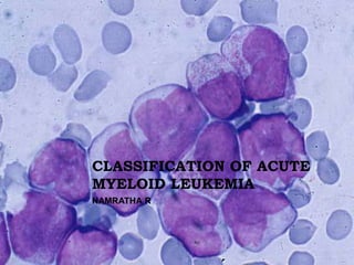
- 1. CLASSIFICATION OF ACUTE MYELOID LEUKEMIA NAMRATHA R
- 2. OVERVIEW CLASSIFICATIONS PROPOSED FOR ACUTE MYELOID LEUKEMIA 2008 WHO CLASSIFICATION- NEW FEATURES MORPHOLOGY,CYTOCHEMISTRY AND IMMUNOPHENOTYPING OF AML
- 3. THE PHENOTYPIC DIAGNOSIS Virchow was the first to use the term “Leukemia” The term "myeloid" was coined by Franz Ernst Christian Neumann in 1869, - first to recognize white blood cells were made in the bone myelos = (bone) marrow) as opposed to the spleen. 1900, the myeloblast- characterized by Otto Naegeli, who divided the leukemias into myeloid and lymphocytic
- 4. ACUTE MYELOID LEUKEMIA A group of relatively well-defined hematopoietic neoplasms involving precursor cells committed to the myeloid line of cellular development (ie, those giving rise to granulocytic, monocytic, erythroid, or megakaryocytic elements).
- 5. Classification is the language of medicine An effort so that everyone speaks the same language All diseases must be classified before being studied and treated Bhargava R, Dalal BI. Two steps forward, one step back: 4th WHO classification of myeloid neoplasms (2008). Indian J Pathol Microbiol 2010;53:391-4
- 6. The purpose of any classification- identify distinct biological entities which differ in aetiology, mechanisms of leukaemogenesis, clinicopathological features, and prognosis. Optimal treatment for such entities differs, their recognition is not only of scientific interest but is also essential for optimal care of patients.
- 7. CLASSIFICATIONS (1) by morphology and cytochemistry supplemented by immunophenotyping, as proposed by the French- American-British (FAB) group (2) by morphology, immunophenotyping and cytogenetics as proposed by the MIC groups (3) by immunophenotyping alone, as proposed by the European Group for the Immunological Classification of Leukemias (EGIL) (4) by the nature of the stem cell or progenitor cell in which the leukaemogenic mutation occurred. Classification of acute leukaemia: the need to incorporate cytogenetic and molecular genetic information Barbara J Bain Clin Pathol 1998;51:420-423
- 8. FAB In 1976 - FAB- a series of papers concerning the classification of acute leukaemia. Initially classification was based on cytology and cytochemistry. Subsequently - immunophenotyping - acute lymphoblastic leukaemia and facilitated the diagnosis of acute megakaryoblastic leukaemia and acute myeloid leukaemia with minimal evidence of myeloid differentiation (MO AML).
- 9. FAB- DEFINING A BLAST CELL Whether immature myeloid cells containing small numbers of granules are classified as blasts is a matter of convention. Myeloblasts rather than promyelocytes. Type I blasts lack granules and have a diffuse chromatin pattern, a high nucleocytoplasmic ratio and usually prominent nucleoli. Type II blasts resemble type I blasts except for the presence of a few azurophilic granules and a somewhat lower nucleocytoplasmic ratio.
- 10. Type II blast cells may contain Auer rods rather than granules; less often they contain large rectangular crystals or large inclusions (pseudo- Chédiak–Higashi inclusions). Other groups have proposed accepting a type III blast, which has more than ‘scanty’ granules but otherwise has no features of a promyelocyte Galton DAG (1962) Contributions of chemotherapy to the study of leukaemia. In: The Scientific Basis of Medicine, Annual Reviews. British Postgraduate Medical Federation, Athlone Press, London, pp. 152–171
- 11. No azuriphilic primary granules. No Auer rod Few (<20) azuriphilic primary granules. Auer rods may be seen. >20 azuriphilic primary granules without a Golgi zone. Auer rods may be seen.
- 12. Two groups of acute leukaemia, 'lymphoblastic' and myeloid Dysmyelopoietic syndromes that may be confused with acute myeloid leukaemia are also considered. The FAB classification requires that peripheral blood and bone marrow films be examined and that differential counts be performed on both. Br J Haematol. 1976 Aug;33(4):451-8. Proposals for the classification of the acute leukaemias. French- American-British (FAB) co-operative group. Bennett JM, Catovsky D, Daniel MT, Flandrin G, Galton DA, Gralnick HR, Sultan C.
- 13. Eight subtypes, M0 through to M7- based on the type of cell from which the leukemia developed and its degree of maturity. 30%* of the total nucleated cells in the marrow- blast cells If the bone marrow shows erythroid predominance (erythroblasts ≥50% of total nucleated cells) and at least 30% of non-erythroid cells are blast cells (lymphocytes, plasma cells and macrophages also being excluded from the differential count of nonerythroid cells) If the characteristic morphological features of acute promyelocytic leukaemia are present
- 14. FOR Some morphological categories recognised by the FAB group, specifically acute hypergranular promyelocytic leukaemia and its variant form (M3 and M3 variant AML) and L3 acute lymphoblastic leukaemia (ALL), were subsequently shown to indeed be biological entities Introduced objectivity Improved uniformity of diagnosis Common language for people studying and treating acute leukemias
- 15. AGAINST 30% or more myeloblasts and promyelocytes in marrow for diagnosis Ambiguity in the interpretations of descriptions Did not recognize the significance of myelodysplastic changes in acute myeloid leukemias or cytogenetic abnormalities
- 16. REVISED CLASSIFICATION-1986 Distinguish between MI and M2 M4 with eosinophilia added Defined the number of marrow cells to be counted to diagnose acute leukemia Definition of erythroleukemia (M6) and its distinction from refractory anemia or refractory anemia with an excess blast cells (RAEB). The Revised French-American-British Classification of Acute Myeloid Leukemia: Is New Better? CLARA D. BLOOMFIELD, M.D.; and RICHARD D. BRUNNING, M.D. Ann Intern Med. 1985;103(4):614-616. doi:10.7326/0003-4819-103-4-614 Acute basophilic leukemia was proposed as a ninth subtype, M8, in 1999. Duchayne E, Demur C, Rubie H, Robert A, Dastugue N (1999). "Diagnosis of acute basophilic leukemia". Leuk Lymphoma
- 17. MIC CLASSIFICATION MIC groups- classification of acute leukaemia on the basis of the FAB morphological classification supplemented by immunophenotype and cytogenetics - a morphological, immunological, cytogenetic (MIC) classification.
- 18. FOR AGAINST Cases with atypical cytogenetic findings or failed cytogenetic can be included Without cytogenetic anomaly but with molecular abnormality Morphological and cytogenetic similarity- differ at molecular level No single definable molecular lesion Accumulations of mutations of oncogenes and cancer supressor genes Eg- AML with alkylating drugs, AML in elderly
- 19. THE EUROPEAN GROUP FOR THE IMMUNOLOGICAL CLASSIFICATION OF LEUKEMIAS (EGIL) Acute leukaemia be classified on the basis of immunophenotype alone. This classification has the strength that it suggests standardised criteria for defining a leukaemia as myeloid, T lineage, B lineage, or biphenotypic.
- 20. Criteria for distinguishing biphenotypic leukaemia from AML with aberrant expression of lymphoid antigens, and from ALL with aberrant expression of myeloid antigens. However, a purely immunologic classification has the disadvantage that discrete entities may fall into one of two categories For example - some cases of AML of FAB M2 subtype associated with t(8;21)(q22;q22) would be classified as "AML of myelomonocytic lineage“ while others would be classified as "AML with lymphoid antigen expression," depending on whether or not a case showed aberrant expression of CD19.
- 21. 2001- WHO A Classification of Tumors of the Hematopoietic and Lymphoid Tissues as part of the 3rd edition of the series, WHO Classification of Tumors. Genetic information with morphologic, cytochemical, immunophenotypic, and clinical information into diagnostic algorithms for the myeloid neoplasms
- 22. Morphologic, genetic, and clinical data is a major theme of the WHO classification In the WHO classification, the blast threshold for the diagnosis of AML is reduced from 30% to 20% blasts in the blood or marrow. Patients with the clonal, recurring cytogenetic abnormalities t(8;21)(q22;q22), inv(16)(p13q22) or t(16;16)(p13;q22), and t(15;17)(q22;q12) should be considered to have AML regardless of the blast percentage
- 24. 2001 CLASSIFICATION OF AML AML with recurrent genetic abnormalities AML with multilineage dysplasia AML and myelodysplastic syndromes, therapy-related AML not otherwise categorized AML of ambiguous lineage
- 25. 20% to 29% blasts - similar clinical features, response to therapy and survival times—as those with 30% or more blasts. Identical profiles of proliferation and apoptosis Poor-risk cytogenetic abnormalities and increased expression of multidrug-resistance glycoproteins
- 26. 20% to 29% blasts - similar clinical features, response to therapy and survival times—as those with 30% or more blasts. Identical profiles of proliferation and apoptosis Poor-risk cytogenetic abnormalities and increased expression of multidrug-resistance glycoproteins
- 27. In addition, data from the International MDS Risk Analysis Workshop indicate that RAEBT is not an indolent disease. In that study, 25% of patients with 20% to 30% blasts evolved to AML in 2 to 3 months, 50% in 3 months, and more than 60% developed AML within 1 year. WHO proposal- 20% to 29% blasts and myelodysplasia will be classified as AML with multilineage dysplasia
- 28. The blast percentage, assessment of degree of maturation and dysplastic abnormalities - 200-cell leukocyte differential performed on a peripheral blood smear and a 500-cell differential performed on marrow Monoblasts and promonocytes and the megakaryoblasts - “blast equivalents” (APL)- blast equivalent is the abnormal promyelocyte.
- 29. Erythroid precursors (erythroblasts) – Not included in the blast count except - “pure” erythroleukemia. Dysplastic micromegakaryocytes are also excluded from the blast percentage. The percentage of CD34 cells should not be considered a substitute for a blast count from the smears or an estimate from the bone marrow biopsy. Although CD34 hematopoietic cells generally are blasts, not all blasts express CD34. Geller RB, Zahurak M, Hurwitz CA, et al. Prognostic importance of immunophenotyping in adults with acute myelocytic leukaemia: the significance of the stem-cell glycoprotein CD34 (My10). Br J Haematol. 1990;76:340- 347.
- 30. 2008 WHO CLASSIFICATION Expanded the genetic disease subtypes of AML- addition of 3 entities- t(9;11), t(6;9),inv(3),t(1;22) Two provisional- AML with NMPI or CEBPA mutation AML with multilineage dysplasia changed to AML with MDS related changes Proliferations associated with Down’s syndrome and blastic plasmacytic dendritic cell neoplasm
- 34. CLINICAL FEATURES Hepatosplenomegaly: Most AML t(1;22) and A Baso. Some AML inv(3) or t(3;3).. DIC : APL, AML t(9;11) CNS: Common in A Monoblastic and A Monocytic,APL with systemic relapse and inv(16) Adenopathy: AML NPM1. Rare in AML inv(3) or t(3;3).
- 35. Hb : Low in many AML. High in AML CEBPA Thrombocytopenia: many AML. Very common in AML t(6;9) and AML t(1;22). Thrombocytosis: highest count of all in AML NPM1. Common in AML inv(3) or t(3;3). Rarely in A Mega (more commonly thrombocytopenia). Platelet morphology: Occasional giant hypogranular platelets in AML inv(3) or t(3;3). Other- High LDH in AML CEBPA
- 36. MORPHOLOGY First step in the evaluation PBS, Aspirate, Trephine biopsy, Particle crush smears Critical for requesting ancillary studies Standard Romanowsky stains Wright unsuitable- granules suboptimal staining Diagnosis of AML with <20% blasts
- 37. MORPHOLOGIC EVALUATION Identification of blasts and blast equivalents Differentiation of blasts and promoncytes from mature monocytes Identification of blast cell features characteristic of a recurrent cytogenetic abnormality
- 38. The percentage of blasts - the percent blasts as a component of all nucleated marrow cells, with the exception of acute erythroleukemia If a myeloid neoplasm is found concomitantly with another hematologic neoplasm—for example, therapy-related AML and plasma cell myeloma— the cells of the nonmyeloid neoplasm should be excluded when blasts are enumerated.
- 39. NON BLAST MORPHOLOGY Monocytosis in AML inv(16) or t(16;16),AML t(9;11), AML with inv(3), AML w M. Topoisomerase therapy related Increase in eos - AML inv(16) or t(16;16), AML t(8;21), AML w M and AML t(8;21), Abnormal eos: AML inv(16) or t(16;16) Basophilia common in AML t(6;9), A Baso, AML w M, t-AML/t-MDS/MPN
- 40. DYSPLASIA MULTILINEAGE AML t(6;9), inv(3) or t(3;3) Cases of AML with MDS. AML NPM1 t-AML/t-MDS/MPN related with alkylating drugs Granulocyte dysplasia- AML with maturation, AMLt(8;21) Increased megakaryocytes with dysplasia- Inv(3) Intense fibrosis- Acute megakaryoblastic leukemia
- 44. AML characterized by a multitude of chromosomal abnormalities and gene mutations- marked differences in responses and survival following chemotherapy A detailed understanding of the molecular changes associated with the chromosomal and genetic abnormalities in AML - rationale for therapy design and biomarker development
- 45. AML WITH RECURRENT GENETIC ABNORMALITIES,” 30% of patients with AML Strong correlation between the genetic findings and the morphology The genetic abnormality can usually be predicted from the microscopic evaluation of the blood and marrow specimens. Distinctive clinical findings and a favorable response to appropriate therapy
- 46. AML with morphologic features suggestive of a specific genetic abnormality- the pathologist should issue a report that indicates the case may belong to a particular genetic category but that more data are required to prove it. The report should indicate what data are needed and whether the studies are in progress or if a new specimen is necessary. A carefully worded report that informs the clinician of what more needs to be done to accurately complete the diagnosis.
- 51. Most common structural aberration in AML 5-12% of AML and one third of cases of AML with maturation Predominantly in younger patients Myeloid sarcomas at presentation
- 53. Auer rods(long and sharp with tapered ends in neutrophils) Promyelocytes, myelocytes and neutrophils with variable dysplasia cytoplasm has a waxy, orange appearance and lacks a granular texture in Romanowsky-stained specimens
- 55. c-KIT mutations - adverse prognostic features Transcripts of the RUNX1-RUNX1T1 detected by RT-PCR even when they have been in remission for many years IPT: CD19 + and CD34 positive blasts in 2/3 of cases Co-expression of lymphoid and myeloid molecules is a well-known feature of acute myeloblastic leukemia (AML) with t(8;21). Subset is CD 56 positive- significantly shorter CR duration and survival
- 57. AML WITH INV(16)(P13.1Q22) OR T(16;16)(P13.1;Q22); CBFB-MYH11 10-20% of AML Morphological features of acute myelomonocytic leukemia with bone marrow eosinophilia (AML-M~EO). May be overlooked on cytogenetics Improved prognosis compared with other AMLs, similar to the t(8;21) AMLs higher WBC and blast counts vs t(8;21)
- 60. All stages of eosinophils in marrow with no significant maturation arrest Immature eosinophils granules at promyelocyte and myelocyte stage- Large, purple violet obscuring cell morphology
- 61. CYTOCHEMISTRY NSE faintly positive The abnormal eosinophils are weakly positive for chloracetate esterase. The blast cells express most of the myeloid- associated antigens, and some cases aberrantly express CD2.
- 63. APL WITH T(15;17)(Q24.1;Q21.1); PML-RARA Chromosomal translocation involving the retinoic acid receptor-alpha gene on chromosome 17(RARA). In 95% of cases of APL, (RARA) is involved in a reciprocal translocation with the promyelocytic leukemia gene (PML) on chromosome 15, a translocation denoted as t(15;17)(q22;q21). This rearrangement is seen in 13 percent of newly diagnosed AML and is highly specific for APL Typical, hypergranular and hypogranular
- 64. The t(15;17)(q22;q21), resulting in a PML/RARα fusion, is the most commonly identified abnormality, but other translocations of RARα on chromosome 17 with PLZF at 11q23, NuMA at 11q13 and NPM at 5q35 may occur. Cases that lack an RARα translocation or have a translocation involving PLZF do not respond to all- trans-retinoic acid and require a different therapeutic approach
- 66. ZBTB16-RARA VARIANT OF APML Predominance of cells that lack the characteristic bi-lobed or folded nuclei with many granules. Increased number of pseudo-Pelger-Huet cells with strong MPO reactivity.
- 68. 11Q23 REARRANGEMENTS IN AML The MLL gene on 11q23 is involved in a number of translocations with different partner chromosomes. The most common translocations - t(9;11)(p21;q23) and the t(11;19)(q23;p13.1); Strong association between AML M5/M4 and deletion and translocations involving 11q23. Two clinical subgroups of patients - one is AML in infants with MLL rearrangement in about 50% of cases; the other group is "secondary leukemia" (sAML) potentially after treatment with DNA topoisomerase II inhibitors.
- 69. M5a in half cases, M4 (20%), M1 or M5b (10% each), M2 (5%) DIC, extramedullary myeloid sarcoma and tissue infiltration (gingiva, skin) Prognosis may be superior to other AML with 11q23 translocations
- 70. ACUTE MYELOID LEUKEMIA WITH T(6;9) (P23;Q34) 0.7-1.8% of AML Chimeric fusion gene between DEK (6p23) and CAN/NUP214 (9q34). Any FAB morphology (except APM). Most common AML with maturation, acute myelomonocytic or megakaryoblastic. Considered by some to be a separate disease entity because of its distinct clinical and morphologic features and poor prognostic implication. Associated with a high frequency of FLT3 gene mutations
- 71. t(6;9) AML and FLT3-ITD mutations - higher white blood cell counts, higher bone marrow blasts, and significantly lower rates of complete remission >20% blasts, multilineage dysplasia with frequent ringed sideroblasts and basophilia (>2% in marrow and peripheral blood)
- 72. Presence of basophilia and myelodysplasia in an AML- FAB-M2 suggest this diagnosis,
- 73. IPT Immunophenotype. No specific immunophenotypic features have been recognized.
- 74. AML WITH INV(3) May present de novo or arise from a prior MDS. M4 was the most common type (11 cases, 37%), followed by M0 (6 cases, 20%), M2 (6 cases, 20%), M1 (3 cases, 10%), M5 (1 case, 3%), and M7 (1 case, 3%). Normal or increased platelet counts A subset of cases has less than 20% blast cells at the time of diagnosis- with features of chronic myelomonocytic leukaemia.
- 75. Atypical megakaryocytes in a background of multilineage dysplasia De novo acute myeloid leukemia with inv(3)(q21q26.2) or t(3;3)(q21;q26.2): a clinicopathologic and cytogenetic study of an entity recently added to the WHO classificationJianlan Sun1, Sergej N Konoplev, Xuemei Wang, Wei Cui, Su S Chen, L Jeffrey Medeiros and Pei Lin,Modern Pathology (2011) 24, 384–389; doi:10.1038/modpathol.2010.210; published online 26 November 2010
- 76. Acute myelomonocytic and dysplastic granulocytes acute myeloid leukemia without maturation showing a dysplastic granulocyte erythroid dysplasia; Trephine biopsy section, numerous dysplastic, and micromegakaryocytes (arrows) are present in a background of increased immature cells
- 77. Blasts are increased in the marrow aspirate. Dysplastic changes can also be noted in the myeloid and erythoid series. Dysplastic megakaryocytes cluster together.
- 78. After the publication of the 3rd edition of the classification in 2001- not only chromosomal rearrangement or numerical abnormalities but also mutated genes- establish the leukemic process and influence its morphologic and clinical features Discovery of new mutations in leukemogenesis paved the way for the genetic characterization of many cases of cytogenetically normal AML. Identification of new leukemic entity and powerful prognostic indicators James Vardiman, University of Chicago Medical Center, Chicago, USA
- 80. AML WITH MUTATED CEBPA(CCAAT/ENHANCER BINDING PROTEIN A (CEBPA) GENE ) CEBPA - transcription factor- development and differentiation of granulocytes from hematopoietic precursors. Newly diagnosed AML - 5% to 9%, with predominance for the M1 and M2 FAB morphologic subtypes. Independent prognostic marker- lower relapse rates and improved overall survival.
- 81. PATHOLOGIC FEATURES OF LEUKEMIC CELLS IN AML WITH MUTATED CEBPA Normal karyotype Many Auer rods seen in blasts in peripheral blood smear or bone marrow aspirate (Auer rods are abnormal, needle-shaped or round, light blue or pink-staining inclusions found in the cytoplasm of leukemic cells.) Aberrant CD7 expression on blasts
- 82. AML WITH MUTATED NPM1 Nucleophosmin is a nucleolar protein found mutated and rearranged in a number of haematological disorder Distinctive biological and clinical characteristics_support its inclusion as a provisional entity in the new WHO classification. High WBC counts Approximately 10% of therapy-related AML are NPM1-mutated. Blasts- Myelomonocytic or monocytic with dysplasia in atleast 2 cell lines
- 83. NPM1 staining pattern most commonly observed - leukaemic cells show both cytoplasmic and nuclear positivity for nucleophosmin the totality of leukaemic cells show an aberrant cytoplasmic expression of nucleophosmin Cells showing nucleus- restricted positivity in the centre of the field represent normal residual haemopoietic cells.
- 84. When the 2008 WHO classification was being prepared, the significance of an NPM1 mutation in the setting of morphologic dysplasia in an AML patient with NK was still unclear. Thus, the new WHO classification presently recommends that cases with overlapping features should be diagnosed as “AML with MD-related changes,” additionally annotating the presence of NPM1 mutation.
- 87. AML WITH MDS RELATED CHANGES >20% blasts in blood or marrow Dysplasia in 2 or more cell lines, generally including megakarocyes Dysplasia > 50% cells of atleast 2 lines in a pre treatment specimen De novo or following MDS or MDS/MPN When MDS precedes MPN- AML evolving from MDS
- 88. Previous, well-documented, history of MDS or MDS/myeloproliferative neoplasm Myelodysplasia-related cytogenetic abnormalities Multilineage dysplasia (ie, detection of dysplasia in 50% or more of cells in 2 or more myeloid lineages in bone marrow and/or peripheral blood smears). Absence of both Prior cytotoxic therapy for unrelated disease Recurrent cytogenetic abnormality
- 89. DYSGRANULOPOIESIS Hypogranulosis Hyposegmented (i.e., pseudo-Pelger Huet forms), or abnormally segmented nuclei. MPO may be aberrant, because patients may develop an acquired MPO deficiency as part of the dysplastic process.
- 92. DYSERYTHROPOIESIS: megaloblastoid forms;nuclear fragmentation and multinucleation karyorrhexis; mitoses within erythroid forms and cytoplasmic vacuolization.
- 94. Ringed sideroblasts, cytoplasmic vacuoles, PAS positivity
- 95. DYSMEGAKARYOPOEISIS Micromegakaryocytes, Better in sections than smears Approximately the size of a metamyelocyte and have few or no nuclear lobulations. mononucleated and multinucleated forms
- 96. S Monolobed nuclei Hypersegmented (osteoclastic appearing) megakaryocytes
- 97. The three-ball pawnbroker's symbol that these megakaryocytes resemble is ancient and may have derived from the insignia of the Medici family or the symbol of Saint Nicholas of Myra.
- 98. AML WITH MDS, THERAPY RELATED -Alkylating agent/radiation -Topoisomerase II inhibitor related
- 101. AML, NOT OTHERWISE SPECIFIED (NOS) 2008 WHO classification - AML, not otherwise specified (NOS) [Arber 2008c, Vardiman 2009]. Cases that fulfill general criteria for AML but lack: a) an AML recurrent cytogenetic or molecular abnormality, b) a link to prior chemotherapy, c) multilineage dysplasia involving the majority of cells, d) MDS-type cytogenetic abnormalities, e) an association with Down syndrome, or f) a history of MDS or MDS/MPN
- 102. AML, MINIMALLY DIFFERENTIATED No evidence of myeloid differentiation by morphology on light microscopy or cytochemistry Associated dysplastic features in erythroid and megakaryocyte lineages may provide indirect evidence that a leukaemia is myeloid, not lymphoid. Immunohistochemistry, flow cytometry, or EM cytochemistry to characterize as myeloid Cell or origin- Stem at earliest stage of myeloid maturation
- 103. PROOF OF MYELOID DIFFERENTIATION The demonstration of ultrastructural features of cells of granulocytic lineage, e.g. characteristic granules The demonstration of MPO activity ultrastructural cytochemistry The demonstration of MPO protein by immunocytochemistry with an anti-MPO monoclonal antibody. The demonstration of other antigens characteristic of myeloid cells by the use of monoclonal antibodies - CD13, CD14, CD15, CD33, CD64, CD65 and CD117 (but without expression of platelet-specific antigens, which would lead to the case being categorized as AML M7). Messenger RNA (mRNA) for MPO has also been
- 104. Medium sized,round to slightly indented nuclei, dispersed chromatin Agranular cytoplasm with varying basophilia No Auer rods 1-2 nucleoli M0 AML is associated with adverse cytogenetic abnormalities and poor prognosis
- 105. CYTOCHEMISTRY Fewer than 3% of blasts are positive for MPO, SBB and CAE Maturing myeloid cells may show peroxidase deficiency or aberrant positivity for both chloroacetate and non-specific esterase
- 106. IPT The blasts must not express CD79a and must express at least two myelomonocytic antigens such as CD13, CD33, CD15, or CD117 to be classified as myeloblasts. IPT- most specific lymphoid markers – CD3 and CD22 – not expressed in M0 AML Expression of less specific lymphoid-associated antigens such as CD2, CD4, CD7, CD10 and CD19, in addition to CD34, human leucocyte antigen DR (HLA- DR) and terminal deoxynucleotidyl transferase (TdT). CD7 is more often expressed than in other FAB categories of AML DDX- ALL, acute megakaryoblastic leukemia, Biphenotypic leukemia, leukemic phase of large cell lymphoma
- 107. ACUTE MYELOID LEUKEMIA WITHOUT MATURATION-M1 15-20% of AML Significant number of blasts(>90% of non erythroid cells) without evidence of maturation to neutrophils <10% of non-erythroid cells in the bone marrow belong to the maturing granulocytic component (promyelocytes to neutrophils). MPO/SBB positivity(>3% of cells) and/or Auer rods
- 108. Typically markedly hypercellular marrow, but normocellular and hypocellular cases occur Very immature cells with few azurophilic cytoplasmic granules Variation in size Irregular/indented nuclei with folding Prominent nucleoli Auer rods
- 109. CYTOCHEMISTRY Minimum of 3% of blasts that are positive for MPO or SBB. Hayhoe and Quaglino - SBB reaction is a more sensitive marker of early granulocyte precursors than MPO. Positive for CAE, - less sensitive than either MPO or SBB Weak or negative reaction for non-specific esterases. In the case of (ANAE) and (ANBE) the reaction is usually negative, whereas in the cases of naphthol AS-D acetate esterase (NASDA) there is usually a weak fluoride-resistant reaction. The PAS reaction- usually negative, but may show a weak diffuse reaction with superimposed fine granular positivity.
- 110. S
- 111. Auer rods give positive MPO and SBB reactions and occasionally weak PAS reactions. The reaction for CAE is usually weak or negative except in M2 AML associated with t(8;21)) in which Auer rods are often positive for CAE
- 112. MOLECULAR DESCRIPTION ● Associated with t(8;21) ● FLT3 ITD in 22% ● FLT3 mutations associated with HLA-DR negative patients
- 113. ACUTE MYELOID LEUKEMIA WITH MATURATION (FAB AML M2) 10% of AML cases; 5% of childhood leukemias Bone marrow maturing granulocytic component (promyelocytes to polymorphonuclear leucocytes) >10% of non-erythroid cells Bone marrow monocytic component (monoblasts to monocytes) <20% of non- erythroid cells and other criteria for M4 not met
- 114. S In contrast to M1 AML, blasts are often predominantly type II. Auer rods frequent(70%) Promyelocytes,myelocyte s and neutrophils>10% of cells Neutrophils-hyper and hyposegmentation,abnor mal nuclear shape Erythroid and megakaryocyte precursors - dyspoiesis and a frank panmyelosis is present in some cases
- 115. A leukemic blast population composed mostly of granulated promyelocytes vs acute promyelocytic leukemia. The latter often shows bilobed nuclei and a more homogeneous blast population compared with cases of AML with differentiation.
- 116. Maturation - along the neutrophil pathway but eosinophilic or basophilic maturation occurs in a minority. - M2Eo or M2Baso. Increased basophils/mast cells- deletion/translocation 12p(11-13) or t(6;9) Erythrophagocytosis- t(8:16) (p11:p13) AML with eosinophilia - M1/M2 t(8;21) Leuk Lymphoma.1995;18 Suppl 1:61-3. AML M1 and M2 with eosinophilia and AML M4Eo: diagnostic and clinical aspects.Löffler H, Gassmann W, Haferlach T.
- 117. S Myeloperoxidase+ blasts and immature neutrophils are intensely positive SBB positive
- 118. The cytochemical reactions in M2 AML - same as M1 AML- reactions are stronger and a higher percentage of cells are positive with MPO and SBB stains. CAE - more often positive in M2 than in M1 AML and reactions are stronger. Auer rods show the same staining characteristics as in M1 AML but are more numerous. A population of neutrophils- which lack SBB and MPO activity. This may be demonstrated cytochemically or by means of an automated differential counter based on the peroxidase reaction, which shows a low mean peroxidase score and an abnormally placed neutrophil
- 119. ACUTE PROMYELOCYTIC LEUKEMIA 5-8% of AML cases Median age 35-40 years, but can occur at any age Typical and microgranular APL- DIC Microgranular APL- Very high leucocyte count with rapid doubling time, The WBC is usually higher in M3V than in M3 AML.
- 120. MORPHOLOGY Examining an adequate bone marrow aspirate important The specimen may clot during attempted aspiration- associated hypercoagulable state, In acute hypergranular promyelocytic leukaemia, the predominant cell is a highly abnormal promyelocyte. In the majority of cases, blasts are fewer than 30% of bone marrow nucleated cells
- 121. Hypergranular APL, the most typical form, showing promyelocytes with cytoplasm packed with purple granules(120-1000nm). Bilobed/kidney shaped nuclei Auer rods, sometimes stacked in bundles of faggots
- 122. A minority of cells - agranular, have sparse granules or have fine red or rust-coloured dust-like granules. Cells that lack granules - lakes of hyaline pink material in the cytoplasm may also be seen. Basophilic differentiation No dysplasia
- 123. MICROGRANULAR APL Fine dustlike granulation(100- 400nm) in the cytoplasm Most of the cells show bilobed or folded nuclei, a – DDX acute myelomonocytic leukemia. Few of the cells - typical features of abnormal promyelocytes the prognosis of M3 variant is somewhat worse than that of M3 AML
- 124. Hyperbasophilic form, - cells with high N/C ratio, and strongly basophilic cytoplasm with either sparse or no granules. Conspicuous cytoplasmatic budding is usually present, recalling the feature of micromegakaryocytes Leukemia.1994 Sep;8(9):1441- 6. Acute promyelocytic leukemia: morphological aspects.Castoldi GL, Liso V, Specchia G, Tomasi P
- 125. AUER RODS OF PROMYELOCYTIC LEUKAEMIA Auer rods in promyelocytic leukaemia differ from those in M1 and M2 AML Larger Auer rods Hexagonal arrangement of tubular structure, specific periodicity- 250mm vs 6-20 of other AML Microfibrils and stellate configurations of rough endoplasmic reticulum - characteristic of M3 AML, particularly M3 variant
- 126. MPO- strongly positive, reaction product covering cytoplasm and nucleus MPO positivity - granules, Auer rods, perinuclear space and some rough endoplasmic reticulum profiles
- 127. Strong SBB and CAE positivity NSE- positive in 25%(The reaction is weaker than in monocytes) The PAS reaction usually shows a cytoplasmic ‘blush’ – a fine diffuse or dust-like positivity M3V AML usually shows similar cytochemical reactions but sometimes the reactions are weaker Cases with basophilic differentiation show metachromatic staining with toluidine blue. The acid phosphatase reaction is strongly positive. Strong lysozyme activity of granules and Auer rods, M3 variant AML activity - weak to moderately strong
- 128. Auer rods in M3 AML are SBB, MPO and CAE positive, whereas in other categories of AML they are usually negative with CAE Weakly PAS positive. SBB, MPO and CAE - core of the rod may be left unstained and occasionally the core is ANAE positive on a mixed esterase stain
- 130. ACUTE MYELOMONOYCTIC LEUKEMIA Proliferation of neutrophil and monocyte precursors 5-12% of AML > 20% blasts, Neutrophils and their precursors, monocytes and their precursors each > 20% of marrow cells Distinguishes from AML with or without maturation in which some monocytes may be present Peripheral blood- monocytes more mature than those in marrow
- 131. Goasguen JE, Bennett JM, Bain BJ, Vallespi T, Brunning R, Mufti GJ for the International Working Group on Morphology of Myelodysplastic Syndrome (IWGM-MDS). Morphological evaluation of monocytes an their precursors. Haematologica 2009;94:994-997. doi 10.3324/haematol.2008.005421
- 134. CYTOCHEMISTRY 3% of cells – MPO positivity, Monoblasts- MPO negative, Promonocytes-scattered positivity Monocytes, monoblasts and promonocytes- NSE positive If morphological criteria met- NSE negativity does not exclude diagnosis Double staining with NSE and naphthol ASD chloracetate esterase or MPO- dual positive cells DDX- Acute monocytic leukemia and AML with maturation
- 137. IPT Variably express myeloid markers- CD13, CD33 Markers of monocytic differentiation- CD14, CD 4, CD11c,CD11b, CD64, CD36 and lysozyme Residual population of less differentiated myeloblasts- CD34
- 138. ACUTE MONOBLASTIC AND ACUTE MONOCYTIC LEUKEMIA 80% of cells are of monocytic lineage- Monoblasts, promonocytes and monocytes Minor neutrophil component< 20% 5-8% of AML, young individuals Infancy- abnormalities of 11q23 Weakness, bleeding and a diffuse erythematous skin rash. High frequency of extramedulary infiltration of the lungs, colon, meninges, lymphnodes, bladder and larynx and gingival hyperplasia. Serum and urinary muramidase levels are often extremely high.
- 140. MORPHOLOGY AND CYTOCHEMISTRY Intense NSE activity 10-20% weak or negative NSE Monoblasts- MPO negative, Promonocytes- scattered positivity Biopsy-Monoblast- positive with antibody to lysozyme Promonocytes- CD68 and lysozyme positive
- 142. HAEMOPHAGOCYTOSIS- T(8;16)(P11;P13) M4, M5a, M5b Possible involvement of a granulo-monocytic precursor Cytoplasmic granules Extramedullary disease Poor prognosis
- 143. ACUTE ERYTHROID LEUKEMIA Predominant erythroid population Two subtypes based on presence and absence of significant myeloid population ERYTHROLEUKEMIA > 50% erythroid precursors in nucleated cell population >20% myeloblasts in non erythroid population PURE ERYTHROID LEUKEMIA(extremely rare) Neoplastic proliferation of immature cells - erythroid population(>80% of marrow cells) with no significant myeloid component
- 145. 1% of AML cases Children to >age 90, usually adults, male predominance 20% of therapy related AML but only 1% of de novo AMLDi Guglielmo syndrome: nonspecific clinical findings of anemia, thrombocytopenia, variable neutropenia Hemophagocytic lymphohistiocytosis: recurrent and specific complication Peripheral smear may have prominent erythroblasts
- 147. Dysplastic erythroid precursors megaloblastoid morphology with bi/multinucleated nuclei, Large multinucleated erythroid cells Dysplasia< 50% of cells
- 149. ERYTHROLEUKEMIA Iron stain- ringed sideroblasts PAS- globular/ diffuse staining MPO/SBB- positive in myeloblasts
- 151. >80%Erythroblasts with deeply basophilic, often agranular cytoplasm with poorly demarcated vacuoles, round nuclei, fine chromatin, and 1 or more nucleoli.
- 152. Block like staining with PAS Negative for SBB, MPO Reactive with alpha napthyl esterase and acid phosphatase IHC- HB, glycophorin
- 153. IPT Myeloblasts- CD33, CD13, CD117 with or without CD34 and HLA DR Erythroblasts- Glycophorin A, Haemoglobin A Gero antibody, CD36 and carbonic anhydrase 1
- 154. ACUTE MEGAKARYOBLASTIC LEUKEMIA Acute leukemia where >50% of blasts are of megakaryocyte lineage Up to 10% of AML in children, 5% or less of adult AML In 1963, Szur and Lewis - pancytopenia, low percentages of blast cells, and intense myelofibrosis but an absence of teardrop red cells, splenomegaly, leukocytosis, and thrombocytosis, the usual features of primary myelofibrosis. They designated the syndrome malignant myelosclerosis IPT- megakaryoblasts indicated the cases were variants of AML rather than of primary myelofibrosis
- 155. ADULTS CHILDREN Pancytopenia with low blast counts in the blood, myelofibrosis, an absence of hepatosplenomegaly, poor response to chemotherapy,and short clinical course. Markedly elevated LDH Acute myelofibrosis Leukocytosis Complete remission and long term survival Germ cell tumors in young boys. Hepatosplenomegaly- prominent abdominal masses- t(1;22)
- 156. Micromegakaryocytes- small cells, with mature cytoplasm and one or two round nuclei with condensed chromatin, Megakaryblastic fragments, dysplastic platelets Circulating hypogranular neutrophils
- 157. Medium to large blasts(12-18micromm) Round slightly irregular to indented nuclei Fine reticular chromatin 1-3 nucleoli Large and small blasts in the same patient Agranular, basophilic cytoplasm with pseudopod formation or blebs
- 158. ACUTE MEGAKARYOBLASTIC LEUKEMIA WITH T(1;22)(P13;Q13); RBM15-MKL1 acute megakaryoblastic leukaemia occurring in infants and young children. Prominent collagenous and reticulin fibrosis Small and large megakaryoblasts , micromegakaryocytes, undifferentiated blasts,
- 159. Blasts may be clustered and spindled and form intertwining bundles resembling metastatic disease cytoplasmic platelet shedding may be a useful clue to the diagnosis in these cases.
- 161. CYTOCHEMISTRY Consistently negative for MPO and SBB They are alpha naphthyl butyrate esterase negative Variable alpha naphythyl acetate esterase activity usually in scattered clumps or granules in the cytoplasm inhibited by flouride (vs diffuse cytoplasmic positivity) PAS staining also varies from negative to focal or granular positivity, to strongly positive staining. Acid phosphatase – localised positive
- 162. IMMUNOPHENOTYPING expression of platelet glycoproteins CD41 and CD61, and less frequently CD42b. positive for CD36. with aberrant expression of CD7. The myeloid markers CD13 and CD33 may be positive, but CD34, CD45, HLA-DR and anti-MPO are negative.
- 163. ACUTE BASOPHILIC LEUKEMIA Primary differentiation is to basophils May represent trasformation of an undetected Philadelphia chromosome positive CML Symptoms related to hyperhistaminemia,skin Pinvolvement, organomegaly, lytic lesions Poorly differentiated acute basophilic leukemias - AML minimally differentiated without confirmation by electron microscopy.
- 164. Medium sized blasts with round to bilobed nuclei, dispersed chromatin and prominent nucleoli Basophilic cytoplasm with coarse basophilic granules which may stain with metachromatic stains Vacuolation of cytoplasm Sparse mature basophils Erythroid dysplasia
- 166. DDX- ● AML- t(6;9) ● ALL with coarse granules ● CML in blast crisis ● Mast cell leukemia APL with basophilic differentiation Diagnosis: may require EM ● Enzyme cytochemistry: MPO+ by EM but negative by light microscopy acid phosphatase+; variable PAS+ in blocks and lakes; Sudan Black B-, non-specific esterase-
- 167. LEUKEMIAS WITH BASOPHILIA ACUTE BASOPHILIC LEUKEMIA Blasts with coarse granules and vacuoules MPO -, TB +, PAS +, AB+ ,CD117 - ACUTE MYELOID LEUKEMIA T(6;9) Multilineage dysplasia Occasional ringed siderblasts ALL WITH COARSE GRANULES At least three clearly defined azurophilic granules (Each 0.5 microns or greater in diameter) in more than 5% blasts and are negative for myeloperoxidase ,SB and NSE CML IN BLAST CRISIS bcr/abl, t(9;22)(q34;q11). History of CML ACUTE MAST CELL LEUKEMIA peroxidase and PAS stains -, chloroesterase strongly +, TB and AB +,CD 117 +
- 168. ACUTE PANMYELOSIS WITH MYELOFIBROSIS Acute panmyeloid proliferation with accompanying fibrosis of the bone marrow Pancytopenia without splenomegaly The prognosis is usually poor. Aspiration- suboptimal specimen Marked increase in reticulin fibres> collagen
- 169. Marked pancytopenia Anisocytosis with minimal poikilocytosis Macrocytes, rare normoblasts Immature neutrophils with rare blasts Atypical platelets
- 170. Hypercellular marrow with erythroblasts, immature granulocytes, megakaryocytes foci of immature haematopoietic cells including blasts
- 171. Megakaryocytes - loose clustering, dislocation towards the endosteal border and appearance of atypical microforms with compact nuclei. increase in reticulin fibrosis. CD34, CD13, CD33 and CD117 positive and negative for megakaryocytic lineage and myeloperoxidase.
- 172. DDX Acute megakaryoblastic leukemia(no prominent changes in granulocytes or erythroid cells) Metastatic tumour with desmoplastic reaction Chronic idiopathic myelofibrosis (more immature cells, dispersed chromatin in hypolobated or monolobate megakaryocytes, no splenomegaly) AML with MDS related changes with fibrosis(lacks high percentage of blasts
- 173. MYELOID SARCOMA A tumour mass of myeloblasts or immature myeloid cells Extramedullary site/bone Precede/concurrent with AML/MDS/MPN Relapse of leukemia in remission FAB classification (1976) – does not specify WHO classification (2001). Included under ‘AML not otherwise categorized’ • WHO Classification (2008). Separate entity in classification of AML Lymphoid tissue: lymph node, tonsil, spleen, thymus Subperiostal bone: skull, orbit (classical presentation), sternum, ribs, vertebrae, pelvis, etc. Skin Soft tissue: muscle, mediastinum, etc. Mucosae: gastrointestinal tract, urinary tract, mouth, larynx, etc. Different organs: breast, kidney, lung, testis, ovary, uterus (cervix), genital tract, prostate Central nervous system and
- 174. A blastic variant with predominance of myeloblasts, An immature variant with a mix of myeloblasts and promyelocytes, A differentiated variant with promyelocytes and more mature granulocytes Intracytoplasmic auer rods-MDS Monoblastic-11q23 Predominantly erythroid/megakaryocyte s/trilineage haematopoesis- MPN FAB M2 specific cytogenetic abnormalities (e.g. t(8;21) or inv(16)) Myeloblasts express T- cell surface markers, CD13, or CD14 high peripheral WBC counts
- 175. diffuse monotonous infiltrate. medium-sized to large blastic cells with ovoid vesicular nuclei with medium-sized or large centrally located nucleoli and dispersed chromatin. cytoplasm scant to moderate. The mitotic count can be high. There may be apoptotic bodies phagocytosed by histiocytes (tingible body macrophages).
- 176. CYTOCHEMISTRY IPT MPO and naphthol ASD CAE positive Monoblastic- NSE positive CD13,CD33,CD117,M PO CD14, CD116, CD11c- monoblastic CD 43 but not CD3
- 177. TRANSIENT MYELOPROLIFERATIVE DISORDER 10% of newborns with Down’s syndrome Resembles congenital acute leukemia Within first days of life with numerous blasts in peripheral blood, more than those in marrow High rate of spontaneous resolution; usually resolves in 2-14 weeks in neonates 20-30% progress to AML-M7 [FAB] (acute megakaryoblastic leukemia) within 3 years Phenotypically normal neonates - mosaic for trisomy 21
- 178. Positive stains ======================================= ================================== ● Platelet antigens CD41 (100%), CD42b (75%), CD61 (75%) ● Also CD7 (93%), CD13 (47%), CD14 (27%) CD34 (89%), CD33 (83%), CD45 (100%), HLA-DR (80%) (Am J Clin Pathol 2001;116:204, Blood 2006;107:4606)
- 179. Thrombocytopenia, marked leucocytosis,hepatosplen omegaly, cardiopulmonary failure, hyperviscosity, splenic necrosis and progressive hepatic fibrosis TAM has morphologic and immunophenotypic features characteristic of megakaryoblastic leukemia
- 180. AML M7 IN DOWN’S SYNDROME Most common type of leukemia in Down’s syndrome Down Syndrome (DS): 150x increased risk of AML compared to non-Down children age 0-4 years; 70% are AML M7 compared to 3-6% in non-Down children DS children ages 0-3 years: ALL vs AML risk is 1:1.2 compared to 4:1 for non-DS children Trisomy 8 Leukemogenesis of AMkL - somatic mutations involving the GATA1 gene GATA1 is a chromosome X linked transcription factor which is essential for erythroid andmegakaryocytic differentiation.
- 181. 12-15 µm Round to irreagular nuclei Basophilic cytoplasm Cytoplasmic blebs Promegakaryocytes, micromegs Dyserythropoeisis Dysgranulopoeisis
- 182. Young children with DS and AML with GATA1 mutation - better response to chemotherapy and better prognosis compared to non-DS with AML Older children with GATA1 mutation have poorer prognosis - AML in non-DS children SBB, MPO and Tdt negative PAS- scattered granular positive May express CD7
- 183. BLASTIC PLASMACYTIC DENDRITIC CELL NEOPLASM The tumor was initially described in 1995 as an acute agranular CD4-positive natural killer (NK) cell leukemia Based on the blastic appearance and CD56 expression, the term "blastic NK cell lymphoma" was used. Subsequently, the term "agranular CD4+CD56+ hematodermic neoplasm/tumor" was coined based on the immunophenotype and a predilection for skin involvement BPDCN - derived from plasmacytoid dendritic cells (type 2 dendritic cells)blastic plasmacytoid dendritic cell neoplasm- 2008 WHO classification of tumors of the hematopoietic and lymphoid tissues
- 184. monomorphic, poorly differentiated, intermediate-sized blasts, resembling those seen in the skin Bone marrow involvement is present in over 80 percent of the patients and diffuse involvement is common. The tumor cells may show microvacuoles along the cell membrane ("pearl necklace" appearance) and pseudopod-like
- 185. The tumor cells express CD4 and CD56 CD123 (interleukin-3 α- chain), BDCA- 2/CD303 (blood dendritic cell antigen- 2), TCL1, and SPIB TdT- 40% CD7 (a T cell antigen) and CD33 (a myeloid antigen) are also expressed relatively frequently
- 186. BIPHENOTYPIC ACUTE LEUKEMIA A single clone of leukemia cells express markers of 2 lineages- myeloid and lymphoid. Two populations of cells or cytologically uniform
- 190. REFERENCES WHO Tumours of haematopoetic and lymphoid tissues Leukemia Diagnosis- Barbara Bain Acute Myeloid Leukemia: Importance of Ancillary Studies in Diagnosis and Classification Daniel A. Arber, MD Acute Myeloid leukemia- Kathryn Foucar, Karyn Reichaard World Health Organization (WHO) classification of the myeloid neoplasms James W. Vardiman, Nancy Lee Harris and Richard D. Brunning Acute Myeloid Leukemia Diagnosis in the 21st Century Bryan L. Betz, PhD; Jay L. Hess, MD, PhD (Arch Pathol Lab Med. 2010;134:1427–1433) Current concerns in haematology 2: Classification of acute leukaemia B Bain, D Catovsky J Clin Patlol