SlideShare a Scribd company logo
1 of 36
Download to read offline
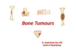
Bone Tumours
Dr. Sanjib Kumar Das, PhD
School of Physiotherapy
INTRODUCTION
• The term ‘bone tumour’ is a broad term used for benign
and malignant neoplasms, as well as ‘tumour-like
conditions’ (e.g., osteochondroma).
• Osteochondroma is the commonest benign tumour of
the bone.
• Metastatic deposits in the bone are commoner than
primary bone tumours. Multiple myeloma is the
commonest primary bone malignancies.
• The nature of a bone tumour can be suspected based on
the type of destruction seen on an X-ray
OSTEOMA
• This is a benign tumour composed of a protruding bone
from the cortical surface.
• The bones involved most often are the skull and facial
bones.
• It may bulge into one of the air sinuses (frontal, ethmoidal
or others), and cause obstruction to the sinus cavity,
leading to pain.
• A simple excision is sufficient and no treatment is
generally required except for cosmetic reasons.
OSTEOCLASTOMA (GIANT CELL TUMOUR)
• Giant cell tumour (GCT) is a common benign
bone tumor but sometimes tends to recur after
local removal.
• These giant cells were mistaken as osteoclasts in
the past, hence the name osteoclastoma.
CLINICAL FEATURES
• The tumour is seen commonly in the age group of
20-40 years i.e., after epiphyseal fusion.
• Knee is commonly affected i.e., lower-end of the
femur and upper-end of the tibia. Lower-end of
the radius is another common site.
• The tumour is located at the epiphysis. It often
reaches almost up to the joint surface.
• Common presenting complaints are swelling and
vague pain.
DIAGNOSIS
The characteristic radiological features of
GCT:
• A solitary, eccentric location, often
subchondral.
• Expansion of the overlying cortex
(expansile lesion).
• ‘Soap-bubble’ appearance
• No calcification within the tumour.
• Cortex may be thinned out or perforated at
places.
TREATMENT
a) Excision:
• Excision of the tumor is the best treatment.
• For sites like the spine, where excision is not possible,
radiotherapy is done.
b) Excision with reconstruction: the defect created by
excision is made up, usually partially, by some reconstructive
procedure.
• For example, in tumors affecting the lower-end of femur,
the affected part is excised, and the defect thus created
made up by one of the reconstructive methods.
TREATMENT
 Arthrodesis by the Turn-o-Plasty procedure :
In this technique, the required length of the
tibia is split into two halves. One half is turned
upside down and fixed with the stump of the
femur left after excising the tumour. A similar
procedure can be used for a tibial lesion by
taking half of the femur.
 Arthrodesis by bridging the gap by fibulae,
taken from same extremity or from the
opposite leg.
 Arthroplasty: The tumor is excised, and the
joint is reconstructed using an autograft
(patella to substitute the articular defect),
allograft (replacing the defect with the
preserved bone of a cadaver), or an artificial
joint (prosthesis).
TREATMENT
c) Curettage with supplementary procedures:
• Curettage performed alone has the disadvantage of a high
recurrence rate because some cells are always left along the walls of
the cavity. Some supplementary procedures used with curettage
reduce recurrence.
• Cryotherapy, where liquid nitrogen is used to produce a freezing
effect and thus kill the residual cells.
• Thermal burning of the residual cells using cauterization of the
walls of the tumor is popular.
• Bone cement can also be used. The cavity is filled with bone
cement.
TREATMENT
d) Amputation: For more aggressive tumors, or
following recurrence, amputation may be necessary.
e) Radiotherapy: Preferred treatment method for GCT
affecting the vertebrae.
OSTEOSARCOMA (OSTEOGENIC SARCOMA)
• An osteosarcoma (second most
common) is a malignant tumor of the
mesenchymal cells, characterized by
formation of bone by the tumor cells.
CLASSIFICATION
a) On the basis of clinical setting, this tumor can be divided into
primary and secondary.
• Primary osteosarcoma, the commoner, occurs in the age group of
15-25 years. It is more malignant than the secondary one. The
secondary osteosarcoma occurs in older age (45 years onwards).
b) On the basis of dominant histo-morphology, an osteosarcoma may
be:
(i) Osteoblastic i.e., with a lot of new bone formation
(ii) Chondroid i.e., with basic cell being a cartilage cell
(iii) Fibroblastic i.e. the basic cell being a fibroblast
(iv) Telangiectatic or Osteolytic type, a predominantly lytic tumor.
• All osteosarcomas are aggressive lesions and
metastasize widely.
• Despite its aggressiveness, osteosarcoma rarely
penetrates the epiphyseal plate.
• The following are the important features:
 Age at onset: These tumors occur between the ages of
15-25 years, constituting the commonest
musculoskeletal tumor at that age.
 Common sites of origin: The lower-end of the femur;
upper-end of the tibia; and upper-end of the humerus.
However, any bone of the body may be affected.
 Gross appearance of the tumor depends upon its
dominant histo-morphology.
 Histologically, these tumors vary; but common to all
is a basically anaplastic mesenchymal with tumor
cells surrounded by osteoid.
EXAMINATION
• The swelling is in the region of the metaphysis. Skin
over the swelling is shiny with prominent veins.
• The swelling is warm and tender.
• Movement at the adjacent joint may be limited mainly
because of the mechanical block by the swelling.
• The tumor may compress the neurovascular structures
of the limb, and produce symptoms due to that.
INVESTIGATIONS
 Radiological examination
• Irregular destruction in the metaphysis. There
is new bone formation in the tumor.
• Periosteal reaction: As the tumour lifts the
periosteum, it incites periosteal reaction.
• Codman’s triangle: A triangular area of
subperiosteal new bone is seen at the tumor-
host cortex junction at the ends of the tumor.
• Sun-ray appearance: As the periosteum is
unable to contain the tumour, the tumour grows
into the overlying soft tissues, giving rise to a
‘sun-ray appearance’ on the X-ray.
INVESTIGATIONS
 Serum alkaline phosphatase (SAP): It is generally
elevated. A rise of SAP after an initial fall after tumor
removal is taken as an indicator of recurrence or metastasis.
 Biopsy: Performed to confirm the diagnosis.
• A small tissue sample obtained by a needle (corebiopsy), or
by fine needle aspiration cytology (FNAC).
 Methods used for precise evaluation of spread of the
tumor locally are: bone scan, CT and MRI scans for finding
the soft tissue spread.
TREATMENT
• It is important to know the extent of involvement of the affected bone by
the tumour for the following reasons:
• To plan amputation surgery: Complete removal of local tumor is of vital
importance in amputation surgery. The amputation is performed taking a
safe margin beyond the tumour (usually 10 cm from the tumor margin).
• To plan a limb saving operation: In cases presenting early, a radical
excision of the tumor is being performed these days (limb saving surgery),
thus avoiding amputation.
• Control of distant macro or micro-metastasis: In the majority of cases,
micro-metastasis has already occurred by the time diagnosis is made.
These are effectively controlled by adjuvant chemotherapy,
immunotherapy etc.
PROGNOSIS
• Without treatment, death occurs within 2 years,
usually within 6 months of detection of metastasis.
• 5-year survival with surgery alone is 20 per cent.
• With surgery and adjuvant chemotherapy, a 5-year
disease free period is reported to be as high as 70 per
cent.
• A primarily lytic type (telangiectatic) osteosarcoma
has the worst prognosis.
EWING’S SARCOMA
• This is highly malignant tumor occurring
between the age of 10-20 years, sometimes up
to 30 years.
PATHOLOGY
 Bones affected: It commonly occurs in long
bones (in two-third cases), mainly in the
femur and tibia.
• About one-third of cases occur in flat bones,
usually in the pelvis and in short bone
calcaneum.
 Site: The tumour may begin anywhere, but
diaphysis of the long bone is the most
common site.
CLINICAL FEATURES
• The tumour occurs between 10-20 years of age.
• The patient presents with pain and swelling.
• There may be a history of trauma preceding onset, but
it is usually incidental.
• Often there is an associated fever, in which case it
may be confused with osteomyelitis.
EXAMINATION
• The swelling is usually located in the diaphysis and
has features suggesting a malignant swelling.
RADIOLOGICAL FEATURES
• In a typical case, there is a lytic
lesion in the medullary zone of the
midshaft of a long bone, with
cortical destruction and new bone
formation in layers – onion-peel
appearance.
• In flat bones, it is primarily a lytic
lesion with hardly any new bone
formation.
TREATMENT
• This is a highly radio-sensitive tumour, melts quickly
but recurs.
• In most cases, distant metastasis has occurred by the
time diagnosis is made.
• Treatment consists of control of local tumour by
radiotherapy and control of metastasis by
chemotherapy.
MULTIPLE MYELOMA
• It is a malignant neoplasm derived from
plasma cells.
PATHOLOGY
• The neoplasm characteristically affects flat bones i.e., the pelvis,
skull, and ribs and irregular vertebrae.
• It may occur as a solitary lesion (plasmacytoma), multiple lesion
(multiple myeloma), extramedullary (myelomatosis).
• The lesions are mostly small and circumscribed.
• The bone is simply replaced by tumor tissue and there is no
reactive new bone formation.
CLINICAL FEATURES
• The tumor affects adults above 40 years of age.
• Men are affected more often than women.
• Usual presentation is that of multiple site involvement.
• Common presenting complaint is increasingly severe
pain in the lumbar and thoracic spine.
• Pathological fractures, especially of the vertebrae and
ribs may result in acute symptoms.
• There may be no swelling or deformity unless a
pathological fracture occurs.
• The patient is weak, and will have loss of weight.
• Neurological symptoms may result if the tumour
presses on the spinal cord or the nerves in the spinal
canal.
INVESTIGATIONS
Radiological examination
• Multiple punched out lesions in the skull and other flat bones.
• Diffuse, severe rarefaction of bones.
• Erosions of the borders of the ribs.
INVESTIGATIONS
 Blood: Low haemoglobin, high ESR, increased serum
calcium, normal alkaline phosphatase.
 Sternal puncture: Myeloma cells may be seen.
 Bone biopsy from the iliac crest, or a CT guided
needle biopsy from the vertebral lesion may show
features suggestive of multiple myeloma.
 Bone scan: This may be required in cases presenting
as solitary bone lesion, where lesions at other sites
may be detected on a bone scan.
TREATMENT
• It consists of control of the tumor by chemotherapy.
• Radiotherapy plays a useful role in cases with
neurological compression.
• Complications like pathological fractures must be
prevented by splinting the affected part by PoP, brace
etc.
OSTEOCHONDROMA
• This is the commonest benign tumour of the bone.
• It is not a true neoplasm since its growth stops with
cessation of growth at the epiphyseal plate.
• It is a result of an aberration at the growth plate, where a
few cells from the plate grow centrifugally as a separate
lump of bone.
• Though the tumour originates at the growth plate, it gets
‘left behind’ as the bone grows in length, and thus comes
to lie at the metaphysis.
CLINICAL PRESENTATION
• The patient, usually around adolescence, presents
with a painless swelling around a joint, usually
around the knee.
• There may be similar swellings in other parts of the
body in case of multiple exostosis
EXAMINATION
• The swelling has all the features of a benign bony swelling. It
may be a sessile or pedunculated swelling.
• Usual location is metaphyseal, but often it comes to lie as far
as the diaphysis.
• There may be signs suggestive of complications secondary to
the swelling. These are:
(i) signs due to compression of the neurovascular bundle of the
limb
(ii) limitation of joint movements due to mechanical block by
the swelling.
EXAMINATION
• Occasionally, the tumour undergoes
malignant transformation
(chondrosarcoma).
• A rapid increase in the size of the
tumour and appearance of pain in a
hitherto painless swelling may be
suggestive of malignant
transformation.
• Diagnosis is made on X-ray where
one can see a bony growth made up
of mature cortical bone.
TREATMENT
• The tumour should be excised.
• The excision includes the periosteum over the
exostosis; since leaving it may result in leaving a few
cartilage cells, which will grow again and cause
recurrence of the swelling.
ENCHONDROMA
• This is a benign tumour consisting of a lobulated
mass of cartilage encapsulated by fibrous tissue.
• The intercellular matrix may undergo mucoid
degeneration.
• The tumour is seen commonly between the ages
of 20-30 years.
• Small bones of the hands and feet are commonly
affected.
• The presenting complaint is a long standing
swelling from one or more phalanges or
metacarpals, without much pain.
• The swelling increases in size very slowly, often
totally replacing the bone.
• An X-ray shows expanding lytic lesions in one
or more bones.
TREATMENT
• The lesion is curetted thoroughly, and the cavity, if it
is big, is filled with bone grafts.
• Prognosis is good.
• Although, chondromas in small bones are not known
to undergo malignant change, those in the long bones
may change into chondrosarcoma.
THANK YOU
Dr. Sanjib Kumar Das, MPT(Musculoskeletal),
Fellow (PhD) NITIE-Ergonomics and Human Factors,
Asst. Prof., School of Physiotherapy,
P.P. Savani University, Surat, India
Mail: sanjib_bpt@yahoo.co.in
Contact No. :+91 8879485847

More Related Content

What's hot

Shoulder fractures around the shoulder
Shoulder fractures around the shoulder Shoulder fractures around the shoulder
Shoulder fractures around the shoulder bibincmc
 
39. tibial plafond (pilon) fractures
39. tibial plafond (pilon) fractures39. tibial plafond (pilon) fractures
39. tibial plafond (pilon) fracturesMuhammad Abdelghani
 
Dr. pl srinivas ug class 1
Dr. pl srinivas ug class 1Dr. pl srinivas ug class 1
Dr. pl srinivas ug class 1varuntandra
 
Metallurgy in orthopaedics
Metallurgy in orthopaedicsMetallurgy in orthopaedics
Metallurgy in orthopaedicsRakshith Kumar
 
Osteochondritis dessicans
Osteochondritis dessicansOsteochondritis dessicans
Osteochondritis dessicansPratikDhabalia
 
General principles of arthroscopy kle, belgaum, dr utkarsh dwivedi
General principles of arthroscopy kle, belgaum, dr utkarsh dwivediGeneral principles of arthroscopy kle, belgaum, dr utkarsh dwivedi
General principles of arthroscopy kle, belgaum, dr utkarsh dwivediUtkarsh Dwivedi
 
Spine mobilization and manipulation 1
Spine mobilization and manipulation 1Spine mobilization and manipulation 1
Spine mobilization and manipulation 1Simba Syed
 
MEDIAN NERVE PALSY AND TENDON TRANSFERS
MEDIAN NERVE PALSY AND TENDON TRANSFERSMEDIAN NERVE PALSY AND TENDON TRANSFERS
MEDIAN NERVE PALSY AND TENDON TRANSFERSBenthungo Tungoe
 
Volkmann¶s ischemic contracture
Volkmann¶s ischemic contractureVolkmann¶s ischemic contracture
Volkmann¶s ischemic contractureSagar Savsani
 
Myositis ossificans
Myositis ossificans Myositis ossificans
Myositis ossificans orthoprince
 
Role of usg in orthopedics
Role of usg in orthopedicsRole of usg in orthopedics
Role of usg in orthopedicsSj Karthik
 
Advances in shoulder surgery and rehabilitation - Len Funk 2012
Advances in shoulder surgery and rehabilitation - Len Funk 2012Advances in shoulder surgery and rehabilitation - Len Funk 2012
Advances in shoulder surgery and rehabilitation - Len Funk 2012Lennard Funk
 

What's hot (20)

Crps
CrpsCrps
Crps
 
Meniscal Injuries
Meniscal InjuriesMeniscal Injuries
Meniscal Injuries
 
Shoulder fractures around the shoulder
Shoulder fractures around the shoulder Shoulder fractures around the shoulder
Shoulder fractures around the shoulder
 
Plates and screws 11
Plates and screws 11Plates and screws 11
Plates and screws 11
 
39. tibial plafond (pilon) fractures
39. tibial plafond (pilon) fractures39. tibial plafond (pilon) fractures
39. tibial plafond (pilon) fractures
 
Dr. pl srinivas ug class 1
Dr. pl srinivas ug class 1Dr. pl srinivas ug class 1
Dr. pl srinivas ug class 1
 
Metallurgy in orthopaedics
Metallurgy in orthopaedicsMetallurgy in orthopaedics
Metallurgy in orthopaedics
 
Osteochondritis dessicans
Osteochondritis dessicansOsteochondritis dessicans
Osteochondritis dessicans
 
General principles of arthroscopy kle, belgaum, dr utkarsh dwivedi
General principles of arthroscopy kle, belgaum, dr utkarsh dwivediGeneral principles of arthroscopy kle, belgaum, dr utkarsh dwivedi
General principles of arthroscopy kle, belgaum, dr utkarsh dwivedi
 
Ra hand
Ra handRa hand
Ra hand
 
Spine mobilization and manipulation 1
Spine mobilization and manipulation 1Spine mobilization and manipulation 1
Spine mobilization and manipulation 1
 
Spinal orthosis
Spinal orthosisSpinal orthosis
Spinal orthosis
 
Implants in orthopaedics
Implants in orthopaedicsImplants in orthopaedics
Implants in orthopaedics
 
MEDIAN NERVE PALSY AND TENDON TRANSFERS
MEDIAN NERVE PALSY AND TENDON TRANSFERSMEDIAN NERVE PALSY AND TENDON TRANSFERS
MEDIAN NERVE PALSY AND TENDON TRANSFERS
 
Volkmann¶s ischemic contracture
Volkmann¶s ischemic contractureVolkmann¶s ischemic contracture
Volkmann¶s ischemic contracture
 
Myositis ossificans
Myositis ossificans Myositis ossificans
Myositis ossificans
 
Role of usg in orthopedics
Role of usg in orthopedicsRole of usg in orthopedics
Role of usg in orthopedics
 
Myositis ossificans
Myositis ossificans Myositis ossificans
Myositis ossificans
 
Advances in shoulder surgery and rehabilitation - Len Funk 2012
Advances in shoulder surgery and rehabilitation - Len Funk 2012Advances in shoulder surgery and rehabilitation - Len Funk 2012
Advances in shoulder surgery and rehabilitation - Len Funk 2012
 
Tendon tranfer
Tendon tranferTendon tranfer
Tendon tranfer
 

Similar to Bone Tumour Types and Treatments

Tumours of the bones &joints
Tumours of the bones &jointsTumours of the bones &joints
Tumours of the bones &jointsDr KAMBLE
 
BONE TUMORS.ppt
BONE TUMORS.pptBONE TUMORS.ppt
BONE TUMORS.pptHOME
 
umy-benignbonetumors-140824003220-phpapp01.pptx
umy-benignbonetumors-140824003220-phpapp01.pptxumy-benignbonetumors-140824003220-phpapp01.pptx
umy-benignbonetumors-140824003220-phpapp01.pptxasdgja
 
Benign bone tumors
Benign bone tumorsBenign bone tumors
Benign bone tumorsUmesh Yadav
 
Osteosarcoma and GCT.pptx
Osteosarcoma and GCT.pptxOsteosarcoma and GCT.pptx
Osteosarcoma and GCT.pptxKarthik MV
 
osteosarcomaandgct-231015051229-2eb956c7 (2).pptx
osteosarcomaandgct-231015051229-2eb956c7 (2).pptxosteosarcomaandgct-231015051229-2eb956c7 (2).pptx
osteosarcomaandgct-231015051229-2eb956c7 (2).pptxKareemElsharkawy6
 
Primary bone tumors
Primary bone tumorsPrimary bone tumors
Primary bone tumorsDpt Memon
 
Swellings of the jaw
Swellings of the jaw Swellings of the jaw
Swellings of the jaw Arjun Shenoy
 
malignantbonetumours-170608150210.pptx
malignantbonetumours-170608150210.pptxmalignantbonetumours-170608150210.pptx
malignantbonetumours-170608150210.pptxasdgja
 
An approach to malignant bone tumors
An approach to malignant bone tumors An approach to malignant bone tumors
An approach to malignant bone tumors Dr.Suhas Basavaiah
 
Bone Tumor (Benign and malignant)
Bone Tumor (Benign and malignant)Bone Tumor (Benign and malignant)
Bone Tumor (Benign and malignant)Nassr ALBarhi
 
Malignant bone tumours
Malignant bone tumoursMalignant bone tumours
Malignant bone tumoursArchana Koshy
 
Bone tumors and tumor-like lesions.ppt
Bone tumors and tumor-like lesions.pptBone tumors and tumor-like lesions.ppt
Bone tumors and tumor-like lesions.pptdrqazi7777
 
Bone tumor 2020 uptodate
Bone tumor 2020 uptodateBone tumor 2020 uptodate
Bone tumor 2020 uptodateAmjadHazim
 

Similar to Bone Tumour Types and Treatments (20)

Tumours of the bones &joints
Tumours of the bones &jointsTumours of the bones &joints
Tumours of the bones &joints
 
BONE TUMORS.ppt
BONE TUMORS.pptBONE TUMORS.ppt
BONE TUMORS.ppt
 
umy-benignbonetumors-140824003220-phpapp01.pptx
umy-benignbonetumors-140824003220-phpapp01.pptxumy-benignbonetumors-140824003220-phpapp01.pptx
umy-benignbonetumors-140824003220-phpapp01.pptx
 
Benign bone tumors
Benign bone tumorsBenign bone tumors
Benign bone tumors
 
Osteosarcoma.pdf
Osteosarcoma.pdfOsteosarcoma.pdf
Osteosarcoma.pdf
 
Ct spine tumors
Ct spine tumorsCt spine tumors
Ct spine tumors
 
Osteosarcoma and GCT.pptx
Osteosarcoma and GCT.pptxOsteosarcoma and GCT.pptx
Osteosarcoma and GCT.pptx
 
Bone Tumor.pptx
Bone Tumor.pptxBone Tumor.pptx
Bone Tumor.pptx
 
osteosarcomaandgct-231015051229-2eb956c7 (2).pptx
osteosarcomaandgct-231015051229-2eb956c7 (2).pptxosteosarcomaandgct-231015051229-2eb956c7 (2).pptx
osteosarcomaandgct-231015051229-2eb956c7 (2).pptx
 
Primary bone tumors
Primary bone tumorsPrimary bone tumors
Primary bone tumors
 
ssgrsir.pptx
ssgrsir.pptxssgrsir.pptx
ssgrsir.pptx
 
Swellings of the jaw
Swellings of the jaw Swellings of the jaw
Swellings of the jaw
 
malignantbonetumours-170608150210.pptx
malignantbonetumours-170608150210.pptxmalignantbonetumours-170608150210.pptx
malignantbonetumours-170608150210.pptx
 
Radiology Malignant bone tumors
Radiology Malignant bone tumorsRadiology Malignant bone tumors
Radiology Malignant bone tumors
 
An approach to malignant bone tumors
An approach to malignant bone tumors An approach to malignant bone tumors
An approach to malignant bone tumors
 
Bone Tumor (Benign and malignant)
Bone Tumor (Benign and malignant)Bone Tumor (Benign and malignant)
Bone Tumor (Benign and malignant)
 
malignantbonetumours-170608150210.pdf
malignantbonetumours-170608150210.pdfmalignantbonetumours-170608150210.pdf
malignantbonetumours-170608150210.pdf
 
Malignant bone tumours
Malignant bone tumoursMalignant bone tumours
Malignant bone tumours
 
Bone tumors and tumor-like lesions.ppt
Bone tumors and tumor-like lesions.pptBone tumors and tumor-like lesions.ppt
Bone tumors and tumor-like lesions.ppt
 
Bone tumor 2020 uptodate
Bone tumor 2020 uptodateBone tumor 2020 uptodate
Bone tumor 2020 uptodate
 

More from Dr. Sanjib Kumar Das (18)

Poliomyelitis & its Physiotherapeutic Management
Poliomyelitis & its Physiotherapeutic ManagementPoliomyelitis & its Physiotherapeutic Management
Poliomyelitis & its Physiotherapeutic Management
 
Research and Publication Pedagogy
Research and Publication PedagogyResearch and Publication Pedagogy
Research and Publication Pedagogy
 
Fractures around shoulder
Fractures around shoulderFractures around shoulder
Fractures around shoulder
 
Arthritis and Rhematic diseases
Arthritis and Rhematic diseasesArthritis and Rhematic diseases
Arthritis and Rhematic diseases
 
Amputation
AmputationAmputation
Amputation
 
General Pharmacology for Physiotherapists
General Pharmacology for PhysiotherapistsGeneral Pharmacology for Physiotherapists
General Pharmacology for Physiotherapists
 
Wheelchairs - Types and Parts
Wheelchairs - Types and PartsWheelchairs - Types and Parts
Wheelchairs - Types and Parts
 
Prosthesis
ProsthesisProsthesis
Prosthesis
 
Ergonomics and Human Factors Fundamentals: An introduction
Ergonomics and Human Factors Fundamentals: An introductionErgonomics and Human Factors Fundamentals: An introduction
Ergonomics and Human Factors Fundamentals: An introduction
 
Orthotics and Splints
Orthotics and SplintsOrthotics and Splints
Orthotics and Splints
 
Safety and Environmental Management- Case Study
Safety and Environmental Management- Case StudySafety and Environmental Management- Case Study
Safety and Environmental Management- Case Study
 
Heat
HeatHeat
Heat
 
Illumination
IlluminationIllumination
Illumination
 
Noise
NoiseNoise
Noise
 
Vibration
VibrationVibration
Vibration
 
Spinal injuries (compiled by Dr Sanjib Kumar Das)
Spinal injuries (compiled by Dr Sanjib Kumar Das)Spinal injuries (compiled by Dr Sanjib Kumar Das)
Spinal injuries (compiled by Dr Sanjib Kumar Das)
 
Hand injuries (compiled by Dr. Sanjib Kumar Das)
Hand injuries (compiled by Dr. Sanjib Kumar Das)Hand injuries (compiled by Dr. Sanjib Kumar Das)
Hand injuries (compiled by Dr. Sanjib Kumar Das)
 
Yoga (compiled by Sanjib Kumar Das)
Yoga (compiled by Sanjib Kumar Das)Yoga (compiled by Sanjib Kumar Das)
Yoga (compiled by Sanjib Kumar Das)
 

Recently uploaded

“Oh GOSH! Reflecting on Hackteria's Collaborative Practices in a Global Do-It...
“Oh GOSH! Reflecting on Hackteria's Collaborative Practices in a Global Do-It...“Oh GOSH! Reflecting on Hackteria's Collaborative Practices in a Global Do-It...
“Oh GOSH! Reflecting on Hackteria's Collaborative Practices in a Global Do-It...Marc Dusseiller Dusjagr
 
Alper Gobel In Media Res Media Component
Alper Gobel In Media Res Media ComponentAlper Gobel In Media Res Media Component
Alper Gobel In Media Res Media ComponentInMediaRes1
 
How to Make a Pirate ship Primary Education.pptx
How to Make a Pirate ship Primary Education.pptxHow to Make a Pirate ship Primary Education.pptx
How to Make a Pirate ship Primary Education.pptxmanuelaromero2013
 
CELL CYCLE Division Science 8 quarter IV.pptx
CELL CYCLE Division Science 8 quarter IV.pptxCELL CYCLE Division Science 8 quarter IV.pptx
CELL CYCLE Division Science 8 quarter IV.pptxJiesonDelaCerna
 
ECONOMIC CONTEXT - PAPER 1 Q3: NEWSPAPERS.pptx
ECONOMIC CONTEXT - PAPER 1 Q3: NEWSPAPERS.pptxECONOMIC CONTEXT - PAPER 1 Q3: NEWSPAPERS.pptx
ECONOMIC CONTEXT - PAPER 1 Q3: NEWSPAPERS.pptxiammrhaywood
 
MARGINALIZATION (Different learners in Marginalized Group
MARGINALIZATION (Different learners in Marginalized GroupMARGINALIZATION (Different learners in Marginalized Group
MARGINALIZATION (Different learners in Marginalized GroupJonathanParaisoCruz
 
Like-prefer-love -hate+verb+ing & silent letters & citizenship text.pdf
Like-prefer-love -hate+verb+ing & silent letters & citizenship text.pdfLike-prefer-love -hate+verb+ing & silent letters & citizenship text.pdf
Like-prefer-love -hate+verb+ing & silent letters & citizenship text.pdfMr Bounab Samir
 
18-04-UA_REPORT_MEDIALITERAСY_INDEX-DM_23-1-final-eng.pdf
18-04-UA_REPORT_MEDIALITERAСY_INDEX-DM_23-1-final-eng.pdf18-04-UA_REPORT_MEDIALITERAСY_INDEX-DM_23-1-final-eng.pdf
18-04-UA_REPORT_MEDIALITERAСY_INDEX-DM_23-1-final-eng.pdfssuser54595a
 
MICROBIOLOGY biochemical test detailed.pptx
MICROBIOLOGY biochemical test detailed.pptxMICROBIOLOGY biochemical test detailed.pptx
MICROBIOLOGY biochemical test detailed.pptxabhijeetpadhi001
 
Full Stack Web Development Course for Beginners
Full Stack Web Development Course  for BeginnersFull Stack Web Development Course  for Beginners
Full Stack Web Development Course for BeginnersSabitha Banu
 
EPANDING THE CONTENT OF AN OUTLINE using notes.pptx
EPANDING THE CONTENT OF AN OUTLINE using notes.pptxEPANDING THE CONTENT OF AN OUTLINE using notes.pptx
EPANDING THE CONTENT OF AN OUTLINE using notes.pptxRaymartEstabillo3
 
POINT- BIOCHEMISTRY SEM 2 ENZYMES UNIT 5.pptx
POINT- BIOCHEMISTRY SEM 2 ENZYMES UNIT 5.pptxPOINT- BIOCHEMISTRY SEM 2 ENZYMES UNIT 5.pptx
POINT- BIOCHEMISTRY SEM 2 ENZYMES UNIT 5.pptxSayali Powar
 
What is Model Inheritance in Odoo 17 ERP
What is Model Inheritance in Odoo 17 ERPWhat is Model Inheritance in Odoo 17 ERP
What is Model Inheritance in Odoo 17 ERPCeline George
 
Final demo Grade 9 for demo Plan dessert.pptx
Final demo Grade 9 for demo Plan dessert.pptxFinal demo Grade 9 for demo Plan dessert.pptx
Final demo Grade 9 for demo Plan dessert.pptxAvyJaneVismanos
 
Introduction to ArtificiaI Intelligence in Higher Education
Introduction to ArtificiaI Intelligence in Higher EducationIntroduction to ArtificiaI Intelligence in Higher Education
Introduction to ArtificiaI Intelligence in Higher Educationpboyjonauth
 
Earth Day Presentation wow hello nice great
Earth Day Presentation wow hello nice greatEarth Day Presentation wow hello nice great
Earth Day Presentation wow hello nice greatYousafMalik24
 
Introduction to AI in Higher Education_draft.pptx
Introduction to AI in Higher Education_draft.pptxIntroduction to AI in Higher Education_draft.pptx
Introduction to AI in Higher Education_draft.pptxpboyjonauth
 
Gas measurement O2,Co2,& ph) 04/2024.pptx
Gas measurement O2,Co2,& ph) 04/2024.pptxGas measurement O2,Co2,& ph) 04/2024.pptx
Gas measurement O2,Co2,& ph) 04/2024.pptxDr.Ibrahim Hassaan
 

Recently uploaded (20)

“Oh GOSH! Reflecting on Hackteria's Collaborative Practices in a Global Do-It...
“Oh GOSH! Reflecting on Hackteria's Collaborative Practices in a Global Do-It...“Oh GOSH! Reflecting on Hackteria's Collaborative Practices in a Global Do-It...
“Oh GOSH! Reflecting on Hackteria's Collaborative Practices in a Global Do-It...
 
Alper Gobel In Media Res Media Component
Alper Gobel In Media Res Media ComponentAlper Gobel In Media Res Media Component
Alper Gobel In Media Res Media Component
 
How to Make a Pirate ship Primary Education.pptx
How to Make a Pirate ship Primary Education.pptxHow to Make a Pirate ship Primary Education.pptx
How to Make a Pirate ship Primary Education.pptx
 
OS-operating systems- ch04 (Threads) ...
OS-operating systems- ch04 (Threads) ...OS-operating systems- ch04 (Threads) ...
OS-operating systems- ch04 (Threads) ...
 
CELL CYCLE Division Science 8 quarter IV.pptx
CELL CYCLE Division Science 8 quarter IV.pptxCELL CYCLE Division Science 8 quarter IV.pptx
CELL CYCLE Division Science 8 quarter IV.pptx
 
ECONOMIC CONTEXT - PAPER 1 Q3: NEWSPAPERS.pptx
ECONOMIC CONTEXT - PAPER 1 Q3: NEWSPAPERS.pptxECONOMIC CONTEXT - PAPER 1 Q3: NEWSPAPERS.pptx
ECONOMIC CONTEXT - PAPER 1 Q3: NEWSPAPERS.pptx
 
MARGINALIZATION (Different learners in Marginalized Group
MARGINALIZATION (Different learners in Marginalized GroupMARGINALIZATION (Different learners in Marginalized Group
MARGINALIZATION (Different learners in Marginalized Group
 
Like-prefer-love -hate+verb+ing & silent letters & citizenship text.pdf
Like-prefer-love -hate+verb+ing & silent letters & citizenship text.pdfLike-prefer-love -hate+verb+ing & silent letters & citizenship text.pdf
Like-prefer-love -hate+verb+ing & silent letters & citizenship text.pdf
 
18-04-UA_REPORT_MEDIALITERAСY_INDEX-DM_23-1-final-eng.pdf
18-04-UA_REPORT_MEDIALITERAСY_INDEX-DM_23-1-final-eng.pdf18-04-UA_REPORT_MEDIALITERAСY_INDEX-DM_23-1-final-eng.pdf
18-04-UA_REPORT_MEDIALITERAСY_INDEX-DM_23-1-final-eng.pdf
 
MICROBIOLOGY biochemical test detailed.pptx
MICROBIOLOGY biochemical test detailed.pptxMICROBIOLOGY biochemical test detailed.pptx
MICROBIOLOGY biochemical test detailed.pptx
 
Full Stack Web Development Course for Beginners
Full Stack Web Development Course  for BeginnersFull Stack Web Development Course  for Beginners
Full Stack Web Development Course for Beginners
 
EPANDING THE CONTENT OF AN OUTLINE using notes.pptx
EPANDING THE CONTENT OF AN OUTLINE using notes.pptxEPANDING THE CONTENT OF AN OUTLINE using notes.pptx
EPANDING THE CONTENT OF AN OUTLINE using notes.pptx
 
POINT- BIOCHEMISTRY SEM 2 ENZYMES UNIT 5.pptx
POINT- BIOCHEMISTRY SEM 2 ENZYMES UNIT 5.pptxPOINT- BIOCHEMISTRY SEM 2 ENZYMES UNIT 5.pptx
POINT- BIOCHEMISTRY SEM 2 ENZYMES UNIT 5.pptx
 
What is Model Inheritance in Odoo 17 ERP
What is Model Inheritance in Odoo 17 ERPWhat is Model Inheritance in Odoo 17 ERP
What is Model Inheritance in Odoo 17 ERP
 
Model Call Girl in Bikash Puri Delhi reach out to us at 🔝9953056974🔝
Model Call Girl in Bikash Puri  Delhi reach out to us at 🔝9953056974🔝Model Call Girl in Bikash Puri  Delhi reach out to us at 🔝9953056974🔝
Model Call Girl in Bikash Puri Delhi reach out to us at 🔝9953056974🔝
 
Final demo Grade 9 for demo Plan dessert.pptx
Final demo Grade 9 for demo Plan dessert.pptxFinal demo Grade 9 for demo Plan dessert.pptx
Final demo Grade 9 for demo Plan dessert.pptx
 
Introduction to ArtificiaI Intelligence in Higher Education
Introduction to ArtificiaI Intelligence in Higher EducationIntroduction to ArtificiaI Intelligence in Higher Education
Introduction to ArtificiaI Intelligence in Higher Education
 
Earth Day Presentation wow hello nice great
Earth Day Presentation wow hello nice greatEarth Day Presentation wow hello nice great
Earth Day Presentation wow hello nice great
 
Introduction to AI in Higher Education_draft.pptx
Introduction to AI in Higher Education_draft.pptxIntroduction to AI in Higher Education_draft.pptx
Introduction to AI in Higher Education_draft.pptx
 
Gas measurement O2,Co2,& ph) 04/2024.pptx
Gas measurement O2,Co2,& ph) 04/2024.pptxGas measurement O2,Co2,& ph) 04/2024.pptx
Gas measurement O2,Co2,& ph) 04/2024.pptx
 

Bone Tumour Types and Treatments

  • 1. Bone Tumours Dr. Sanjib Kumar Das, PhD School of Physiotherapy
  • 2. INTRODUCTION • The term ‘bone tumour’ is a broad term used for benign and malignant neoplasms, as well as ‘tumour-like conditions’ (e.g., osteochondroma). • Osteochondroma is the commonest benign tumour of the bone. • Metastatic deposits in the bone are commoner than primary bone tumours. Multiple myeloma is the commonest primary bone malignancies. • The nature of a bone tumour can be suspected based on the type of destruction seen on an X-ray
  • 3. OSTEOMA • This is a benign tumour composed of a protruding bone from the cortical surface. • The bones involved most often are the skull and facial bones. • It may bulge into one of the air sinuses (frontal, ethmoidal or others), and cause obstruction to the sinus cavity, leading to pain. • A simple excision is sufficient and no treatment is generally required except for cosmetic reasons.
  • 4. OSTEOCLASTOMA (GIANT CELL TUMOUR) • Giant cell tumour (GCT) is a common benign bone tumor but sometimes tends to recur after local removal. • These giant cells were mistaken as osteoclasts in the past, hence the name osteoclastoma.
  • 5. CLINICAL FEATURES • The tumour is seen commonly in the age group of 20-40 years i.e., after epiphyseal fusion. • Knee is commonly affected i.e., lower-end of the femur and upper-end of the tibia. Lower-end of the radius is another common site. • The tumour is located at the epiphysis. It often reaches almost up to the joint surface. • Common presenting complaints are swelling and vague pain.
  • 6. DIAGNOSIS The characteristic radiological features of GCT: • A solitary, eccentric location, often subchondral. • Expansion of the overlying cortex (expansile lesion). • ‘Soap-bubble’ appearance • No calcification within the tumour. • Cortex may be thinned out or perforated at places.
  • 7. TREATMENT a) Excision: • Excision of the tumor is the best treatment. • For sites like the spine, where excision is not possible, radiotherapy is done. b) Excision with reconstruction: the defect created by excision is made up, usually partially, by some reconstructive procedure. • For example, in tumors affecting the lower-end of femur, the affected part is excised, and the defect thus created made up by one of the reconstructive methods.
  • 8. TREATMENT  Arthrodesis by the Turn-o-Plasty procedure : In this technique, the required length of the tibia is split into two halves. One half is turned upside down and fixed with the stump of the femur left after excising the tumour. A similar procedure can be used for a tibial lesion by taking half of the femur.  Arthrodesis by bridging the gap by fibulae, taken from same extremity or from the opposite leg.  Arthroplasty: The tumor is excised, and the joint is reconstructed using an autograft (patella to substitute the articular defect), allograft (replacing the defect with the preserved bone of a cadaver), or an artificial joint (prosthesis).
  • 9. TREATMENT c) Curettage with supplementary procedures: • Curettage performed alone has the disadvantage of a high recurrence rate because some cells are always left along the walls of the cavity. Some supplementary procedures used with curettage reduce recurrence. • Cryotherapy, where liquid nitrogen is used to produce a freezing effect and thus kill the residual cells. • Thermal burning of the residual cells using cauterization of the walls of the tumor is popular. • Bone cement can also be used. The cavity is filled with bone cement.
  • 10. TREATMENT d) Amputation: For more aggressive tumors, or following recurrence, amputation may be necessary. e) Radiotherapy: Preferred treatment method for GCT affecting the vertebrae.
  • 11. OSTEOSARCOMA (OSTEOGENIC SARCOMA) • An osteosarcoma (second most common) is a malignant tumor of the mesenchymal cells, characterized by formation of bone by the tumor cells.
  • 12. CLASSIFICATION a) On the basis of clinical setting, this tumor can be divided into primary and secondary. • Primary osteosarcoma, the commoner, occurs in the age group of 15-25 years. It is more malignant than the secondary one. The secondary osteosarcoma occurs in older age (45 years onwards). b) On the basis of dominant histo-morphology, an osteosarcoma may be: (i) Osteoblastic i.e., with a lot of new bone formation (ii) Chondroid i.e., with basic cell being a cartilage cell (iii) Fibroblastic i.e. the basic cell being a fibroblast (iv) Telangiectatic or Osteolytic type, a predominantly lytic tumor.
  • 13. • All osteosarcomas are aggressive lesions and metastasize widely. • Despite its aggressiveness, osteosarcoma rarely penetrates the epiphyseal plate. • The following are the important features:  Age at onset: These tumors occur between the ages of 15-25 years, constituting the commonest musculoskeletal tumor at that age.  Common sites of origin: The lower-end of the femur; upper-end of the tibia; and upper-end of the humerus. However, any bone of the body may be affected.
  • 14.  Gross appearance of the tumor depends upon its dominant histo-morphology.  Histologically, these tumors vary; but common to all is a basically anaplastic mesenchymal with tumor cells surrounded by osteoid.
  • 15. EXAMINATION • The swelling is in the region of the metaphysis. Skin over the swelling is shiny with prominent veins. • The swelling is warm and tender. • Movement at the adjacent joint may be limited mainly because of the mechanical block by the swelling. • The tumor may compress the neurovascular structures of the limb, and produce symptoms due to that.
  • 16. INVESTIGATIONS  Radiological examination • Irregular destruction in the metaphysis. There is new bone formation in the tumor. • Periosteal reaction: As the tumour lifts the periosteum, it incites periosteal reaction. • Codman’s triangle: A triangular area of subperiosteal new bone is seen at the tumor- host cortex junction at the ends of the tumor. • Sun-ray appearance: As the periosteum is unable to contain the tumour, the tumour grows into the overlying soft tissues, giving rise to a ‘sun-ray appearance’ on the X-ray.
  • 17. INVESTIGATIONS  Serum alkaline phosphatase (SAP): It is generally elevated. A rise of SAP after an initial fall after tumor removal is taken as an indicator of recurrence or metastasis.  Biopsy: Performed to confirm the diagnosis. • A small tissue sample obtained by a needle (corebiopsy), or by fine needle aspiration cytology (FNAC).  Methods used for precise evaluation of spread of the tumor locally are: bone scan, CT and MRI scans for finding the soft tissue spread.
  • 18. TREATMENT • It is important to know the extent of involvement of the affected bone by the tumour for the following reasons: • To plan amputation surgery: Complete removal of local tumor is of vital importance in amputation surgery. The amputation is performed taking a safe margin beyond the tumour (usually 10 cm from the tumor margin). • To plan a limb saving operation: In cases presenting early, a radical excision of the tumor is being performed these days (limb saving surgery), thus avoiding amputation. • Control of distant macro or micro-metastasis: In the majority of cases, micro-metastasis has already occurred by the time diagnosis is made. These are effectively controlled by adjuvant chemotherapy, immunotherapy etc.
  • 19. PROGNOSIS • Without treatment, death occurs within 2 years, usually within 6 months of detection of metastasis. • 5-year survival with surgery alone is 20 per cent. • With surgery and adjuvant chemotherapy, a 5-year disease free period is reported to be as high as 70 per cent. • A primarily lytic type (telangiectatic) osteosarcoma has the worst prognosis.
  • 20. EWING’S SARCOMA • This is highly malignant tumor occurring between the age of 10-20 years, sometimes up to 30 years. PATHOLOGY  Bones affected: It commonly occurs in long bones (in two-third cases), mainly in the femur and tibia. • About one-third of cases occur in flat bones, usually in the pelvis and in short bone calcaneum.  Site: The tumour may begin anywhere, but diaphysis of the long bone is the most common site.
  • 21. CLINICAL FEATURES • The tumour occurs between 10-20 years of age. • The patient presents with pain and swelling. • There may be a history of trauma preceding onset, but it is usually incidental. • Often there is an associated fever, in which case it may be confused with osteomyelitis. EXAMINATION • The swelling is usually located in the diaphysis and has features suggesting a malignant swelling.
  • 22. RADIOLOGICAL FEATURES • In a typical case, there is a lytic lesion in the medullary zone of the midshaft of a long bone, with cortical destruction and new bone formation in layers – onion-peel appearance. • In flat bones, it is primarily a lytic lesion with hardly any new bone formation.
  • 23. TREATMENT • This is a highly radio-sensitive tumour, melts quickly but recurs. • In most cases, distant metastasis has occurred by the time diagnosis is made. • Treatment consists of control of local tumour by radiotherapy and control of metastasis by chemotherapy.
  • 24. MULTIPLE MYELOMA • It is a malignant neoplasm derived from plasma cells. PATHOLOGY • The neoplasm characteristically affects flat bones i.e., the pelvis, skull, and ribs and irregular vertebrae. • It may occur as a solitary lesion (plasmacytoma), multiple lesion (multiple myeloma), extramedullary (myelomatosis). • The lesions are mostly small and circumscribed. • The bone is simply replaced by tumor tissue and there is no reactive new bone formation.
  • 25. CLINICAL FEATURES • The tumor affects adults above 40 years of age. • Men are affected more often than women. • Usual presentation is that of multiple site involvement. • Common presenting complaint is increasingly severe pain in the lumbar and thoracic spine. • Pathological fractures, especially of the vertebrae and ribs may result in acute symptoms. • There may be no swelling or deformity unless a pathological fracture occurs. • The patient is weak, and will have loss of weight. • Neurological symptoms may result if the tumour presses on the spinal cord or the nerves in the spinal canal.
  • 26. INVESTIGATIONS Radiological examination • Multiple punched out lesions in the skull and other flat bones. • Diffuse, severe rarefaction of bones. • Erosions of the borders of the ribs.
  • 27. INVESTIGATIONS  Blood: Low haemoglobin, high ESR, increased serum calcium, normal alkaline phosphatase.  Sternal puncture: Myeloma cells may be seen.  Bone biopsy from the iliac crest, or a CT guided needle biopsy from the vertebral lesion may show features suggestive of multiple myeloma.  Bone scan: This may be required in cases presenting as solitary bone lesion, where lesions at other sites may be detected on a bone scan.
  • 28. TREATMENT • It consists of control of the tumor by chemotherapy. • Radiotherapy plays a useful role in cases with neurological compression. • Complications like pathological fractures must be prevented by splinting the affected part by PoP, brace etc.
  • 29. OSTEOCHONDROMA • This is the commonest benign tumour of the bone. • It is not a true neoplasm since its growth stops with cessation of growth at the epiphyseal plate. • It is a result of an aberration at the growth plate, where a few cells from the plate grow centrifugally as a separate lump of bone. • Though the tumour originates at the growth plate, it gets ‘left behind’ as the bone grows in length, and thus comes to lie at the metaphysis.
  • 30. CLINICAL PRESENTATION • The patient, usually around adolescence, presents with a painless swelling around a joint, usually around the knee. • There may be similar swellings in other parts of the body in case of multiple exostosis
  • 31. EXAMINATION • The swelling has all the features of a benign bony swelling. It may be a sessile or pedunculated swelling. • Usual location is metaphyseal, but often it comes to lie as far as the diaphysis. • There may be signs suggestive of complications secondary to the swelling. These are: (i) signs due to compression of the neurovascular bundle of the limb (ii) limitation of joint movements due to mechanical block by the swelling.
  • 32. EXAMINATION • Occasionally, the tumour undergoes malignant transformation (chondrosarcoma). • A rapid increase in the size of the tumour and appearance of pain in a hitherto painless swelling may be suggestive of malignant transformation. • Diagnosis is made on X-ray where one can see a bony growth made up of mature cortical bone.
  • 33. TREATMENT • The tumour should be excised. • The excision includes the periosteum over the exostosis; since leaving it may result in leaving a few cartilage cells, which will grow again and cause recurrence of the swelling.
  • 34. ENCHONDROMA • This is a benign tumour consisting of a lobulated mass of cartilage encapsulated by fibrous tissue. • The intercellular matrix may undergo mucoid degeneration. • The tumour is seen commonly between the ages of 20-30 years. • Small bones of the hands and feet are commonly affected. • The presenting complaint is a long standing swelling from one or more phalanges or metacarpals, without much pain. • The swelling increases in size very slowly, often totally replacing the bone. • An X-ray shows expanding lytic lesions in one or more bones.
  • 35. TREATMENT • The lesion is curetted thoroughly, and the cavity, if it is big, is filled with bone grafts. • Prognosis is good. • Although, chondromas in small bones are not known to undergo malignant change, those in the long bones may change into chondrosarcoma.
  • 36. THANK YOU Dr. Sanjib Kumar Das, MPT(Musculoskeletal), Fellow (PhD) NITIE-Ergonomics and Human Factors, Asst. Prof., School of Physiotherapy, P.P. Savani University, Surat, India Mail: sanjib_bpt@yahoo.co.in Contact No. :+91 8879485847