
Recommended
More Related Content
Similar to Bone tumors and tumor-like lesions.ppt
Similar to Bone tumors and tumor-like lesions.ppt (20)
More from drqazi7777
Recently uploaded
Recently uploaded (20)
Bone tumors and tumor-like lesions.ppt
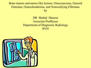
- 1. Bone tumors and tumor-like lesions: Osteosarcoma, Osteoid Osteoma, Osteochondroma, and Nonossifying Fibromas by DR Shahid Manzur Associate Proffessor Department of Diagnostic Radiology BVH
- 2. Introduction • Bone tumors are diverse in their gross and morphologic features and range from benign to rapidly fatal. This diversity makes it critical to accurately diagnose and stage tumors. • Timely, accurate diagnosis allows appropriate treatment so that the patients can not only survive, but also maintain optimal function of the affected body parts.
- 3. Classification • Most bone tumors are classified according to the normal cell or tissue of origin. Lesions that do not have normal tissue counterparts are grouped according to their distinct clinicopathologic features. • Overall, matrix-producing and fibrous tumors are the most common. Among the benign tumors, osteochondroma and fibrous cortical defect are most frequent. Excluding malignant neoplasms of marrow origin, osteosarcoma is the most common primary cancer of bone, followed by chondrosarcoma and Ewing sarcoma.
- 4. Epidemiology • The precise incidence of different bone tumors is not known because many benign lesions are not biopsied. Benign tumors outnumber malignant tumors by at least several hundredfold. • Benign tumors have their greatest frequency within the first three decades of life, whereas malignant tumors are much more common in the elderly. • In the United States, about 2,100 new cases of bone sarcoma are diagnosed annually, and approximately 1,300 deaths from bone sarcoma occur each year.
- 5. Clinical presentation • Clinically, bone tumors present in various ways. The more common benign lesions are frequently asymptomatic and are detected as incidental findings. Many tumors, however, produce pain or are noticed as a slow-growing mass. Sometimes, the first hint of a tumor's presence is a sudden pathologic fracture.
- 6. • Radiographic analysis plays an important role in diagnosing bone tumors. In addition to providing the exact location and extent of the tumor, imaging studies can detect features that help limit the differential diagnosis and give clues to the aggressiveness of the tumor. Ultimately, in most instances, biopsy and histologic study are necessary. Diagnosis
- 7. Osteosarcoma • Osteosarcoma is defined as a malignant mesenchymal tumor in which the cancerous cells produce bone matrix. Osteosarcomas occur in all age groups but have a bimodal age distribution; 75% occur in patients younger than age 20. • In adolescence, and about half of them arise in the metaphysis around the knee, either in the distal femur or proximal tibia. These are the sites of greatest skeletal growth activity. In persons over age 25, the incidence in flat bones and long bones is almost equal.
- 8. Major sites of origin of osteosarcomas. The numbers are approximate percentages.
- 9. Osteosarcoma • Osteosarcomas typically present as painful and progressively enlarging masses. Sometimes a sudden fracture of the bone is the first symptom. • Grossly, osteosarcomas are bulky tumors that are gritty, gray-white, and often contain areas of hemorrhage and cystic degeneration. The formation of bone by the tumor cells is characteristic of osteosarcoma. Osteosarcoma of the upper end of the tibia. The tan-white tumor fills most of the medullary cavity of the metaphysis and proximal diaphysis. It has infiltrated through the cortex, lifted the periosteum, and formed soft tissue masses on both sides of the bone.
- 10. Osteosarcoma • Radiographs of the primary tumor usually show a large, destructive, mixed lytic and blastic mass. The tumor frequently breaks through the cortex and lifts the periosteum, resulting in reactive periosteal bone formation. The triangular shadow between the cortex and raised ends of periosteum is known radiographically as Codman triangle and is characteristic, but not diagnostic of this tumor. Distal femoral osteosarcoma with prominent bone formation extending into the soft tissues. The periosteum, which has been lifted, has laid down a proximal triangular shell of reactive bone known as a Codman triangle (arrow).
- 11. Central osteosarcoma. A, A destructive lesion is seen in the metaphysis on this anteroposterior view of the knee in a young teenager with pain. B, A magnetic resonance scan of both legs shows the soft tissue extent of the tumor (arrows). Osteosarcoma
- 12. Osteoid Osteoma • Osteoid osteomas are bone tumors less than 2 cm in greatest dimension and usually occur in patients in their teens and twenties. In fact, 75% of patients are under age 25. • Osteoid osteomas can arise in any bone but have a predilection for the appendicular skeleton. 50% of cases involve the femur or tibia, where they commonly arise in the cortex. • Osteoid osteomas are painful lesions. The pain is caused by excess prostaglandin E2 which is produced by the proliferating osteoblasts. It characteristically occurs at night and is dramatically relieved by aspirin.
- 13. Osteoid Osteoma • Osteoid osteoma. A lateral view (A) of the proximal tibia shows a very dense lesion in the posterior cortex. A darker central area contains a white nidus. This lesion in a 20-year-old man caused pain in this area, relieved by aspirin. B, A nuclear medicine bone scan in a different patient with an osteoid osteoma in the left lower tibia shows increased activity (arrows) at the site of the lesion.
- 14. Osteoid Osteoma • Osteoid osteomas, especially those that arise beneath the periosteum, usually elicit a tremendous amount of reactive bone formation that encircles the lesion. The actual tumor, known as the nidus, manifests radiographically as a small round lucency that is variably mineralized Specimen radiograph of intracortical osteoid osteoma. The round radiolucency with central mineralization represents the lesion and is surrounded by abundant reactive bone that has massively thickened the cortex.
- 15. Osteoid Osteoma • Osteoid osteomas’ are considered benign and are normally treated by conservative surgery. However there is a possibility of malignant transformation. This is rare except when treated with radiation, which promotes this complication.
- 16. Osteochondroma • Osteochondroma, also known as an exostosis, is a benign cartilage-capped outgrowth that is attached to the underlying skeleton by a bony stalk. It is a relatively common lesion and can be solitary or multiple. • Multiple osteochondromas become apparent during childhood but solitary osteochondromas are usually not diagnosed until late adolescence . • Men are affected 3X more often than women. • They arise from the metaphysis near the growth plate of long tubular bones, especially the knee.
- 17. Osteochondroma • Clinically, osteochondromas present as slow-growing masses, which can be painful if they impinge on a nerve or if the stalk is fractured. In many cases, they are detected as an incidental finding. Osteochondroma. On this lateral view of the ankle, a benign osteochondroma is seen projecting posteriorly on a stalk. The end (arrows) is often covered with a cartilaginous cap. These lesions always occur near a joint but point away from it.
- 18. Fibrous Cortical Defect and Nonossifying Fibroma • Fibrous cortical defects are extremely common, found in 30% to 50% of all children older than age 2 years. They are believed to be developmental defects rather than neoplasms. • The vast majority arise in the metaphysis of the distal femur and proximal tibia, and almost one half are bilateral or multiple. • Fibrous cortical defects are small and those that grow to 5 or 6 cm in size are called nonossifying fibromas.
- 19. Fibrous Cortical Defect and Nonossifying Fibroma • Fibrous cortical defects are asymptomatic and are usually detected on x-ray as an incidental finding. The vast majority have limited growth potential and undergo spontaneous resolution within several years. • The few that progressively enlarge into nonossifying fibromas usually show up in adolescence. They may present with pathologic fracture and then require biopsy to exclude other types of tumors.
- 20. Fibrous Cortical Defect and Nonossifying Fibroma • Both fibrous cortical defects and nonossifying fibromas produce elongated, sharply demarcated radiolucencies that are surrounded by a thin zone of sclerosis. Nonossifying fibromas of the distal tibial metaphysis, producing an eccentric lobulated radiolucency surrounded by a sclerotic margin.