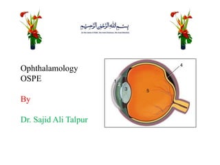
Ophthalmology OSPE guide
•Download as PPTX, PDF•
14 likes•970 views

Ophthalamology ospe for undergradute level. Read, download and share it.

Recommended
More Related Content
What's hot
What's hot (20)
Similar to Ophthalmology OSPE guide
Similar to Ophthalmology OSPE guide (20)
More from Dr. Sajid Ali Talpur
More from Dr. Sajid Ali Talpur (20)
Recently uploaded
Recently uploaded (20)
Ophthalmology OSPE guide
- 1. Ophthalamology OSPE By Dr. Sajid Ali Talpur
- 2. 1. Identify the condition 2. Name the cardinal sign in the diagnosis of congenital glaucoma. 3. How will you distinguish between megalocornea and corneal buphthalamos ? 4. What are haab’s striae?
- 3. 1. Congenital Glaucoma 2. Enlargement of corneal diameter 3. In buphthalamos raised IOP and glaucomatous cupping are present which are absent in megalocornea. 4. Haab’s striae are healed breaks in the decemet’s membrane which appear as curvilinear lines
- 4. 1. Identify the condition 2. Which type of cataract develops as a result of complication of the iridocyclitis? 3. In which condition snow banking or snow ball formation is seen? 4. Name the pathognomonic sign of acute iridocylcitis
- 5. 1. Chronic Iridocyclitis 2. Complicated cataract 3. Pars planitis 4. Keratic precipitates
- 6. 1. Identify the condition 2. Give various symptoms 3. Define anterior synechae 4. How will differentiate between macula and leucoma
- 7. 1. Corneal ulcer (Hypopyon corneal ulcer) 2. Pain, blurred vision, lacrimation, photophobia, halos, redness of the eye 3. The adhesion between peripheral part of iris with cornea is called anterior synechae 4. macula is semidense opacity which results from scarring involving anterior half of corneal stroma whereas leukoma is dense opacity resulting from scarring involving more than half of corneal stroma.
- 8. 1. What is the pathogenesis of Diabetic retinopathy? 2. What are hard exudated? 3. In which stage of Diabetic retinopathy Pan photocoagulation is required? 4. Does burnt out stage of Diabetic retinopathy require Laser? Explain with reason
- 9. 1. The essential lesion in Diabetic retinopathy is microangiopathy that has the features of microvascular leakage and occlusion 2. These are exudates composed of mainly lipids and have yellow waxy appearance with distinctly clear margins. 3. Proliferative stage 4. No, Because the vascular component has already regressed.
- 10. 1. Identify the condition 2. Which serotypes of adenovirus cause epidemic keratoconjuctivitis? 3. In which type of conjuctivitis umblicated nodules are seen? 4. Define trachoma.
- 11. 1. Conjuctivitis (viral conjuctivitis) 2. Serotypes 8 & 19 3. Molluscum contagiosum conjuctivitis 4. It is chronic bilateral cicatrical keratoconjuctivitis caused by chlamydia
- 12. 1. Identify the condition 2. What are the complications scleritis 3. Which type of scleritis is the most common? 4. Give any three differences between scleritis and episcleritis.
- 13. 1. Episcleritis 2. Corneal lesions, anterior uveitis, glaucoma, cataract, staphyloma. 3. Anterior necrotizing scleritis 4.episcleritis is charachterized by sudden onset, no pain but discomfort and vision unaffected while scleritis is charachterized by gradual onset, mild to moderate pain and vision frequently affected.
- 14. 1. Name the structures 1,2 and 3 2. What structures are present in uveal tissue? 3. Define anterior segment 4. What is the composition of tear film?
- 15. 1. Cornea, iris and lens 2. Choroid, cilliary body and iris 3. It is a small cavity in eyeball located between the posterior surface of cornea and anterior surface of lens filled with aquous humor. 4. It consists of superficial lipids, middle aquous and inner mucin.
- 16. 1. Identify the condition 2. What are its types 3. Give its any three complications 4. How will you treat?
- 17. 1. Blepharitis (ulcerative blepharitis) 2. Anterior blepharitis (ulcerative and squamous) and posterior blepharitis 3. trichiasis, poliosis and madarosis 4. Lid hyegiene, antibiotic ointments, steroid drops and artificial tears.
- 18. 1. Identify the condition. 2. Define papiloedema 3. How will you differentiate between pappilitis and retrobulbar neuritis? 4. What is the effect of papiloedema on vision and pupillary light reflex?
- 19. 1. Optic neuritis (pappilitis) 2. it is optic disc swelling that is caused by increased intracranial pressure. The swelling is usually bilateral and can occur over a period of hours to weeks. 3. Pappilitis is the inflammation of intraocular part of optic nerve whereas retrobulbar neuritis is the inflammation of intraorbital part of optic nerve in which O.Disc is spared. 4. Both of them are normal.