
Recommended
More Related Content
Similar to cataract.pptx
Similar to cataract.pptx (20)
Recently uploaded
Recently uploaded (20)
cataract.pptx
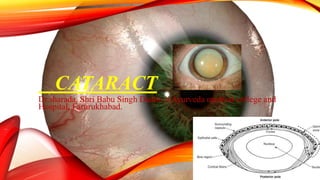
- 1. CATARACT Dr.sharada, Shri Babu Singh Daddu ji Ayurveda medical college and Hospital, Farurukhabad.
- 2. B. Morphological classification 1. Capsular cataract. It involves the capsule and may be: i. Anterior capsular cataract ii. Posterior capsular cataract 2. Subcapsular cataract. It involves the superficial part of the cortex (just below the capsule) and includes: i. Anterior subcapsular cataract ii. Posterior subcapsular cataract 3. Cortical cataract. It involves the major part of the cortex. 4. Supranuclear cataract. It involves only the deeper parts of cortex (just outside the nucleus). 5. Nuclear cataract. It involves the nucleus of the crystalline lens. 6. Polar cataract. It involves the capsule and superficial part of the cortex in the polar region only and may be: i. Anterior polar cataract ii. Posterior polar cataract
- 3. DEFINITION • Development of an opacity in the lens is known as cataract • Classification A. Etiological classification • I. Congenital and developmental cataract • II. Acquired cataract 1. Senile cataract 2. Traumatic cataract 3. Complicated cataract 4. Metabolic cataract 5. Electric cataract 6. Radiational cataract.
- 6. CONGENITAL AND DEVELOPMENTAL CATARACTS • CONGENITAL : • When the disturbance occurs before birth, the child is born with a congenital cataract. (opacity is limited to either embryonic or foetal nucleus.) • Developmental cataract : • may occur from infancy to adolescence.(infantile or adult nucleus, deeper parts of cortex or capsule)
- 7. ETIOLOGY I. Heredity. II. Maternal factors 1. Malnutrition during pregnancy 2. Infections. Maternal infections like rubella are associated with cataract in 50 percent of cases. III. Fetal or infantile factors 1. Deficient oxygenation (anoxia) owing to placental haemorrhage. 2. Metabolic disorders of the foetus or infant such as galactosemia,
- 8. CLINICAL TYPES CONGENITAL AND DEVELOPMENTAL CATARACTS I. Congenital capsular cataracts 1. Anterior capsular cataract 2. Posterior capsular cataract II. Polar cataracts 1. Anterior polar cataract 2. Posterior polar cataract III. Nuclear cataract 1. Cataracta centralis pulverulenta 2. Total nuclear cataract. iV. Generalized cataracts 1. Coronary cataract 2. Blue dot cataract. 3. Total congenital cataract.
- 9. I. Congenital capsular cataracts 1. Anterior capsular cataracts are visually insignificant. 2. Posterior capsular cataracts are rare II. Polar cataracts 1. Anterior polar cataract. It involves the central part of the anterior capsule and the adjoining superficial-most cortex 2. Posterior polar cataract. It is a very common lens anomaly and consists of a small circular circumscribed opacity involving the posterior pole.
- 10. III. Nuclear cataracts i. Cataracta centralis pulverulenta (Embryonic nuclear cataract). • It has dominant genetic trait and occurs due to inhibition of the lens development at a very early stage and thus, involves the embryonic nucleus. • The condition is bilateral and is characterised by a small rounded opacity lying exactly in the centre of the lens. • The opacity has a powdery appearance (pulverulenta) ii. Total nuclear cataract. • It usually involves the embryonic and fetal nucleus and sometimes infantile nucleus as well. • It is characterized by a dense chalky white central opacity seriously impairing vision. • The opacities are usually bilateral
- 12. GENERALIZED CATARACTS 1. Coronary cataract • involving either the adolescent nucleus or deeper layer of the cortex. • the opacities are situated peripherally, vision is usually unaffected.
- 13. 2. Blue dot cataract. • It is also called cataractapunctata-caerulea. • opacities are in the form of rounded bluish dots situated in the peripheral part of adolescent nucleus and deeper layer of the cortex. 3. Total congenital cataract. • The child is born with a dense white nuclear cataract. • It is a progressive type of cataract.
- 15. DIFFERENTIAL DIAGNOSIS • Leukocoria need to be differentiated from various other conditions presenting with leukocoria such as • Retinoblastoma, • Retinopathy of prematurity,
- 16. ACQUIRED CATARACT SENILE CATARACT • Called as ‘age-related cataract’ • commonest type • The condition is usually bilateral Etiology 1. Heredity. 2. Ultraviolet irradiations 3. Dietary factors. 4. Dehydrational crisis 5. Smoking
- 17. MECHANISM OF LOSS OF TRANSPARENCY • With increasing age Decrease in function of active transport pump mechanism of lens Reduced oxidative stress Reversal of NA , K ratio Decreased level of aminio acids Hydration of lens fibres Decreased synthesis of protein in lens fibres Denaturation of lens proteins, opacification of lens fibres
- 18. • Stages of maturation Stage of lamellar separation Stage of incipient catarct Immature catarat Mature cataract Hypermature cataract
- 19. • Demarcation of cortical fibres owing to their separation by fluid(stage of lamellar separation) Opacity starts at periphery and extends centrally, visual disturbences in later stages(stage of incipient ,cuneiniform cataract);; Opacification further progress, becomes more diffuse and irregular; lens appears – greyish white( Stage of IMC) Opacification becomes complete, lens appears – pearly white.( mature cataract/ripe) Mature cataract left in situ Hypermature cataract( lens- milky white)
- 21. CLINICAL FEATURES OF CATARACT •Glare •Coloured halos • back spots •Blurring of visison •Deteriotion of visison •Pain less progressive vision loss •Frequent cshnage of glasses
- 22. SIGNS OF CATARCT examination immature mature Hypermature Viasual acuity 6/9- CF + HM TO PL + PL+ COLOR OF LENS DIATANT DIRECT ophthalmoscope GrEYISH white Multiple dark areas against red fundal glow Pearly white No red glow Milky white No red glow
- 23. NON SURGICAL MANAGEMENT OF CATARACT • Adequate control of diabetes mellitus, when discovered. • Removal of cataractogenic drugs such as corticosteroids. • Removal of irradiation (infrared or X-rays) may also delay or prevent cataract formation. •Early and adequate treatment of ocular diseases like uveitis may prevent occurrence of complicated cataract.
- 24. SURGICAL MANAGEMENT I. General medical examination of the patient to exclude the presence of serious systemic diseases especially: diabetes mellitus; hypertension and cardiac problems; obstructive lung disorders and any potential source of infection in the body such as septic gums, urinary tract infection etc. II. Ocular examination. • Visula acuity • pupils –RAPD • cornea- note about any scarring,KPs • IOP • Fundus examination • Examination sac , conjunvtiva
- 25. MANUAL SMALL INCISION CATARACT SURGERY(SICS) • anaesthesia • Conjuctival flap and exposure of sclera by sharp scissors along the limbus • Haemostasis by cautery • Sclerocornealn tunnel incision with the cresent knife • Side port entry corneal incision is made at 9 o clock position • Anterior capsulotomy is done • hydrodessiection
- 26. • Prolapse of nucleus(cataract) is completed • Removal of nucleus (cataract) is done • Aspirate the remaining cortex material by side port entry • IOL implantion • Injecting of RL through side port entry • Wound closure
- 27. POST OP INSTRUCTION • Not to rub the eye • Exposure of eye to dust , smoke • Use protective goggles • No head bath for two wks • Use of prescribed eye drops • To report immediately if patient notice redness, oain , reduced vision
- 28. • Complications Preoperative operative Post op Anxiety Nausea Gastritis SRM – laceration Ex.bleding Irregualr incision Hyphema Iris prolapse Ant.uveitis Bactrerial endophthalmitis Cornael abrasion during anaesthesia Perforation of globe during anaesthesia Injury to cornea , lens Vitreous loss Nucleus drop