Microbial Examination of Milk .pptx
•Download as PPTX, PDF•
0 likes•295 views

Microbial examination of milk: SPC, MBRT method

Recommended
Recommended
More Related Content
What's hot
What's hot (20)
Similar to Microbial Examination of Milk .pptx
Similar to Microbial Examination of Milk .pptx (20)
Recently uploaded
Recently uploaded (20)
Microbial Examination of Milk .pptx
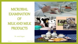
- 1. MICROBIAL EXAMINATION OF MILK AND MILK PRODUCTS Dr. smriti bajpai 9/19/2021 Dr Smriti Bajpai, Department of Microbiology (BVC, Secunderabad) 1
- 2. Microbial Examination of Milk 1. Direct Microscopic Count (DMC) 2. Standard Plate Count (SPC) 3. Dye (Methylene Blue, Resazurin) ReductionTest 9/19/2021 Dr Smriti Bajpai, Department of Microbiology (BVC, Secunderabad) 2
- 3. Direct Microscopic Count Principle: The DMC method enables rapid enumeration of bacterial cells along with their study of morphology of the total bacterial count in milk and cream with minimum equipment. It consists of examination of stained films of a measured volume of milk or milk product (0.01 ml) spread over 1 eq. cm area and dried on a glass slide under microscope. Somatic cells, shapes and arrangement of bacterial cells present in films can be easily and rapidly visualized and recorded. The microbial morphology and arrangement give the clue to possible cause of high count while high somatic cell indicates udder infection e.g. mastitis. For determination of average number of bacterial cells or clumps of cells about 5 to 50 microscopic fields are scanned (fewer the number of cells, more fields to be scanned). The diameter of a field is measured with the help of a stage micrometer to calculate microscopic factor (MF). The DMC/ml is then calculated as follows: DMC/ml=N x MF Where N= Average number of cells per field MF= Microscopic Factor (Microscopic field is an area of the field observed through the microscope) MF= Area of Smear/Area of Microscopic x 1/Volume of milk (0.01 ml)=10,000/3.1416 x r2 This technique is very useful for screening of milk supplies on the receiving platform of a dairy plant as well as for grading of milk. 9/19/2021 Dr Smriti Bajpai, Department of Microbiology (BVC, Secunderabad) 3
- 4. Requirements Test tubes Milk samples (Raw milk, Pasteurized milk, Boiled milk etc.) Hemocytometer Cover slip Micropipette with tips Methylene blue Microscope Oil immersion 9/19/2021 Dr Smriti Bajpai, Department of Microbiology (BVC, Secunderabad) 4
- 5. Methodology Preparation of Slide: A standard film of 1 sq. cm. of 0.01 ml of given milk sample was made on a clear slide. Fix the smear by air drying. Do not heat-fix the slide on direct flame followed by washing of slide with xylene to remove excess fat. Slide was then stained with methylene blue. Then slide was observed under microscope and the number of micro organisms per field were calculated. Microscopic examination Examine under the oil immersion objective and count the number of micro- organisms (individual or clumps of cells) in a number of fields of the film. The fields for counting the bacterial cells are selected at random. The number of microscopic fields occurring in one square centimeter area of the smear is very high. Different milk samples were observed in the same manner. 9/19/2021 Dr Smriti Bajpai, Department of Microbiology (BVC, Secunderabad) 5
- 6. Observation • Calculate the average number of clumps per field and multiply by the microscopic factor to get the DMC per milliliter of milk ( example- N=5 (no of organism observed per field R=0.08 mm Area= 0.02 sq. mm Milk film area 1 sq cm =100 sq. mm In 1 field area of milk =0.02 sq. mm So number of fields (MF) in 100 mm=5000 fields If 1 field has 4 organisms 9/19/2021 Dr Smriti Bajpai, Department of Microbiology (BVC, Secunderabad) 6
- 7. Result By using formula DMC/ml=N x MF DMC= 4 x5000= 20,000 organisms per sq mm. 0.01 ml suspension contain 20,000 organisms So 1 ml of suspension contain 20,00,000 organisms/ ml. Table 17.2 Grading of milk based on DMC (BIS standards) Advantages of Direct Microscopic Count • Rapid, Simple and easy method requiring minimum equipment. • Morphology of the bacteria can be observed as they counted. • Very dense suspensions can be counted if they are diluted appropriately. 9/19/2021 Dr Smriti Bajpai, Department of Microbiology (BVC, Secunderabad) 7 However, the limitation of this method is that both dead as well as viable cells are counted.
- 8. Standard Plate Count (SPC) The standard plate count (SPC) is suitable for estimating bacterial populations in most types of dairy products, and it is a reference method specified in the Grade A Pasteurized Milk Ordinance to be used to examine raw and pasteurized milk. This procedure is also recommended for application in detecting sources of contamination by testing line-samples taken at successive stages in the processing. Principle The test employs a serial dilution technique for easy quantification of the micro-organisms. The appropriate dilutions of the milk sample are mixed with a sterile nutrient medium that can support the growth of the micro-organisms, when incubated at a suitable temperature. Each bacterial colony that develops on the plate is presumed to have grown from one bacterium or clump of bacteria in the inoculums. The total number of colonies counted on the plates multiplied by the dilution factor to represent the number of viable micro- organisms present in the sample tested. 9/19/2021 Dr Smriti Bajpai, Department of Microbiology (BVC, Secunderabad) 8
- 9. Requirements 9/19/2021 Dr Smriti Bajpai, Department of Microbiology (BVC, Secunderabad) 9 o Milk samples o Test tubes o Pipettes o Petri dishes o Burner o Incubator o Colony counter o Nutrient agar media
- 10. Methodology Sample preparation: • Mark each plate with sample number, dilution, and other desired information before making dilutions. Dilution of samples: • Make serial dilutions of different milk samples. Plating and Incubation: • Equal portions of each dilution is poured in to a petri plate followed by addition of nutrient agar medium, a technique known as pour plate method. • The medium is allowed to solidify after mixing the contents by gentle rotation of the plate. • The organisms present in the sample are expected to be trapped in the agar gel. • The plates are subsequently incubated at 37 C for 48 to 72 hours. 9/19/2021 Dr Smriti Bajpai, Department of Microbiology (BVC, Secunderabad) 10
- 11. Observation • In principle each organism is expected to take up a separate position in the medium and grow in to a mass of cells of a size sufficient enough to be counted by naked eyes, recognized as a colony forming unit (cfu). • Hence, a colony count performed at this stage represents number of viable bacteria present in the given volume of milk sample. • Determination of microbiological quality of milk and milk products invariably involves performing different plate counts by colony counter. 9/19/2021 Dr Smriti Bajpai, Department of Microbiology (BVC, Secunderabad) 11
- 12. Result 9/19/2021 Dr Smriti Bajpai, Department of Microbiology (BVC, Secunderabad) 12 Calculating and recording of microbial counts by colony counter. Interpreting microbial counts Grading of milk based on standard plate count test (BIS Standards) Advantage of SPC • Enumeration of only viable microbes. • Cultural and morphological differentiation based on colony characteristics. • Suitable for determination of quality of milk samples like pasteurized milk and high grade raw milk with low bacterial number. • Useful for pasteurized and for line testing at various stages of processing.
- 13. Dye (Methylene Blue, Resazurin) ReductionTest • Dye reduction tests are based on qualitative analysis. • There are certain dyes (i.e. Methylene Blue, Resazurin), which act as oxidation-reduction indicator. • Bacteria consume dissolved oxygen during their growth in milk and consequently reduce the OH to a level at which these dyes are reduced and get decolorized. • Such dyes can be employed to assess the biochemical activity of bacteria and thus estimate number of bacteria indirectly. • The greater the number of bacteria in milk, the quicker will the oxygen be consumed, and in turn the color will disappear. • Thus, the time of reduction is taken as a measure of the number of microorganisms in milk. • Although, it is likely that it is more truly a measure of the total metabolic reactions proceeding at the cell surface of the bacteria. • The test is useful in assessing the bacteriological quality of milk by determination of the time taken for the reduction of methylene blue in milk indicated by its color change. 9/19/2021 Dr Smriti Bajpai, Department of Microbiology (BVC, Secunderabad) 13
- 14. Principle • Oxidation reduction potential of a substrate may be defined generally as the chemical process in which the substrate either loses or gains electrons. • When an element or compound loses electrons the substrate is said to be oxidized, while a substrate that gains electrons becomes reduced. • Milk, as it exists in the udder has a sufficiently low redox potential to reduce the methylene blue immediately. • The processes like milking, cooling, dumping etc. raise the oxidation reduction potential of milk to +0.3V, because of the incorporation of atmospheric oxygen. • At this particular O-R potential, methylene blue is in oxidized state. • When bacterial cells multiply in milk these, consume dissolved oxygen and as more and more oxygen is used and gets depleted, the dye starts acting as electron acceptor instead of oxygen. • As the oxidation reduction potential decreases from + 0.06 to0.01 V, methylene blue gets reduced. • One atom of hydrogen is taken up by the double bonded nitrogen of the dye that converts it into colorless state. • The greater is the number of microorganisms in milk, the greater is the metabolic activity and the faster is the reduction of dye (methylene blue, resazurin). 9/19/2021 Dr Smriti Bajpai, Department of Microbiology (BVC, Secunderabad) 14
- 15. Requirements • Milk samples • Pipette • Burner • Test tubes • Dye Methylene blue (0.01%) Resazurin (0.01%) 9/19/2021 Dr Smriti Bajpai, Department of Microbiology (BVC, Secunderabad) 15
- 16. Methodology The samples of milk are mixed thoroughly. Take 10 ml of milk into a test tube and add 1 ml of standard dye (methylene blue/ resazurin) solution. Invert the test tube to mix the milk and methylene blue solution. Place the test tube in a thermostatically maintained water bath at 37.5 C and note down the time of incubation. 9/19/2021 Dr Smriti Bajpai, Department of Microbiology (BVC, Secunderabad) 16
- 17. Observation • Observe the test tubes after 30 min for decolorization reduction of dye. • If there is no decolourization the tubes are inverted once and transferred to the water bath for further incubation. • After 30 min, continue to observe for the reduction of dye at an interval of every one-hour. • The milk shall be regarded as decolorized, when the entire column of milk is completely decolorized or is decolorized up to 5 mm of the surface. 9/19/2021 Dr Smriti Bajpai, Department of Microbiology (BVC, Secunderabad) 17
- 18. Result 9/19/2021 Dr Smriti Bajpai, Department of Microbiology (BVC, Secunderabad) 18 The quality of raw milk is adjusted by making the following observations Grading of milk based on MBRT as per BIS standard MBRTime (hr) Quality of raw milk 5 and above Very good 3 and 4 Good 1 and 2 Fair 1/2 and below Poor