2. Introduction
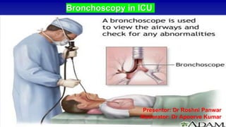
• Bronchoscopy is an important tool for pulmonary and critical care physicians to
diagnose and treat various pulmonary diseases in
• Plays an imp role in ICU due to its safety and portability.
• Is of utmost importance in critically ill patients who cannot be transported to the
remote imaging or diagnostic units.
• Though it is considered as low-risk procedure in healthy patients with mild
systemic diseases but can be challenging in patients with pre existing hypoxic
respiratory failure.
3. FOB has three main components
–Handle
–Insertion
cord
–Universal
cord
4. FOB – handle
• Eyepiece
– For viewing
– Diopter adjustment ring for focusing
• Bending lever
– Located at the back of the handle
– Maneuvered by thumb
– To move the tip in one plane
• Suction button
– Manipulated by index finger
• Access port to suction channel
5. Insertion cord
• Light transmission bundle
• Image transmission bundles
• Suction channel
– Suction
– Spray
– Brush, biopsy
– Insufflate oxygen
• Bending control wires
• Held together in a stainless steel mesh &wrapped in a water-impermeable
plastic coating
• Outer diameter of the insertion cord
• Only the distal 6 cm of the FOB is designed for maximal bending
6. Universal cord
• Light transmission bundle
• Electric wiring for automatic photography system
• Light source connector
– Light guide
– Air vent connector
• ETO
• Air leak test
– Electrical contacts for photography
7. Cleaning and Disinfection
• Prevent damage
• Prevent disease transmission
• Universal precautions
• Wash immediately after each use
• Flush working channel with water to remove secretions before they dry
• Inspect for defects that might allow water to penetrate inside FOB
• Remove suction and biopsy ports and place in disfinfectant solution
• TB or transmissible disease – ETO sterilisation - 24h
8. Diagnostic
• Pneumonia (bact. / vira/ fungal)
• Unexplained infiltrates on chest xray
• Suspected foreign body
• Hemoptysis
• Identify: site of bleeding
site of tracheal injury/ tef
etiology (benign/malignant)
• Airway obstruction
• Post trauma
Therapeutic
• Foreign body removal
• Mucus plugging
• Atlectasis
• Tumour debridement from airway
obstruction
• Stent placement
• Control bleeding (endobronchial
blockers/ cryotherapy/ argon plasma
coagulation
Indications of Bronchoscopy
9. Contraindications for Bronchoscopy
• Avoid BAL in pts. with Pa02 <75mmhg, Pa02/Fi02 <150 or Spo2 <90% with supplemental oxygen.
• Avoid transbronchial biopsy in pt with severe thrombocytopenian (platelet count <50k)
• Avoid NIV in patients with:
• high FiO2 requirement/ resp failure
• Facial deformity
• Upper GI bleeding
• Upper airway obstruction
• Unprotected airway
• Altered mental status
• Acute coronary syndrome
• Hemodynamic instability
• Respiratory/ cardiac arrest
• decompensated cardiac failure
10. How to plan bronchoscopy
• Verbal and written informed consent.
• Explain about indication, procedure and potential complications to the patients/ guardian
• Relatives should be explained about possible need of endotracheal intubation during/after procedure
for no-intubated patients.
• NBM: 8 hrs in elective bronchoscopy for solids, 6 hours for milk/other fluids, 2hrs for clear fluids.
• Pts safety to be kept in mind:
• Safety of doctor: TB, MDR, TB, H1N1, HIV, Hep B, SARS Cov-2,
• Timing: usefullness
– Antibioitic start/change
– Soon after platelet/ FFP transfusion
– Stopping clopidogrel
– 6 hrs after heparin or 12 hrs after LMWX or 3 days after oral coagulants
• Site selection:
– Most diseased side or if both equally involved then side with icd insitu
– Most involved lobe/ segment on chest xray/ ct
11. Ventilator settings
• Increase Fio2 to 100 % few mins before procedure.
• Just at onset of bronchoscopy reduce PEEP to 0/ reduce to half.
• Pressure control mode (High PIP)
• Remove closed suction in case present
• Attatch catheter mount with bronchoscopy port to the ETT.
• Nebulize before bronchoscopy
• In c/o desaturation remove FOB wait for recovery.
• Antisecretory agents
12. Risk and Precautions
• High risk procedure for critically ill patients especially one with
metabolic derrangements
• Bleeding diathesis
• Correct all electrolyte disturbances and coagulopathies before
procedure
• Factors such as PaO2 <70mmhg with FiO2 >70, PEEP >10
cm H2O, bronchospasm, unstable arrhytjmia, raised ICP, MI,
13. Monitoring
• Bronchoscopy is ass. with a change in lung compliance, increased MAP, mean PAP, and CO2
levels
• Hypoxia and tachycardia are a result of BAL in several studies,
• Bronchoscopy with BAL is associated with a worsened oxygenation, with an 80–86% reduction in
PaO2/FiO2 ratio from baseline.
• To avoid this, increase the FiO2 to 100% prior to starting the bronchoscopy.
• If persistent desturation below 88%, withdraw bronchoscope from airway/ increase PEEP/ FiO2.
• Resume bronchoscopy when FiO2 >95%
• Monitor: NIBP/ IBP
• Pulse oximetry
• EtCO2
• ICP montitoring in c/o Head iniury patients
• Emergency cart and high risk medications for resucitation should be kept ready
14. Sedation in Bronchoscopy
• Offers high success rate and lesser chances of failed procedure
• Intubated patients require increased iv sedation temporarily to facilitate
bronchoscopy.
• Non-intubated hypoxic patients pose increased risk of sedation related worsening
of respiratory drive and hypoxia during procedure.
• Sedation with low dose fentanyl and intermittant oropofol are safer options.
• Topical anaesthesia
• Lidocaine can be used as spray-as-you-go (1-2% lidocaine) or as nebulization for
delivering lidocaine to the airway unless contraindicated
• Excessive dose may cause CNS/ CVS toxicity.
15. Bronchoscopy with NIV.
• It constitutes a cornerstone in treatment of acute resp. failure of various etiologies.
• Reduces risk of intubation in patients with hypoxemic resp. failure
• NIV can prevent resp deterioration in spont. breathing hypoxemic patients
• NIV helps compensate for resistance and extra work for breathing created by
bronchoscopy during procedure.
• A CPAP machine + oxygen is considered superior to oxygen alone during
bronchoscopy in pts with hypoxia.
• Reduces the need of mechanical ventilation post-bronchoscopy.