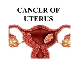
2. DEFINITION
Uterine cancer also known as endometrial
cancer and it involves a malignant growth
that originates in the lining of the uterus - the
endometrium.
3. INCIDENCE
• Higher amongst USA and lower in India
and Japan.
• The high incidence within a few decades of
menopause is associated with excessive
exposure to estrogen.
4. ETIOLOGY
• Estrogen – persistent stimulation of endometrium with
estrogen is the single most important factor for the
development of endometrial cancer.
• Age – 60 years of age
• Parity – unmarried and nulliparous women
• Late menopause – after 52 years
• Corpus cancer syndrome – encompasses obesity,
hypertension, diabetes.
5. • Estrogen stimulation that occurs in conditions such as
ovarian tumor, PCOD, and estrogen replacement
therapy in postmenopausal women.
• Tamoxifen used for the treatment of breast cancer
• Family history or personal history of colon, ovarian or
breast cancer increases the risk of endometrial cancer.
• Uterine fibroid
6. RISK FACTORS
• Early menarche
• Endometrial polyps or other benign growths of uterus lining
• H/O irregular and excessive premenopausal bleeding
• High intake of animal fat
• Pelvic radiation therapy
7.
8. CLASSIFICATION
• Carcinoma
most endometrial cancers are adenocarcinomas meaning
that they originate from the single glandular epithelial cells that
line the endometrium.
• Sarcoma
originate in the non-glandular connective tissue of the
endometrium.
9. • Carcinosarcoma
this is a rare uterine cancer that contains
cancerous cells of both glandular and sarcomatous
appearance.
10. SIGNS & SYMPTOMS
• Vaginal bleeding and/or spotting in postmenopausal women
• Abnormal uterine bleeding and abnormal menstrual
periods
• Bleeding between normal periods in premenopausal women.
Extremely long, heavy or frequent episodes of bleeding may
indicate premalignant changes.
11. • Anemia caused by chronic blood loss
• Lower abdominal pain or cramping
• Thin white or clear vaginal discharge in postmenopausal
women
• Abdominal mass
• Pain during sexual intercourse
12. DIAGNOSIS
• History & PE
• Pap smear
• Endometrial biopsy
• Transvaginal ultrasound
• Hysteroscopy
• Fractional curettage to detect the extent of the disease
13. STAGING
• Stage I – tumor confined to endometrium
• Stage II – endocervical glandular involvement
• Stage III – Tumor invades serosa or adnexa
• Stage IV – tumor invasion to bladder and/or bowel
mucosa