The document discusses the anatomy and biomechanics of the foot and ankle complex. It notes that the foot is made up of 28 bones, 33 joints, over 100 ligaments, and controlled by 13 extrinsic and 21 intrinsic muscles. The foot is divided into the rearfoot, midfoot, and forefoot. Key bones of the foot include the talus, calcaneus, navicular, and cuboid bones. The ankle joint is a synovial hinge joint that allows dorsiflexion and plantar flexion. Other important joints of the foot include the subtalar and transverse tarsal joints.
Biomechanics of Foot and Ankle complex, CP orthotic management &Tone reducing...Fiona Verma
Biomechanics of Foot and ankle complex along with common foot pathology like flatfeet has been discussed.
Types of Flatfeet, pathophysiology & its biomechanics negative impact on gait with Orthotic treatment has been discussed.
Types of CP (hemiplegia and diplegia spastic CP ), its gait patterns and appropriate orthotic management around the ankle and foot complex in child with spastic cp has been discussed including various tone reducing AFOs and Neurophysiology AFOs.
Newton's laws of motion are three physical laws that, together, laid the foundation for classical mechanics. They describe the relationship between a body and the forces acting upon it, and its motion in response to those forces
Newton's laws of motion are three physical laws that, together, laid the foundation for classical mechanics. They describe the relationship between a body and the forces acting upon it, and its motion in response to those forces
this PPT contain detailed kinetics & kinematics of ankle joint & all joints of foot complex, muscles of ankle & foot complex, plantar arches & weight distribution during standing.
Synthetic Fiber Construction in lab .pptxPavel ( NSTU)
Synthetic fiber production is a fascinating and complex field that blends chemistry, engineering, and environmental science. By understanding these aspects, students can gain a comprehensive view of synthetic fiber production, its impact on society and the environment, and the potential for future innovations. Synthetic fibers play a crucial role in modern society, impacting various aspects of daily life, industry, and the environment. ynthetic fibers are integral to modern life, offering a range of benefits from cost-effectiveness and versatility to innovative applications and performance characteristics. While they pose environmental challenges, ongoing research and development aim to create more sustainable and eco-friendly alternatives. Understanding the importance of synthetic fibers helps in appreciating their role in the economy, industry, and daily life, while also emphasizing the need for sustainable practices and innovation.
The French Revolution, which began in 1789, was a period of radical social and political upheaval in France. It marked the decline of absolute monarchies, the rise of secular and democratic republics, and the eventual rise of Napoleon Bonaparte. This revolutionary period is crucial in understanding the transition from feudalism to modernity in Europe.
For more information, visit-www.vavaclasses.com
Palestine last event orientationfvgnh .pptxRaedMohamed3
An EFL lesson about the current events in Palestine. It is intended to be for intermediate students who wish to increase their listening skills through a short lesson in power point.
Biological screening of herbal drugs: Introduction and Need for
Phyto-Pharmacological Screening, New Strategies for evaluating
Natural Products, In vitro evaluation techniques for Antioxidants, Antimicrobial and Anticancer drugs. In vivo evaluation techniques
for Anti-inflammatory, Antiulcer, Anticancer, Wound healing, Antidiabetic, Hepatoprotective, Cardio protective, Diuretics and
Antifertility, Toxicity studies as per OECD guidelines
Macroeconomics- Movie Location
This will be used as part of your Personal Professional Portfolio once graded.
Objective:
Prepare a presentation or a paper using research, basic comparative analysis, data organization and application of economic information. You will make an informed assessment of an economic climate outside of the United States to accomplish an entertainment industry objective.
2024.06.01 Introducing a competency framework for languag learning materials ...Sandy Millin
http://sandymillin.wordpress.com/iateflwebinar2024
Published classroom materials form the basis of syllabuses, drive teacher professional development, and have a potentially huge influence on learners, teachers and education systems. All teachers also create their own materials, whether a few sentences on a blackboard, a highly-structured fully-realised online course, or anything in between. Despite this, the knowledge and skills needed to create effective language learning materials are rarely part of teacher training, and are mostly learnt by trial and error.
Knowledge and skills frameworks, generally called competency frameworks, for ELT teachers, trainers and managers have existed for a few years now. However, until I created one for my MA dissertation, there wasn’t one drawing together what we need to know and do to be able to effectively produce language learning materials.
This webinar will introduce you to my framework, highlighting the key competencies I identified from my research. It will also show how anybody involved in language teaching (any language, not just English!), teacher training, managing schools or developing language learning materials can benefit from using the framework.
Honest Reviews of Tim Han LMA Course Program.pptxtimhan337
Personal development courses are widely available today, with each one promising life-changing outcomes. Tim Han’s Life Mastery Achievers (LMA) Course has drawn a lot of interest. In addition to offering my frank assessment of Success Insider’s LMA Course, this piece examines the course’s effects via a variety of Tim Han LMA course reviews and Success Insider comments.
How to Make a Field invisible in Odoo 17Celine George
It is possible to hide or invisible some fields in odoo. Commonly using “invisible” attribute in the field definition to invisible the fields. This slide will show how to make a field invisible in odoo 17.
Francesca Gottschalk - How can education support child empowerment.pptxEduSkills OECD
Francesca Gottschalk from the OECD’s Centre for Educational Research and Innovation presents at the Ask an Expert Webinar: How can education support child empowerment?
Read| The latest issue of The Challenger is here! We are thrilled to announce that our school paper has qualified for the NATIONAL SCHOOLS PRESS CONFERENCE (NSPC) 2024. Thank you for your unwavering support and trust. Dive into the stories that made us stand out!
2. Ankle/foot complex is structurally analogous with
wrist/hand of the upper extremity but it has a
distinct differences to optimizes its primary role to
bear weight.
Providing a stable base of support in the variety of
weight bearing postures without undue muscular
activity and energy expenditure.
Acting as a rigid lever for effective push-off during
gait
Monday, February 13, 2023 Dechasa Imiru 2
3. …
Dampening rotations imposed by the more proximal
joints of the lower limbs
Being flexible enough to absorb the shock of the
superimposed body weight as the foot hits the
ground.
Permitting the foot to conform to a wide range of
changing
To serve these functions foot is made up of series of
small bones and designed in a form of elastic arches or
springs.
Monday, February 13, 2023 Dechasa Imiru 3
4. .
The foot and ankle complex meets these diverse
requirements through the integrated
movements its 28 bones, 33 joints, more 100
ligaments, controlled by 13 extrinsic and 21
intrinsic muscles.
The foot and ankle complex meets these diverse
requirements through the integrated
movements its 28 bones, 33 joints, more 100
ligaments, controlled by 13 extrinsic and 21
intrinsic muscles.
The foot is subdivided into the rear
foot, mid foot, and forefoot.
The foot is subdivided into the rear
foot, mid foot, and forefoot.
.
Monday, February 13, 2023 Dechasa Imiru 4
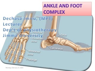
5. TALUS: Most proximal tarsal bone, Dome(trochlear),
Prominent head forwards/medially, Neck & 3 facets.
CALCANEUS: Largest tarsals, attaches to Achilles,
forms sinus tarsi with talus, sustentaculum talus
NAVICULAR: means “ship”, its concave hull with head
of talus, 3 facet with 3 cuneiform, Tibialis posterior
muscle attaches.
MEDIAL, INTERMEDIATE, & LATERAL CUNEIFORM:
Acts as “spacer” between Navicular & 3 medial MT
bones, forms transverse arch, joint with cuboid.
CUBOID: six surfaces, joint with 4th & 5th metatarsal,
Calcaneum, Lat cuneiform, Navicular.
Monday, February 13, 2023 Dechasa Imiru 5
6. Proximal and distal tibio fibular joints
The talocrural, or ankle, joint
The talo-calcaneal, or sub talar joint
The talo-navicular
The calcaneo-cuboid joints
The five tarso-metatarsal joints
Five meta-tarsophalangeal joints
Nine inter-phalangeal joints.
Note : Talus is included in ankle & infoot
Proximal and distal tibio fibular joints
The talocrural, or ankle, joint
The talo-calcaneal, or sub talar joint
The talo-navicular
The calcaneo-cuboid joints
The five tarso-metatarsal joints
Five meta-tarsophalangeal joints
Nine inter-phalangeal joints.
Note : Talus is included in ankle & infoot
Monday, February 13, 2023 Dechasa Imiru 6
7. HINDFOOT/rear foot (posterior segment),
composed of the talus and calcaneus.
MIDFOOT (middle segment), composed of the
navicular, cuboid, and three cuneiforms.
FOREFOOT (anterior segment), composed of the
metatarsals and the phalanges.
NOTE :Talus is an extremely
important bone, has an
essential role to play in local
kinesiology and kinesiology
of lower extremity.
Monday, February 13, 2023 Dechasa Imiru 7
9. The three motions of the ankle/foot complex that
approximate cardinal planes and axes are
Dorsiflexion/ Plantar flexion
Dorsiflexion and plantar flexion are motions that occur
(appx) in the sagittal plane around a coronal axis (x-axis).
Dorsiflexion decreases the angle between the leg and the
dorsum of the foot, whereas plantar flexion increases this
angle.
Inversion/eversion
Occurs (appx) in the frontal plane around a longitudinal
(antero-posterior [A-P]) axis that runs through the length
of the foot.
Inversion plantar surface turn towards midline.
The three motions of the ankle/foot complex that
approximate cardinal planes and axes are
Dorsiflexion/ Plantar flexion
Dorsiflexion and plantar flexion are motions that occur
(appx) in the sagittal plane around a coronal axis (x-axis).
Dorsiflexion decreases the angle between the leg and the
dorsum of the foot, whereas plantar flexion increases this
angle.
Inversion/eversion
Occurs (appx) in the frontal plane around a longitudinal
(antero-posterior [A-P]) axis that runs through the length
of the foot.
Inversion plantar surface turn towards midline.
Monday, February 13, 2023 Dechasa Imiru 9
10. Abduction/ adduction
Occur (appx) in the transverse plane around a vertical axis.
Abduction is when the distal aspect of a segment moves away from
the midline of the body, Adduction is opposite.
Pronation/supination of foot(TRIPLANAR MOTION)
Motions that occur around an axis that lies at an angle to each of
the axes for “cardinal” motion.
Pronation is motion about an axis that results in coupled motions
of dorsiflexion, eversion, and abduction.
Supination is a motion about an axis that results in coupled motions
of plantar flexion, inversion, and adduction.
Note: Pathological motions like Calcaneo valgus, Calcaneo varus
Abduction/ adduction
Occur (appx) in the transverse plane around a vertical axis.
Abduction is when the distal aspect of a segment moves away from
the midline of the body, Adduction is opposite.
Pronation/supination of foot(TRIPLANAR MOTION)
Motions that occur around an axis that lies at an angle to each of
the axes for “cardinal” motion.
Pronation is motion about an axis that results in coupled motions
of dorsiflexion, eversion, and abduction.
Supination is a motion about an axis that results in coupled motions
of plantar flexion, inversion, and adduction.
Note: Pathological motions like Calcaneo valgus, Calcaneo varus
Monday, February 13, 2023 Dechasa Imiru 10
11. The term ankle refers specifically to the
talocrural joint.
Ankle is a synovial hinge joint, have a single
oblique axis with one degree of freedom around
which the motions of dorsiflexion/ plantar
flexion .
Monday, February 13, 2023 Dechasa Imiru 11
12. composed of the concave surface of the distal
tibia and fibular malleoli.
3 facets forms a continuous concave surface.
The structure of the distal tibia and the malleoli
resembles & referred as “Adjustable mortise”
composed of the concave surface of the distal
tibia and fibular malleoli.
3 facets forms a continuous concave surface.
The structure of the distal tibia and the malleoli
resembles & referred as “Adjustable mortise”
Monday, February 13, 2023 Dechasa Imiru 12
13. The mortise of the ankle is adjustable, relying on the
proximal and distal tibiofibular joints to both permit
and control the changes in the mortise.
The proximal and distal tibiofibular joints functions
exclusively to serve ankle joint.
Proximal tibiofibular joint is a plane synovial joint
formed by the articulation of the head of the fibula
with the postero-lateral aspect of the tibia.
Distal tibiofibular joint is a syndesmosis, or fibrous
union, between the concave facet of the tibia and the
convex facet of the fibula.
Fusion of the tibiofibular joints may impair normal ankle
function by limiting the ability of the talus to move within
the ankle mortise.
Monday, February 13, 2023 Dechasa Imiru 13
14. Body of the talus forms the distal articulation
of the ankle joint.
Body of the talus has three articular surfaces: a
large lateral (fibular) facet, a smaller medial
(tibial) facet, and a trochlear (superior) facet.
The wedge shaped trochlear surface is wider
anteriorly than posteriorly, so provides more
stability in dorsiflexion.
Monday, February 13, 2023 Dechasa Imiru 14
15. Capsule of the ankle joint is fairly thin and
especially weak anteriorly and posteriorly.
Stability of the ankle depends on an intact
ligamentous structure.
Capsule of the ankle joint is fairly thin and
especially weak anteriorly and posteriorly.
Stability of the ankle depends on an intact
ligamentous structure.
Ligaments of Proximal & Distal tibio-fibular joint
Crural tibio-fibular interosseous ligament
The anterior and posterior tibio-fibular ligaments
The tibiofibular interosseous membrane
Two other major ligaments maintain contact &
congruence of the mortise and talus and control
medial-lateral joint
stability, also provide key support for subtalar joint.
1.Medial collateral ligament (MCL)
2.Lateral collateral ligament (LCL)
Monday, February 13, 2023 Dechasa Imiru 15
16. Deltoid ligament is a fan-shaped.(Extremely strong)
It arise from the borders of the tibial malleolus and insert
on the navicular bone anteriorly and on the talus and
calcaneus distally and posteriorly. It checks calcaneal
eversion, medial distractions, extremes
Deltoid ligament is a fan-shaped.(Extremely strong)
It arise from the borders of the tibial malleolus and insert
on the navicular bone anteriorly and on the talus and
calcaneus distally and posteriorly. It checks calcaneal
eversion, medial distractions, extremes
Monday, February 13, 2023 Dechasa Imiru 16
17. is composed of three separate bands
Anterior and posterior talo-fibular ligaments and the
longer calcaneo- fibular ligament.
LCL helps control varus stresses that result in lateral
distraction of the joint ,Calcaneal inversion, Extreme ROM.
is composed of three separate bands
Anterior and posterior talo-fibular ligaments and the
longer calcaneo- fibular ligament.
LCL helps control varus stresses that result in lateral
distraction of the joint ,Calcaneal inversion, Extreme ROM.
Monday, February 13, 2023 Dechasa Imiru 17
18. Inferior extensor retinaculum
Superior peroneal
retinaculum
Superior extensor retinacula
Inferior peroneal retinacula
Inferior extensor retinaculum
Superior peroneal
retinaculum
Superior extensor retinacula
Inferior peroneal retinacula
Monday, February 13, 2023 Dechasa Imiru 18
19. Dorsiflexion:10 to 20 degrees
Plantar flexion: 20 to 50 degrees
The enhanced stability at the ankle joint in
dorsiflexion allows the ankle to withstand
compression forces of as much as 45% of
body weight.
Loose packed position of the ankle joint is
in plantar flexion when only the relatively
narrow posterior body of the talus is in
contact with the mortise.(Ankle is instable)
Isolated talus ROM yields
lower ranges in Dorsi &
plantar flexions
Isolated talus ROM yields
lower ranges in Dorsi &
plantar flexions
High incidence of ankle
sprains reported in Plantar
flexion
High incidence of ankle
sprains reported in Plantar
flexion
Monday, February 13, 2023 Dechasa Imiru 19
20. Dorsiflexion is limited with the knee in extension
than with the knee in flexion because the
gastrocnemius muscle is lengthened over two joints
when the knee is extended.
Tension in the tibialis anterior, extensor hallucis longus,
and extensor digitorum longus muscles is the primary
limit to plantar flexion.
Tibialis posterior, flexor hallucis longus, and flexor
digitorum longus muscles stabilize medial aspect of
the ankle.
Peroneus longus and peroneus brevis muscles protect
the lateral aspect.
Monday, February 13, 2023 Dechasa Imiru 20
21. Talocalcaneal, or subtalar joint is a composite joint
formed by three separate plane articulations
between the talus superiorly and the calcaneus
inferiorly.
Triplanar movement around a single joint axis.
Function at the weight bearing subtalar joint is critical
for dampening the rotational forces imposed by the
body weight while maintaining contact of the foot
with the supporting surface.
Subtalar articular surfaces, although smaller than
those of the ankle joint surfaces, showed a similar
proportion of contact across surfaces, given the
contact area this joint rarely undergoes degeneration
Monday, February 13, 2023 Dechasa Imiru 21
22. Between the posterior articulation and the anterior
and medial articulations, there is a bony tunnel
formed by a sulcus (concave groove) in the inferior
talus and superior calcaneus. This funnel-shaped
tunnel, known as the tarsal canal, runs obliquely
across the foot. Its large end (the sinus tarsi) lies just
anterior to the fibular malleolus & small lies posterior
to tibial malleolus called Sustentaculum tali
Monday, February 13, 2023 Dechasa Imiru 22
23. Sub talar joint is a stable joint it rarely dislocates
LIGAMENTS
Interosseous talo-calcaneal ligament
Calcaneo fibular ligament
Lateral talo-calcaneal ligament
Cervical ligament
LIGAMENTS
Interosseous talo-calcaneal ligament
Calcaneo fibular ligament
Lateral talo-calcaneal ligament
Cervical ligament
Monday, February 13, 2023 Dechasa Imiru 23
24. In reality, the subtalar axis lies about halfway between being
longitudinal and being vertical
In reality, the subtalar axis lies about halfway between being
longitudinal and being vertical
Motion of the talus on the calcaneus, therefore, is a
complex twisting or screw like motion.
Single oblique joint axis, producing the motion of
supination/pronation.
Consequently, pronation/supination includes about equal
magnitudes of eversion/inversion & abduction/adduction
Subtalar joint supination is a normal foot motion, a foot
that appears fixed in this position is called a “supinated”
or cavus foot. Foot which is fixed in pronation is called
pes planus or flat foot.
Monday, February 13, 2023 Dechasa Imiru 24
25. Range of subtalar supination and pronation is
difficult to determine objectively because of the
triplanar nature of the movement
Calcaneal inversion/eversion component of
subtalar motion is relatively easy to measure.
Calcaneal eversion (valgus) : 5° to 10 °
Calcaneal inversion :20° to 30 °
Calcaneal inversion/eversion component of
subtalar motion is relatively easy to measure.
Calcaneal eversion (valgus) : 5° to 10 °
Calcaneal inversion :20° to 30 °
Monday, February 13, 2023 Dechasa Imiru 25
26. Transverse tarsal joint, also called the midtarsal or Chopart
joint.
It’s a compound joint formed by
The 2 joints form “S” shaped joint line which horizontally
separates rear foot from mid & fore foot.
Navicular & cuboid bones are relatively fixed in wt bearing
Transverse tarsal joint, also called the midtarsal or Chopart
joint.
It’s a compound joint formed by
The 2 joints form “S” shaped joint line which horizontally
separates rear foot from mid & fore foot.
Navicular & cuboid bones are relatively fixed in wt bearing
Monday, February 13, 2023 Dechasa Imiru 26
27. Proximally: Head of talus,
Distally: Concave posterior aspect of navicular bone.
Proximally: Head of talus,
Distally: Concave posterior aspect of navicular bone.
Plantar calcaneonavicular ligament
(spring ligament)
Bifurcate ligaments
Dorsal talo navicular ligament
Anterior edge of deltiod ligament
Dorsal calcaneocuboid ligament
Plantar calcaneocuboid
Long plantar ligaments
LONG PLANTAR LIGAMENT
makes a significant contribution
both to transverse tarsal joint
stability and to related support
of the lateral longitudinal arch
of the foot
Monday, February 13, 2023 Dechasa Imiru 27
28. The longitudinal and oblique axes for the transverse tarsal
joint indicate a function similar to that of the subtalar joint.
When subtalar joint is fully supinated and locked (bony
surfaces are drawn together),the transverse tarsal joint is
also carried into full .
Supination locks not only the subtalar joint but also the
transverse tarsal joint
When the subtalar joint is pronated and loose- packed, the
transverse tarsal joint is also mobile and loose-packed.
The longitudinal and oblique axes for the transverse tarsal
joint indicate a function similar to that of the subtalar joint.
When subtalar joint is fully supinated and locked (bony
surfaces are drawn together),the transverse tarsal joint is
also carried into full .
Supination locks not only the subtalar joint but also the
transverse tarsal joint
When the subtalar joint is pronated and loose- packed, the
transverse tarsal joint is also mobile and loose-packed.
Monday, February 13, 2023 Dechasa Imiru 28
30. Tarsometatarsal-TMT joints are plane synovial joints
formed by the distal row of tarsal bones and the bases
of the metatarsals.
The motions of the TMT joints are interdependent,
as are the motions of the CMC joints in the hand.
TMT joints attempt to regulate position of the
metatarsals and phalanges (the forefoot) in relation
to the weight-bearing surface (Forefoot adjustment).
Supination Twist
Pronation Twist
Tarsometatarsal-TMT joints are plane synovial joints
formed by the distal row of tarsal bones and the bases
of the metatarsals.
The motions of the TMT joints are interdependent,
as are the motions of the CMC joints in the hand.
TMT joints attempt to regulate position of the
metatarsals and phalanges (the forefoot) in relation
to the weight-bearing surface (Forefoot adjustment).
Supination Twist
Pronation Twist
Monday, February 13, 2023 Dechasa Imiru 30
31. The five metatarsophalangeal (MTP) joints are
condyloid synovial joints with two degrees of freedom:
extension/flexion or dorsiflexion/plantar flexion) and
abduction/adduction.
MTP joints are formed proximally by the convex heads
of the metatarsals and distally by the concave bases of
the proximal phalanges.
During the late stance phase of walking, toe extension
at the MTP joints permits the foot to pass over the toes.
Stability of the MTP joints is provided by a joint
capsule, plantar plates, collateral ligaments, and the
deep transverse metatarsal ligament
The five metatarsophalangeal (MTP) joints are
condyloid synovial joints with two degrees of freedom:
extension/flexion or dorsiflexion/plantar flexion) and
abduction/adduction.
MTP joints are formed proximally by the convex heads
of the metatarsals and distally by the concave bases of
the proximal phalanges.
During the late stance phase of walking, toe extension
at the MTP joints permits the foot to pass over the toes.
Stability of the MTP joints is provided by a joint
capsule, plantar plates, collateral ligaments, and the
deep transverse metatarsal ligament
Monday, February 13, 2023 Dechasa Imiru 31
32. The metatarsal break derives its name from the
hinge or “break” that occurs at the MTP joints as the
heel rises and the metatarsal heads and toes remain
weight bearing.
The metatarsal break occurs as MTP extension
around a single oblique axis that lies through the
second to fifth metatarsal heads.
Limited extension ROM at the first MTP joint will
interfere with the metatarsal break and is known as
hallux rigidus.
An increase in this normal valgus angulation of the
1st MTP joint is referred to as hallux valgus.
The metatarsal break derives its name from the
hinge or “break” that occurs at the MTP joints as the
heel rises and the metatarsal heads and toes remain
weight bearing.
The metatarsal break occurs as MTP extension
around a single oblique axis that lies through the
second to fifth metatarsal heads.
Limited extension ROM at the first MTP joint will
interfere with the metatarsal break and is known as
hallux rigidus.
An increase in this normal valgus angulation of the
1st MTP joint is referred to as hallux valgus.
Monday, February 13, 2023 Dechasa Imiru 32
33. The foot typically is characterized as having three
arches:
Medial longitudinal arches
Lateral longitudinal arches
Transverse arch
The foot typically is characterized as having three
arches:
Medial longitudinal arches
Lateral longitudinal arches
Transverse arch
Monday, February 13, 2023 Dechasa Imiru 33
34. FUNCTIONS OF THE ARCHES
-weight distributed equally through the anterior and the
posterior part of the foot
-heads of five metatarsals posses six weight bearing points
Plantar concavity prevents compression of neurovascular
structures of the foot
Arched foot is dynamic and pliable
Invertors and evertors help in shifting weight distribution
Normal Foot
Monday, February 13, 2023 Dechasa Imiru 34
35. medial longitudinal arch
lateral longitudinal arch
plantar ligaments, plantar
aponeurosis bear maximum stress
muscles are active
windlass action of plantar
aponeurosis
medial longitudinal arch
lateral longitudinal arch
plantar ligaments, plantar
aponeurosis bear maximum stress
muscles are active
windlass action of plantar
aponeurosis
Monday, February 13, 2023 Dechasa Imiru 35
37. o trochlear surface of talus
o heads of medial three
metatarsals
o medial tubercle of calcaneus
o head of talus (keystone)
o Resiliency/elasticity
Monday, February 13, 2023 Dechasa Imiru 37
38. FACTORS MAINTINING MEDIAL ARCH
Shape of bones
- wedge shaped bones
- keystone (head of talus)
Staples
- plantar ligaments
- most important plantar
calcaneonavicular (spring ligament)
Tie beam
- plantar aponeurosis, abductor hallucis,
flexor hallucis longus and brevis tendon,
medial part of flexor digitorum longus
and brevis
Slings
- tibialis anterior tendon, deltoid
ligament and tibialis posterior tendon
Monday, February 13, 2023 Dechasa Imiru 38
39. LATERAL LONGITUDNAL ARCH
Summit
- subtalar joint
Anterior pillar
- head of fourth and fifth
metatarsals
Posterior pillar
- medial tubercle of calcaneus
Vulnerable part of arch
- calcaneocuboid joint
Characteristic feature of arch
- rigidity
Summit
- subtalar joint
Anterior pillar
- head of fourth and fifth
metatarsals
Posterior pillar
- medial tubercle of calcaneus
Vulnerable part of arch
- calcaneocuboid joint
Characteristic feature of arch
- rigidity
Monday, February 13, 2023 Dechasa Imiru 39
40. formed by heads of the five
metatarsal bones
is complete
formed by greater parts of
tarsus & metatarsus
is incomplete
only the lateral end comes in
contact with the ground
formed by heads of the five
metatarsal bones
is complete
formed by greater parts of
tarsus & metatarsus
is incomplete
only the lateral end comes in
contact with the ground
Monday, February 13, 2023 Dechasa Imiru 40
41. The medial arch is the higher of
the two longitudinal arches. It
is made up of the calcaneus,
the talus, the navicular, the
three cuneiforms, and the first,
second, and third metatarsals.
The chief characteristic of this
arch is its elasticity
The lateral arch is the flatter of the
two longitudinal arches and lies on
the ground in the standing position.
It is composed of the calcaneus, the
cuboid, and the fourth and fifth
metatarsals.
The lateral arch is the flatter of the
two longitudinal arches and lies on
the ground in the standing position.
It is composed of the calcaneus, the
cuboid, and the fourth and fifth
metatarsals.
The transverse arch is located in the coronal
plane of the foot.
The transverse arches are strengthened by
the interosseous, plantar, and dorsal
ligaments, by the short muscles of the first
and fifth toes
Monday, February 13, 2023 Dechasa Imiru 41
43. PES CAVUS
deformity characterised by an abnormally high
medial longitudinal arch
PES PLANUS
Monday, February 13, 2023 Dechasa Imiru 43
44. Medial longitudinal arch of the foot is not formed by-
A. Cuboid
B. Calcaneus
C. Talus
D. Navicular
The keystone of the lateral longitudinal arch is
A. Navicular
B. Lateral Cuneiform
C. Calcaneum
D. Cuboid
Monday, February 13, 2023 Dechasa Imiru 44
45. A. Peroneus Brevis
B. Peroneus Longus
C. Tibialis Anterior
D. Tibialis Posterior
A. Peroneus Brevis
B. Peroneus Longus
C. Tibialis Anterior
D. Tibialis Posterior
Monday, February 13, 2023 Dechasa Imiru 45