SlideShare a Scribd company logo
1 of 255
Chapter 22 Musculoskeletal System
STRUCTURE AND FUNCTION
The musculoskeletal system consists of the body's bones, joints, and muscles. Humans need
this system (1) for support to stand erect and (2) for movement. The musculoskeletal system
also functions (3) to encase and protect the inner vital organs (e.g., brain, spinal cord, heart),
(4) to produce the red blood cells in the bone marrow (hematopoiesis), and (5) as a reservoir
for storage of essential minerals, such as calcium and phosphorus in the bones.
COMPONENTS OF THE MUSCULOSKELETAL SYSTEM
The skeleton is the bony framework of the body. It has 206 bones, which support the body
like the posts and beams of a building. Bone and cartilage are specialized forms of
connective tissue. Bone is hard, rigid, and very dense. Its cells are continually turning over
and remodeling. The joint (or articulation) is the place of union of two or more bones.
Joints are the functional units of the musculoskeletal system because they permit the
mobility needed for activities of daily living.
Nonsynovial or Synovial Joints
In nonsynovial joints, the bones are united by fibrous tissue or cartilage and are
immovable (e.g., the sutures in the skull) or only slightly movable (e.g., the vertebrae).
Synovial joints are freely movable because they have bones that are separated from each
other and are enclosed in a joint cavity (Fig. 22-1). This cavity is filled with a lubricant,
or synovial fluid. Just like grease on gears, synovial fluid allows sliding of opposing
surfaces, and this sliding permits movement.
Fig. 22-1
In synovial joints, a layer of resilient cartilage covers the surface of opposing bones.
Cartilage is avascular; it receives nourishment from synovial fluid that circulates during
joint movement. It is a very stable connective tissue with a slow cell turnover. It has a
tough, firm consistency, yet is flexible. This cartilage cushions the bones and gives a
smooth surface to facilitate movement.
The joint is surrounded by a fibrous capsule and is supported by ligaments. Ligaments
are fibrous bands running directly from one bone to another that strengthen the joint and
help prevent movement in undesirable directions. A bursa is an enclosed sac filled with
viscous synovial fluid, much like a joint. Bursae are located in areas of potential friction
(e.g., subacromial bursa of the shoulder, prepatellar bursa of the knee) and help muscles
and tendons glide smoothly over bone.
Muscles
Muscles account for 40% to 50% of the body's weight. When they contract, they produce
movement. Muscles are of three types: skeletal, smooth, and cardiac. This chapter is
concerned with skeletal, or voluntary, muscles, those under conscious control.
Fig. 22-2
Each skeletal muscle is composed of bundles of muscle fibers, or fasciculi. The skeletal
muscle is attached to bone by a tendon-a strong fibrous cord. Skeletal muscles produce
the following movements (Fig. 22-2):
1. Flexion—bending a limb at a joint
2. Extension—straightening a limb at a joint
3. Abduction—moving a limb away from the midline of the body
4. Adduction—moving a limb toward the midline of the body
5. Pronation—turning the forearm so that the palm is down
6. Supination—turning the forearm so that the palm is up
7. Circumduction—moving the arm in a circle around the shoulder
8. Inversion—moving the sole of the foot inward at the ankle
9. Eversion—moving the sole of the foot outward at the ankle
10. Rotation—moving the head around a central axis
11. Protraction—moving a body part forward and parallel to the ground
12. Retraction—moving a body part backward and parallel to the ground
13. Elevation—raising a body part
14. Depression—lowering a body part
Fig. 22-3
Temporomandibular Joint
The temporomandibular joint (TMJ) is the articulation of the mandible and the temporal
bone (Fig. 22-3). You can feel it in the depression anterior to the tragus of the ear. The
TMJ permits jaw function for speaking and chewing. The joint allows three motions: (1)
hinge action to open and close the jaws, (2) gliding action for protrusion and retraction,
and (3) gliding for side-to-side movement of the lower jaw.
Spine
The vertebrae are 33 connecting bones stacked in a vertical column (Fig. 22-4). You can
feel their spinous processes in a furrow down the midline of the back. The furrow has
paravertebral muscles mounded on either side down to the sacrum, where it flattens.
Humans have 7 cervical, 12 thoracic, 5 lumbar, 5 sacral, and 3 to 4 coccygeal vertebrae.
The following surface landmarks will orient you to their levels:
• The spinous processes of C7 and T1 are prominent at the base of the neck.
• The inferior angle of the scapula normally is at the level of the interspace between
T7 and T8.
• An imaginary line connecting the highest point on each iliac crest crosses L4.
• An imaginary line joining the two symmetric dimples that overlie the posterior
superior iliac spines crosses the sacrum.
Fig. 22-4
Fig. 22-5
Fig. 22-6
A lateral view shows that the vertebral column has four curves (a double-S shape) (Fig.
22-5). The cervical and lumbar curves are concave (inward or anterior), and the thoracic
and sacrococcygeal curves are convex. The balanced or compensatory nature of these
curves, together with the resilient intervertebral disks, allows the spine to absorb a great
deal of shock.
The intervertebral disks are elastic fibrocartilaginous plates that constitute one fourth of
the length of the column (Fig. 22-6). Each disk center has a nucleus pulposus, made of
soft, semifluid, mucoid material that has the consistency of toothpaste in the young adult.
The disks cushion the spine like a shock absorber and help it move. As the spine moves,
the elasticity of the disks allows compression on one side, with compensatory expansion
on the other. Sometimes compression can be too great. The disk then can rupture and the
nucleus pulposus can herniate out of the vertebral column, compressing on the spinal
nerves and causing pain.
The unique structure of the spine enables both upright posture and flexibility for motion.
The motions of the vertebral column are flexion (bending forward), extension (bending
back), abduction (to either side), and rotation.
Fig. 22-7
Shoulder
The glenohumeral joint is the articulation of the humerus with the glenoid fossa of the
scapula (Fig. 22-7). Its ball-and-socket action allows great mobility of the arm on many
axes. The joint is enclosed by a group of four powerful muscles and tendons that support
and stabilize it. Together these are called the rotator cuff of the shoulder. The large
subacromial bursa helps during abduction of the arm, so that the greater tubercle of the
humerus moves easily under the acromion process of the scapula.
Fig. 22-8
The bones of the shoulder have palpable landmarks to guide your examination (Fig. 22-
8). The scapula and the clavicle connect to form the shoulder girdle. You can feel the
bump of the scapula's acromion process at the very top of the shoulder. Move your
fingers in a small circle outward, down, and around. The next bump is the greater
tubercle of the humerus a few centimeters down and laterally, and from that the
coracoid process of the scapula is a few centimeters medially. These surround the deeply
situated joint.
Fig. 22-9
Fig. 22-10
Elbow
The elbow joint contains the three bony articulations of the humerus, radius, and ulna of
the forearm (Fig. 22-9). Its hinge action moves the forearm (radius and ulna) on one
plane, allowing flexion and extension. The olecranon bursa lies between the olecranon
process and the skin.
Palpable landmarks are the medial and lateral epicondyles of the humerus and the large
olecranon process of the ulna in between them. The sensitive ulnar nerve runs between
the olecranon process and the medial epicondyle.
The radius and ulna articulate with each other at two radioulnar joints, one at the elbow
and one at the wrist. These move together to permit pronation and supination of the hand
and forearm.
Wrist and Carpals
Of the body's 206 bones, over half are in the hands and feet. The wrist or radiocarpal
joint is the articulation of the radius (on the thumb side) and a row of carpal bones (Fig.
22-10). Its condyloid action permits movement in two planes at right angles: flexion and
extension, and side-to-side deviation. You can feel the groove of this joint on the dorsum
of the wrist.
The midcarpal joint is the articulation between the two parallel rows of carpal bones. It
allows flexion, extension, and some rotation. The metacarpophalangeal and the
interphalangeal joints permit finger flexion and extension. The flexor tendons of the
wrist and hand are enclosed in synovial sheaths.
Fig. 22-11
Hip
The hip joint is the articulation between the acetabulum and the head of the femur (Fig.
22-11). As in the shoulder, ball-and-socket action permits a wide range of motion on
many axes. The hip has somewhat less range of motion (ROM) than the shoulder, but it
has more stability as befits its weight-bearing function. Hip stability is due to powerful
muscles that spread over the joint, a strong fibrous articular capsule, and the very deep
insertion of the head of the femur. Three bursae facilitate movement.
Fig. 22-12
Palpation of these bony landmarks will guide your examination. You can feel the entire
iliac crest, from the anterior superior iliac spine to the posterior. The ischial tuberosity
lies under the gluteus maximus muscle and is palpable when the hip is flexed. The
greater trochanter of the femur is normally the width of the person's palm below the
iliac crest and halfway between the anterior superior iliac spine and the ischial tuberosity.
Feel it when the person is standing, in a flat depression on the upper lateral side of the
thigh.
Knee
The knee joint is the articulation of three bones—the femur, the tibia, and the patella
(kneecap)—in one common articular cavity (Fig. 22-12). It is the largest joint in the body
and is complex. It is a hinge joint, permitting flexion and extension of the lower leg on a
single plane.
The knee's synovial membrane is the largest in the body. It forms a sac at the superior
border of the patella, called the suprapatellar pouch, which extends up as much as 6 cm
behind the quadriceps muscle. Two wedge-shaped cartilages, called the medial and
lateral menisci, cushion the tibia and femur. The joint is stabilized by two sets of
ligaments. The cruciate ligaments (not shown) crisscross within the knee; they give
anterior and posterior stability and help control rotation. The collateral ligaments
connect the joint at both sides; they give medial and lateral stability and prevent
dislocation. Numerous bursae prevent friction. One, the prepatellar bursa, lies between
the patella and the skin. The infrapatellar fat pad is a small, triangular fat pad below the
patella behind the patellar ligament.
Landmarks of the knee joint start with the large quadriceps muscle, which you can feel
on your anterior and lateral thigh (Fig. 22-13). The muscle's four heads merge into a
common tendon that continues down to enclose the round bony patella. Then the tendon
inserts down on the tibial tuberosity, which you can feel as a bony prominence in the
midline. Move to the sides and a bit superiorly and note the lateral and medial condyles
of the tibia. Superior to these on either side of the patella are the medial and lateral
epicondyles of the femur.
Fig. 22-13
Fig. 22-14
Ankle and Foot
The ankle or tibiotalar joint is the articulation of the tibia, fibula, and talus (Fig. 22-14).
It is a hinge joint, limited to flexion (dorsiflexion) and extension (plantar flexion) on one
plane. Landmarks are two bony prominences on either side—the medial malleolus and
the lateral malleolus. Strong, tight medial and lateral ligaments extend from each
malleolus onto the foot. These help the lateral stability of the ankle joint, although they
may be torn in eversion or inversion sprains of the ankle.
Joints distal to the ankle give additional mobility to the foot. The subtalar joint permits
inversion and eversion of the foot. The foot has a longitudinal arch, with weight-bearing
distributed between the parts that touch the ground—the heads of the metatarsals and the
calcaneus (heel).
DEVELOPMENTAL CARE
Infants and Children
By 3 months' gestation, the fetus has formed a “scale model” of the skeleton that is
made up of cartilage. During succeeding months in utero, the cartilage ossifies into true
bone and starts to grow. Bone growth continues after birth—rapidly during infancy and
then steadily during childhood—until adolescence, when both boys and girls undergo a
rapid growth spurt.
Long bones grow in two dimensions. They increase in width or diameter by deposition
of new bony tissue around the shafts. Lengthening occurs at the epiphyses, or growth
plates. These specialized growth centers are transverse disks located at the ends of long
bone. Any trauma or infection at this location puts the growing child at risk for bone
deformity. This longitudinal growth continues until closure of the epiphyses; the last
closure occurs at about age 20 years.
Skeletal contour changes are apparent at the vertebral column. At birth the spine has a
single C-shaped curve. At 3 to 4 months, raising the baby's head from prone position
develops the anterior curve in the cervical neck region. From 1 year to 18 months,
standing erect develops the anterior curve in the lumbar region.
Although the skeleton contributes to linear growth, muscles and fat are significant for
weight increase. Individual muscle fibers grow through childhood, but growth is
marked during the adolescent growth spurt. Then muscles respond to increased
secretion of growth hormone, to adrenal androgens, and in boys, to further stimulation
by testosterone. Muscles vary in size and strength in different people. This is due to
genetic programming, nutrition, and exercise. All through life, muscles increase with
use and atrophy with disuse.
The Pregnant Female
Increased levels of circulating hormones (estrogen, relaxin from the corpus luteum, and
corticosteroids) cause increased mobility in the joints. Increased mobility in the
sacroiliac, sacrococcygeal, and symphysis pubis joints in the pelvis contributes to the
noticeable changes in maternal posture. The most characteristic change is progressive
lordosis, which compensates for the enlarging fetus; otherwise, the center of balance
would shift forward. Lordosis compensates by shifting the weight farther back on the
lower extremities. This shift in balance in turn creates strain on the low back muscles,
which in some women is felt as low back pain during late pregnancy.
Anterior flexion of the neck and slumping of the shoulder girdle are other postural
changes that compensate for the lordosis. These upper back changes may put pressure
on the ulnar and median nerves during the third trimester. Nerve pressure creates
aching, numbness, and weakness in the upper extremities in some women.
The Aging Adult
With aging, loss of bone matrix (resorption) occurs more rapidly than new bone growth
(deposition). The net effect is a loss of bone density, or osteoporosis. Although some
degree of osteoporosis is nearly universal, females have it more than males, and whites
more than blacks.
Postural changes are evident with aging, and decreased height is the most noticeable.
Long bones do not shorten with age. Decreased height is due to shortening of the
vertebral column. This is caused by loss of water content and thinning of the
intervertebral disks, which occurs more in the middle years. Also, decreased height is
caused by a decrease in height of individual vertebrae, which occurs in later years from
osteoporosis.
Both men and women can expect a progressive decrease in height beginning at age 40
years in males and age 43 years in females, although this is not significant until age 60
years (Cline et al, 1989). A greater decrease occurs in the 70s and 80s as a result of
osteoporotic collapse of the vertebrae. The result is a shortening of the trunk and
comparatively long extremities. Other postural changes are kyphosis, and a backward
head tilt to compensate for the kyphosis, and a slight flexion of hips and knees.
The distribution of subcutaneous fat changes through life. Usually, men and women
gain weight in their 40s and 50s. The contour is different, even if the weight is the same
as when younger. They begin to lose fat in the face and deposit it in abdomen and hips.
In the 80s and 90s, fat further decreases in the periphery, which is especially noticeable
in the forearms and apparent over the abdomen and hips.
Loss of subcutaneous fat leaves bony prominences more marked (e.g., tips of vertebrae,
ribs, iliac crests), and body hollows deeper (e.g., cheeks, axillae). An absolute loss in
muscle mass occurs; some muscles decrease in size, and some atrophy, producing
weakness. The contour of muscles becomes more prominent, and muscle bundles and
tendons feel more distinct.
It has become more apparent that lifestyle affects musculoskeletal changes. A sedentary
lifestyle hastens musculoskeletal changes of aging. However, physical exercise
increases skeletal mass. This helps prevent or delay osteoporosis. Physical activity
delays or prevents bone loss in postmenopausal and older women (Ebrahim et al, 1997;
Gregg et al, 1998).
CROSS-CULTURAL CARE
The long bones of blacks are significantly longer, narrower, and denser than those of
whites (Overfield, 1995). Measurement of bone density by race and gender reveals that
black males have the densest bones, thus accounting for the relatively low incidence of
osteoporosis in this population. Bone density in the Chinese, Japanese, and Inuits is
below that of white Americans (Overfield, 1995).
Curvature of the long bones varies widely among culturally diverse groups. American
Indians have anteriorly convex femurs, blacks have markedly straight femurs, and in
whites the femoral curvature is intermediate. This characteristic is related to both genetics
and body weight. Thin blacks and whites have less curvature than average, whereas obese
blacks and whites display increased curvatures. It is possible that the heavier density of
the bones of blacks helps to protect them from increased curvature from obesity.
TABLE 22-1 Biocultural Variations in the Musculoskeletal
System
Bone
Variations
Frontal
Thicker in black males than in white males.
Parietal occiput
Thicker in white males than in black males.
Palate
Tori (protuberances) along the suture line of the hard palate; problematic for
denture wearers. Incidence: blacks— 20%; whites—24%; Asians—up to 50%;
American Indians—up to 50%.
Mandible
Tori (protuberances) on the lingual surface of the mandible near the canine and
premolar teeth; problematic for denture wearers; most common in Asians and
American Indians; incidence exceeds 50% in some Inuit groups.
Humerus
Torsion or rotation of proximal end with muscle pull; whites have a greater
incidence than blacks; torsion in blacks is symmetric; torsion in whites greater
on the right side than on the left side.
Radius
Length at the wrist variable.
Ulna
Ulna or radius may be longer. Equal length in Swedes—61%; in Chinese—
16%. Ulna longer than radius in Swedes—16%; in Chinese—48%. Radius
longer than ulna in Swedes—23%; in Chinese—10%.
Vertebrae
Twenty-four vertebrae (cervical, thoracic, lumbar) are found in 85% to 93% of
all people. Racial and gender differences reveal 23 or 25 vertebrae in select
groups; 11% of black females have 23, and 12% of Inuit and American Indian
males have 25. Increased number is related to lower back pain and lordosis.
Pelvis
Hip width is 1.6 cm (0.6 in) smaller in black women than in white women;
Asian women have significantly smaller pelvises.
Femur
The femur has a convex anterior curve in American Indians, is straight in
blacks, and has an intermediate curve in whites.
Second tarsal
Second toe longer than the great toe—incidence: whites—8%-34%; blacks—
8%-12%; Vietnamese—31%; Melanesians—21%—57%. Clinical significance
for joggers and athletes, who reported increased foot problems.
Height
White males are 1.27 cm (0.5 in) taller than black males and 7.6 cm (2.9 in)
taller than Asian males. White females have the same height as black females.
Asian females are 4.14 cm (1.6 in) shorter than white or black females.
Composition of long bones
Longer, narrower, and denser in blacks than whites; bone density in whites is
greater than in Chinese, Japanese, and Inuits. Osteoporosis incidence is lowest
in black males; highest in white females.
Muscle
Variations
Peroneus tertius
Responsible for dorsiflexion of foot—muscle absent in 3%-10% of Asians,
American Indians, and whites; in 10%-15% of blacks; and in 24% of Berbers
(Sahara desert). No clinical significance because the tibialis anterior also
dorsiflexes the foot.
Palmaris longus
Responsible for wrist flexion—muscle absent in 12%-20% of whites; in 2%-
12% of American Indians; in 5% of blacks; and in 3% of Asians. No clinical
significance because three other muscles are also responsible for flexion.
Data from Overfield T: Biologic variation in health and illness: Race, age, and
sex differences, ed 2, New York, 1995, CRC Press; Andrews MM, Boyle JS,
editors: Transcultural concepts in nursing care, ed 4, Philadelphia, 2003,
Lippincott Williams & Wilkins.
Table 22-1 summarizes reported biocultural variations occurring in the musculoskeletal
system.
SUBJECTIVE DATA
1. Joints
Pain
Stiffness
Swelling, heat, redness
Limitation of movement
2. Muscles
Pain (cramps)
Weakness
3. Bones
Pain
Deformity
Trauma (fractures, sprains, dislocations)
4. Functional assessment (activities of daily living [ADLs])
5. Self-care behaviors
Examiner Asks
1. Joints.
• Any problems with your joints? Any pain?
• Location: Which joints? On one side or both sides?
• Quality: What does the pain feel like: aching, stiff, sharp or dull, shooting?
Severity: How strong is the pain?
• Onset: When did this pain start?
• Timing: What time of day does the pain occur? How long does it last? How often
does it occur?
• Is the pain aggravated by movement, rest, position, weather? Is the pain relieved
by rest, medications, application of heat or ice?
• Is the pain associated with chills, fever, recent sore throat, trauma, repetitive
activity?
• Any stiffness in your joints?
• Any swelling, heat, redness in the joints?
• Any limitation of movement in any joint? Which joint?
• Which activities give you problems? (See Functional Assessment, p. 640.)
Rationale
Joint pain and loss of function are the most common musculoskeletal concerns that
prompt a person to seek care.
Rheumatoid arthritis (RA) involves symmetric joints; other musculoskeletal illnesses
involve isolated or unilateral joints.
Exquisitely tender with acute inflammation.
RA pain is worse in morning when arising; osteoarthritis is worse later in the day;
tendinitis is worse in morning, improves during the day.
Movement increases most joint pain except in RA, in which movement decreases pain.
Joint pain 10 to 14 days after an untreated strep throat suggests rheumatic fever. Joint
injury occurs from trauma, repetitive motion.
RA stiffness occurs in morning and after rest periods.
Suggests acute inflammation.
Decreased ROM may be due to joint injury to cartilage or capsule, or to muscle
contracture.
2. Muscles.
• Any problems in the muscles, such as any pain or cramping? Which muscles?
• If in calf muscles: Is the pain with walking? Does it go away with rest?
• Are your muscle aches associated with fever, chills, the “flu”?
• Any weakness in muscles?
• Location: Where is the weakness? How long have you noticed weakness?
• Do the muscles look smaller there?
Rationale
Myalgia is usually felt as cramping or aching.
Suggests intermittent claudication (see Chapter 20).
Viral illness often includes myalgia.
Weakness may involve musculoskeletal or neurologic systems (see Chapters 20 and 23).
Atrophy.
3. Bones.
• Any bone pain? Is the pain affected by movement?
• Any deformity of any bone or joint? Is the deformity due to injury or trauma?
Does the deformity affect ROM?
• Any accidents or trauma ever affected the bones or joints: fractures, joint strain,
sprain, dislocation? Which ones?
• When did this occur? What treatment was given? Any problems or limitations
now as a result?
• Any back pain? In which part of your back? Is pain felt anywhere else, like
shooting down leg?
• Any numbness and tingling? Any limping?
Rationale
Fracture causes sharp pain that increases with movement. Other bone pain usually feels
“dull” and “deep” and is unrelated to movement.
4. Functional assessment (ADL). Do your joint (muscle, bone) problems create any limits
on your usual activities of daily living (ADLs)? Which ones? (Note: Ask about each
category; if the person answers “yes,” ask specifically about each activity in category.)
• Bathing—getting in and out of the tub, turning faucets?
• Toileting—urinating, moving bowels, able to get self on/off toilet, wipe self?
• Dressing—doing buttons, zipper, fasten opening behind neck, pulling dress or
sweater over head, pulling up pants, tying shoes, getting shoes that fit?
• Grooming—shaving, brushing teeth, brushing or fixing hair, applying makeup?
• Eating—preparing meals, pouring liquids, cutting up foods, bringing food to
mouth, drinking?
• Mobility—walking, walking up or down stairs, getting in/out of bed, getting out
of house?
• Communicating—talking, using phone, writing?
Rationale
Functional assessment screens the safety of independent living, the need for home
health services, and quality of life (see Chapter 30).
Assess any self-care deficit.
Impaired physical mobility.
Impaired verbal communication.
5. Self-care behaviors. Any occupational hazards that could affect the muscles and joints?
Does your work involve heavy lifting? Or any repetitive motion or chronic stress to joints?
Any efforts to alleviate these?
• Tell me about your exercise program. Describe the type of exercise, frequency,
the warm-up program.
• Any pain during exercise? How do you treat it?
• Have you had any recent weight gain? Please describe your usual daily diet. (Note
the person's usual caloric intake, all four food groups, daily amount of protein, calcium.)
• Are you taking any medications for musculoskeletal system: aspirin, anti-
inflammatory, muscle relaxant, pain reliever?
• If person has chronic disability or crippling illness: How has your illness affected
Your interaction with family
Your interaction with friends
The way you view yourself
Rationale
Assess risk for back pain or carpal tunnel syndrome.
Self-care behaviors.
Assess for
• Self-esteem disturbance
• Loss of independence
• Body image disturbance
• Role performance disturbance
• Social isolation
Additional History for Infants and Children
1. Were you told about any trauma to infant during labor and delivery? Did the baby
come head first? Was there a need for forceps?
2. Did the baby need resuscitation?
3. Were the baby's motor milestones achieved at about the same time as siblings or
age-mates?
4. Has your child ever broken any bones? Any dislocations? How were these
treated?
5. Have you ever noticed any bone deformity? Spinal curvature? Unusual shape of
toes or feet? At what age? Have you ever sought treatment for any of these?
Rationale
Traumatic delivery increases risk for fractures, (e.g., humerus, clavicle).
Period of anoxia may result in hypotonia of muscles.
Additional History for Adolescents
1. Involved in any sports at school or after school? How frequently (times per
week)?
2. Do you use any special equipment? Does any training program exist for your
sport?
3. What is the nature of your daily warm-up?
4. What do you do if you get hurt?
5. How does your sport fit in with other school demands and other activities?
Rationale
Assess safety of sport for child. Note if child's height and weight are adequate for the
particular sport (e.g., football).
Use of safety equipment and presence of adult supervision decrease risk of sports
injuries.
Lack of adequate warm-up increases risk of sports injury.
Students may not report injury or pain for fear of limiting participation in sport.
Additional History for the Aging Adult
Use the functional assessment history questions in Chapter 5 (pp. 82 to 84) to elicit any
loss of function, self-care deficit, or safety risk that may occur as a process of aging or
musculoskeletal illness. (Review the complete functional assessment in Chapter 30.)
1. Any change in weakness over the past months or years?
2. Any increase in falls or stumbling over the past months or years?
3. Do you use any mobility aids to help you get around: cane, walker?
OBJECTIVE DATA
PREPARATION
The purpose of the musculoskeletal examination is to assess function for ADL and to
screen for any abnormalities. You already will have considerable data regarding ADL
through the history. Note additional ADL data as the person goes through the motions
necessary for an examination: gait, posture, how the person sits in a chair, raises from
chair, takes off jacket, manipulates small object such as a pen, raises from supine.
EQUIPMENT NEEDED
Tape measure
Goniometer, to measure joint angles
Skin marking pen
A screening musculoskeletal examination suffices for most people:
• Inspection and palpation of joints integrated with each body region
• Observation of ROM as person proceeds through motions described earlier
• Age-specific screening measures, such as Ortolani's sign for infants or scoliosis
screening for adolescents
A complete musculoskeletal examination, as described in this chapter, is appropriate for
persons with articular disease, a history of musculoskeletal symptoms, or any problems
with ADLs.
Make the person comfortable before and throughout the examination. Drape for full
visualization of the body part you are examining without needlessly exposing the person.
Take an orderly approach—head to toe, proximal to distal.
Support each joint at rest. Muscles must be soft and relaxed to assess the joints under them
accurately. Take care when examining any inflamed area where rough manipulation could
cause pain and muscle spasm. To avoid this, use firm support, gentle movement, and gentle
return to a relaxed state.
Compare corresponding paired joints. Expect symmetry of structure and function and
normal parameters for that joint.
Normal Range of Findings
ORDER OF THE EXAMINATION
Inspection
Note the size and contour of the joint. Inspect the skin and tissues over the joints for
color, swelling, and any masses or deformity. Presence of swelling is significant and
signals joint irritation.
Abnormal Findings
Swelling may be excess joint fluid (effusion), thickening of the synovial lining,
inflammation of surrounding soft tissue (bursae, tendons) or bony enlargement.
Deformities include dislocation (one or more bones in a joint being out of position),
subluxation (partial dislocation of a joint), contracture (shortening of a muscle
leading to limited ROM of joint), or ankylosis (stiffness or fixation of a joint).
Palpation
Palpate each joint, including its skin for temperature, its muscles, bony articulations, and
area of joint capsule. Notice any heat, tenderness, swelling, or masses. Joints normally
are not tender to palpation. If any tenderness does occur, try to localize it to specific
anatomic structures (e.g., skin, muscles, bursae, ligaments, tendons, fat pads, or joint
capsule).
The synovial membrane normally is not palpable. When thickened, it feels “doughy” or
“boggy.” A small amount of fluid is present in the normal joint, but it is not palpable.
Abnormal Findings
Palpable fluid is abnormal. Because fluid is contained in an enclosed sac, if you push
on one side of the sac, the fluid will shift and cause a visible bulging on another side.
Range of Motion (ROM)
Ask for active ROM while stabilizing the body area proximal to that being moved.
Familiarize yourself with the type of each joint and its normal ROM so that you can
recognize limitations. If you see a limitation, gently attempt passive motion. Anchor the
joint with one hand while your other hand slowly moves it to its limit. The normal ranges
of active and passive motion should be the same.
If any limitation or any increase in ROM occurs, use a goniometer to measure the angles
precisely (Fig. 22-15). First extend the joint to neutral or 0 degrees. Center the 0 point of
the goniometer on the joint. Keep the fixed arm of the goniometer on the 0 line and use
the movable arm to measure; then flex the joint and measure through the goniometer to
determine the angle of greatest flexion.
Fig. 22-15
Joint motion normally causes no tenderness, pain, or crepitation. Do not confuse
crepitation with the normal discrete “crack” heard as a tendon or ligament slips over bone
during motion, such as when you do a knee bend.
Abnormal Findings
Crepitation is an audible and palpable crunching or grating that accompanies
movement. It occurs when the articular surfaces in the joints are roughened, as with
rheumatoid arthritis (see Table 22-2, p. 643).
Muscle Testing
Test the strength of the prime mover muscle groups for each joint. Repeat the motions
you elicited for active ROM. Now ask the person to flex and hold as you apply opposing
force. Muscle strength should be equal bilaterally and should fully resist your opposing
force. (Note: Muscle status and joint status are interdependent and should be interpreted
together. Chapter 23 discusses the examination of muscles for size and development,
tone, and presence of tenderness.)
A wide variability of strength exists among people. You may wish to use a grading
system from no voluntary movement to full strength, as shown.
Grade
Description
% Normal
Assessment
5
Full ROM against gravity, full resistance
100
Normal
4
Full ROM against gravity, some resistance
75
Good
3
Full ROM with gravity
50
Fair
2
Full ROM with gravity eliminated (passive motion)
25
Poor
1
Slight contraction
10
Trace
0
No contraction
0
Zero
TEMPOROMANDIBULAR JOINT
With the person seated, inspect the area just anterior to the ear. Place the tips of your first
two fingers in front of each ear and ask the person to open and close the mouth. Drop your
fingers into the depressed area over the joint, and note smooth motion of the mandible. An
audible and palpable snap or click occurs in many healthy people as the mouth opens (Fig.
22-16). Then ask the person to:
Abnormal Findings
Swelling looks like a round bulge over the joint, although it must be moderate or marked
to be visible.
Crepitus and pain occur with temporomandibular joint dysfunction.
Fig. 22-16
INSTRUCTIONS TO PERSON
MOTION AND EXPECTED RANGE
• Open mouth maximally.
• Partially open mouth, protrude lower jaw, and move it side to side.
• Stick out lower jaw.
Vertical motion. You can measure the space between the upper and lower
incisors. Normal is 3 to 6 cm, or three fingers inserted sideways.
Lateral motion. Normal extent is 1 to 2 cm (Fig. 22-17).
Protrude without deviation.
Fig. 22-17
Abnormal Findings
Lateral motion may be lost earlier and more significantly than vertical.
Palpate the contracted temporalis and masseter muscles as the person clenches the teeth.
Compare right and left sides for size, firmness, and strength. Ask the person to move the
jaw forward and laterally against your resistance, and to open mouth against your
resistance. This also tests the integrity of cranial nerve V (trigeminal).
CERVICAL SPINE
Inspect the alignment of head and neck. The spine should be straight and the head erect.
Palpate the spinous processes and the sternomastoid, trapezius, and paravertebral muscles.
They should feel firm, with no muscle spasm or tenderness.
Abnormal Findings
Head tilted to one side.
Asymmetry of muscles.
Tenderness and hard muscles with muscle spasm.
Ask the person to follow these motions (Fig. 22-18):*
INSTRUCTIONS TO PERSON
MOTION AND EXPECTED RANGE
• Touch chin to chest.
• Lift the chin toward the ceiling.
• Touch each ear toward the corresponding shoulder. Do not lift up the shoulder.
• Turn the chin toward each shoulder.
Flexion of 45 degrees (Fig. 22-18, A).
Hyperextension of 55 degrees.
Lateral bending of 40 degrees (Fig. 22-18, B).
Rotation of 70 degrees (Fig. 22-18, C).
Abnormal Findings
Limited ROM.
Pain with movement.
Fig. 22-18
Repeat the motions while applying opposing force. The person normally can maintain
flexion against your full resistance. This also tests integrity of cranial nerve XI (spinal).
Abnormal Findings
The person cannot hold flexion.
* Do not attempt if you suspect neck trauma.
UPPER EXTREMITY
Shoulders
Inspect and compare both shoulders posteriorly and anteriorly. Check the size and
contour of the joint and compare shoulders for equality of bony landmarks. Normally, no
redness, muscular atrophy, deformity, or swelling is present. Check the anterior aspect of
the joint capsule and the subacromial bursa for abnormal swelling.
Abnormal Findings
Redness.
Inequality of bony landmarks.
Atrophy, shows as lack of fullness.
Dislocated shoulder loses the normal rounded shape and looks flattened laterally.
Swelling from excess fluid is best seen anteriorly. Considerable fluid must be present to
cause a visible distention because the capsule normally is so loose (see Table 22-3).
If the person reports any shoulder pain, ask that he or she point to the spot with the hand
of the unaffected side. Be aware that shoulder pain may be from local causes or it may be
referred pain from a hiatal hernia or a cardiac or pleural condition, which could be
potentially serious. Pain from a local cause is reproducible during the examination by
palpation or motion.
Abnormal Findings
Swelling of subacromial bursa is localized under deltoid muscle and may be
accentuated when the person tries to abduct the arm.
While standing in front of the person, palpate both shoulders, noting any muscular spasm
or atrophy, swelling, heat, or tenderness. Start at the clavicle and methodically explore
the acromioclavicular joint, scapula, greater tubercle of the humerus, area of the
subacromial bursa, the biceps groove, and the anterior aspect of the glenohumeral joint.
Palpate the pyramid-shaped axilla; no adenopathy or masses should be present.
Abnormal Findings
Swelling.
Hard muscles with muscle spasm.
Tenderness or pain.
Test ROM by asking the person to perform four motions (Fig. 22-19). Cup one hand
over the shoulder during ROM to note any crepitation; normally none is present.
INSTRUCTIONS TO PERSON
MOTION AND EXPECTED RANGE
• With arms at sides and elbows extended, move both arms forward and up in
wide vertical arcs, then move them back.
• Rotate arms internally behind back, place back of hands as high as possible
toward the scapulae.
Forward flexion of 180 degrees. Hyperextension up to 50 degrees (Fig. 22-19,
A).
Internal rotation of 90 degrees (Fig. 22-19, B).
Abnormal Findings
Limited ROM.
Asymmetry.
Pain with motion.
Crepitus with motion.
Rotator cuff lesions may cause limited ROM, pain, and muscle spasm during abduction,
whereas forward flexion stays fairly normal.
Fig. 22-19
Test the strength of the shoulder muscles by asking the person to shrug the shoulders, flex
forward and up, and abduct against your resistance. The shoulder shrug also tests the
integrity of cranial nerve XI, the spinal accessory.
Elbow
Inspect the size and contour of the elbow in both flexed and extended positions. Look for
any deformity, redness, or swelling. Check the olecranon bursa and the normally present
hollows on either side of the olecranon process for abnormal swelling.
Abnormal Findings
Subluxation of the elbow shows the forearm dislocated posteriorly.
Swelling and redness of olecranon bursa are localized and easy to observe because of
the close proximity of the bursa to skin.
Effusion or synovial thickening shows first as a bulge or fullness in groove on either
side of the olecranon process, and it occurs with gouty arthritis.
Palpate with the elbow flexed about 70 degrees and as relaxed as possible (Fig. 22-20).
Use your left hand to support the person's left forearm and palpate the extensor surface of
the elbow—the olecranon process and the medial and lateral epicondyles of humerus—
with your right thumb and fingers.
Abnormal Findings
Epicondyles, head of radius, and tendons are common sites of inflammation and local
tenderness, or “tennis elbow.”
Fig. 22-20
With your thumb in the lateral groove and your index and middle fingers in the medial
groove, palpate either side of the olecranon process using varying pressure. Normally,
present tissues and fat pads feel fairly solid. Check for any synovial thickening, swelling,
nodules, or tenderness.
Abnormal Findings
Soft, boggy, or fluctuant swelling in both grooves occurs with synovial thickening or
effusion.
Local heat or redness (signs of inflammation) can extend beyond synovial membrane.
Palpate the area of the olecranon bursa for heat, swelling, tenderness, consistency, or
nodules.
Abnormal Findings
Subcutaneous nodules are raised, firm, and nontender, and overlying skin moves
freely. Common sites are in the olecranon bursa and along extensor surface of the ulna.
These nodules occur with RA (see Table 22-4).
Test ROM by asking the person to:
INSTRUCTIONS TO PERSON
MOTION AND EXPECTED RANGE
• Bend and straighten the elbow (Fig. 22-21).
• Movement of 90 degrees in pronation and supination (Fig. 22-22).
Flexion of 150 to 160 degrees; extension at 0. Some healthy people lack 5 to 10
degrees of full extension, and others have 5 to 10 degrees of hyperextension.
Hold the hand midway; then touch front and back sides of hand to table.
Fig. 22-21
Fig. 22-22
While testing muscle strength, stabilize the person's arm with one hand (Fig. 22-23).
Have the person flex the elbow against your resistance applied just proximal to the wrist.
Then ask the person to extend the elbow against your resistance.
Fig. 22-23
Wrist and Hand
Inspect the hands and wrists on the dorsal and palmar sides, noting position, contour, and
shape. The normal functional position of the hand shows the wrist in slight extension.
This way the fingers can flex efficiently, and the thumb can oppose them for grip and
manipulation. The fingers lie straight in the same axis as the forearm. Normally, no
swelling or redness, deformity, or nodules are present.
Abnormal Findings
Subluxation of wrist.
Ulnar deviation; fingers list to ulnar side.
Ankylosis; wrist in extreme flexion.
Dupuytren's contracture; flexion contracture of finger(s).
The skin looks smooth with knuckle wrinkles present and no swelling or lesions. Muscles
are full, with the palm showing a rounded mound proximal to the thumb (the thenar
eminence) and a smaller rounded mound proximal to the little finger.
Abnormal Findings
Swan-neck or boutonnière deformity in fingers.
Atrophy of the thenar eminence (see Table 22-5).
Palpate each joint in the wrist and hands. Facing the person, support the hand with your
fingers under it and palpate the wrist firmly with both your thumbs on its dorsum (Fig.
22-24). Make sure the person's wrist is relaxed and in straight alignment. Move your
palpating thumbs side to side to identify the normal depressed areas that overlie the joint
space. Use gentle but firm pressure. Normally the joint surfaces feel smooth, with no
swelling, bogginess, nodules, or tenderness.
Abnormal Findings
Ganglion in wrist.
Synovial swelling on dorsum.
Generalized swelling.
Tenderness.
Fig. 22-24
Palpate the metacarpophalangeal joints with your thumbs, just distal to and on either side
of the knuckle (Fig. 22-25).
Fig. 22-25
Use your thumb and index finger in a pinching motion to palpate the sides of the
interphalangeal joints (Fig. 22-26). Normally, no synovial thickening, tenderness,
warmth, or nodules are present.
Abnormal Findings
Heberden's and Bouchard's nodules are hard and nontender and occur with
osteoarthritis (see Table 22-5).
Fig. 22-26
Test ROM (Fig. 22-27) by asking the person to:
INSTRUCTIONS TO PERSON
MOTION AND EXPECTED RANGE
• Bend the hand up at the wrist.
• Bend hand down at the wrist.
• Bend the fingers up and down at metacarpophalangeal joints.
• With palms flat on table, turn them outward and in.
• Spread fingers apart; make a fist.
• Touch the thumb to each finger and to the base of little finger.
Hyperextension of 70 degrees (Fig. 22-27, A).
Palmar flexion of 90 degrees.
Flexion of 90 degrees. Hyperextension of 30 degrees (Fig. 22-27, B).
Ulnar deviation of 50-60 degrees, and radial deviation of 20 degrees (Fig. 22-
27, C).
Abduction of 20 degrees; fist tight. The responses should be equal bilaterally
(Fig. 22-27, D, E).
The person is able to perform, and the responses are equal bilaterally (Fig. 22-
27, F).
Abnormal Findings
Loss of ROM here is the most common and most significant functional loss of the
wrist.
Limited motion.
Pain on movement.
Fig. 22-27
For muscle testing, position the person's forearm supinated (palm up) and resting on a
table (Fig. 22-28). Stabilize by holding your hand at the person's midforearm. Ask the
person to flex the wrist against your resistance at the palm.
Fig. 22-28
Phalen's Test.
Ask the person to hold both hands back to back while flexing the wrists 90 degrees.
Acute flexion of the wrist for 60 seconds produces no symptoms in the normal hand
(Fig. 22-29).
Abnormal Findings
Phalen's test reproduces numbness and burning in a person with carpal tunnel
syndrome (see Table 22-5).
Tinel's Sign.
Direct percussion of the location of the median nerve at the wrist produces no
symptoms in the normal hand (Fig. 22-30).
Abnormal Findings
In carpal tunnel syndrome, percussion of the median nerve produces burning and
tingling along its distribution, which is a positive Tinel's sign.
Fig. 22-29
Fig. 22-30
LOWER EXTREMITY
Hip
Wait to inspect the hip joint together with the spine a bit later in the examination as the
person stands. At that time, note symmetric levels of iliac crests, gluteal folds, and
equally sized buttocks. A smooth, even gait reflects equal leg lengths and functional hip
motion.
Help the person into a supine position and palpate the hip joints. The joints should feel
stable and symmetric, with no tenderness or crepitance.
Fig. 22-31
Abnormal Findings
Pain with palpation.
Crepitation.
Assess ROM (Fig. 22-31) by asking the person to:
INSTRUCTIONS TO PERSON
MOTION AND EXPECTED RANGE
• Raise each leg with knee extended.
• Bend each knee up to the chest while keeping the other leg straight.
• Flex knee and hip to 90 degrees. Stabilize by holding the thigh with one hand
and the ankle with the other hand. Swing the foot outward. Swing the foot
inward. (Foot and thigh move in opposite directions.)
• Swing leg laterally, then medially, with knee straight. Stabilize pelvis by
pushing down on the opposite anterior superior iliac spine.
• When standing (later in examination), swing straight leg back behind body.
Stabilize pelvis to eliminate exaggerated lumbar lordosis. The most efficient
way is to ask person to bend over the table and to support the trunk on the table.
Or the person can lie prone on the table.
Hip flexion of 90 degrees (Fig. 22-31, A).
Hip flexion of 120 degrees. The opposite thigh should remain on the table (Fig.
22-31, B).
Internal rotation of 40 degrees. External rotation of 45 degrees (Fig. 22-31, C).
Abduction of 40 to 45 degrees. Adduction of 20 to 30 degrees (Fig. 22-31, D).
Hyperextension of 15 degrees when stabilized.
Abnormal Findings
Limited motion.
Pain with motion.
Flexion flattens the lumbar spine; if this reveals a flexion deformity in the opposite hip,
it represents a positive Thomas test.
Limited internal rotation of hip is an early and reliable sign of hip disease.
Limitation of abduction of the hip while supine is the most common motion
dysfunction found in hip disease.
Knee
The person should remain supine with legs extended, although some examiners prefer the
knees to be flexed and dangling for inspection. The skin normally looks smooth, with
even coloring and no lesions.
Abnormal Findings
Shiny and atrophic skin.
Swelling or inflammation (see Table 22-6).
Lesions (e.g., psoriasis).
Inspect lower leg alignment. The lower leg should extend in the same axis as the thigh.
Abnormal Findings
Angulation deformity:
• Genu varum (bowlegs) (see p. 636)
• Genu valgum (knock knees)
• Flexion contracture
Inspect the knee's shape and contour. Normally, distinct concavities, or hollows, are
present on either side of the patella. Check them for any sign of fullness or swelling. Note
other locations, such as the prepatellar bursa and the suprapatellar pouch, for any
abnormal swelling.
Abnormal Findings
Hollows disappear; then they may bulge with synovial thickening or effusion.
Check the quadriceps muscle in the anterior thigh for any atrophy. Because it is the prime
mover of knee extension, this muscle is important for joint stability during weight
bearing.
Abnormal Findings
Atrophy occurs with disuse or chronic disorders. First, it appears in the medial part of
the muscle, although it is difficult to note because the vastus medialis is relatively
small.
Enhance palpation with the knee in the supine position with complete relaxation of the
quadriceps muscle. Start high on the anterior thigh, about 10 cm above the patella.
Palpate with your left thumb and fingers in a grasping fashion (Fig. 22-32). Proceed
down toward the knee, exploring the region of the suprapatellar pouch. Note the
consistency of the tissues. The muscles and soft tissues should feel solid, and the joint
should feel smooth, with no warmth, tenderness, thickening, or nodularity.
Abnormal Findings
Feels fluctuant or boggy with synovitis of suprapatellar pouch.
When swelling occurs, you need to distinguish whether it is due to soft tissue swelling or
increased fluid in the joint. The tests for the bulge sign and ballottement of the patella aid
this assessment.
Fig. 22-32
Bulge Sign.
For swelling in the suprapatellar pouch, the bulge sign confirms the presence of small
amounts of fluid as you try to move the fluid from one side of the joint to the other.
Firmly stroke up on the medial aspect of the knee two or three times to displace any
fluid (Fig. 22-33, A). Tap the lateral aspect (Fig. 22-33, B). Watch the medial side in the
hollow for a distinct bulge from a fluid wave. Normally, none is present.
Fig. 22-33 Bulge sign.
Abnormal Findings
The bulge sign occurs with very small amounts of effusion, 4 to 8 ml, from fluid
flowing across the joint (Fig. 22-33, C).
Ballottement of the Patella.
This test is reliable when larger amounts of fluid are present. Use your left hand to
compress the suprapatellar pouch to move any fluid into the knee joint. With your right
hand, push the patella sharply against the femur. If no fluid is present, the patella is
already snug against the femur (Fig. 22-34, A).
Fig. 22-34 Ballottement.
Abnormal Findings
If fluid has collected, your tap on the patella moves it through the fluid, and you will
hear a tap as the patella bumps up on the femoral condyles (Fig. 22-34, B).
Continue palpation and explore the tibiofemoral joint (Fig. 22-35). Note smooth joint
margins and absence of pain. Palpate the infrapatellar fat pad and the patella. Check for
crepitus by holding your hand on the patella as the knee is flexed and extended. Some
crepitus in an otherwise asymptomatic knee is not uncommon.
Fig. 22-35
Abnormal Findings
Irregular bony margins occur with osteoarthritis.
Pain at joint line.
Pronounced crepitus is significant and it occurs with degenerative diseases of the
knee.
Check ROM (Fig. 22-36) by asking the person to:
INSTRUCTIONS TO PERSON
MOTION AND EXPECTED RANGE
• Bend each knee.
• Extend each knee.
• Check knee ROM during ambulation.
Flexion of 130 to 150 degrees.
A straight line of 0 degrees in some persons; a hyperextension of 15 degrees
in others.
Fig. 22-36
Abnormal Findings
Limited ROM.
Contracture.
Pain with motion.
Limp.
Sudden locking—the person is unable to extend the knee fully. This usually occurs
with a painful and audible “pop” or “click.” Sudden buckling, or “giving way,”
occurs with ligament injury, which causes weakness and instability.
Check muscle strength by asking the person to maintain knee flexion while you
oppose by trying to pull the leg forward. Muscle extension is demonstrated by the
person's success in rising from a seated position in a low chair or by rising from a squat
without using the hands for support.
Special Test for Meniscal Tears
McMurray's Test.
Perform this test when the person has reported a history of trauma followed by
locking, giving way, or local pain in the knee. Position the person supine as you stand
on the affected side. Hold the heel and flex the knee and hip. Place your other hand
on the knee with fingers on the medial side. Rotate the leg in and out to loosen the
joint. Externally rotate the leg and push a valgus (inward) stress on the knee. Then
slowly extend the knee. Normally the leg extends smoothly with no pain (Fig. 22-37).
Fig. 22-37 McMurray's test.
Abnormal Findings
If you hear or feel a “click,” McMurray's test is positive for a torn meniscus.
Ankle and Foot
Inspect while the person is in a sitting, non–weight-bearing position, as well as when
standing and walking. Compare both feet, noting position of feet and toes, contour of
joints, and skin characteristics. The foot should align with the long axis of the lower leg;
an imaginary line would fall from midpatella to between the first and second toes.
Weight-bearing should fall on the middle of the foot, from the heel, along the midfoot, to
between the second and third toes. Most feet have a longitudinal arch, although that can
vary normally from “flat feet” to a high instep.
The toes point straight forward and lie flat. The ankles (malleoli) are smooth bony
prominences. Normally the skin is smooth, with even coloring and no lesions. Note the
locations of any calluses or bursal reactions because they reveal areas of abnormal
friction. Examining well-worn shoes helps assess areas of wear and accommodation.
Abnormal Findings
Hallux valgus (see Table 22-7).
Hammertoes. Claw toes.
Swelling or inflammation.
Calluses. Ulcers.
Support the ankle by grasping the heel with your fingers while palpating with your
thumbs (Fig. 22-38). Explore the joint spaces. They should feel smooth and depressed,
with no fullness, swelling, or tenderness.
Fig. 22-38
Abnormal Findings
Swelling or inflammation.
Tenderness.
Palpate the metatarsophalangeal joints between your thumb on the dorsum and your
fingers on the plantar surface (Fig. 22-39).
Fig. 22-39
Abnormal Findings
Swelling or inflammation; tenderness.
Using a pinching motion of your thumb and forefinger, palpate the interphalangeal joints
on the medial and lateral sides of the toes.
Test ROM (Fig. 22-40) by asking the person to:
INSTRUCTIONS TO PERSON
MOTION AND EXPECTED RANGE
• Point toes toward the floor.
• Point toes toward your nose.
• Turn soles of feet out, then in. (Stabilize the ankle with one hand, hold heel
with the other to test the subtalar joint.)
• Flex and straighten toes.
Plantar flexion of 45 degrees.
Dorsiflexion of 20 degrees (Fig. 22-40, A).
Eversion of 20 degrees.
Inversion of 30 degrees (Fig. 22-40, B).
Abnormal Findings
Limited ROM.
Pain with motion.
Assess muscle strength by asking the person to maintain dorsiflexion and plantar flexion
against your resistance.
Fig. 22-40
Abnormal Findings
Unable to hold flexion.
SPINE
The person should be standing, draped in a gown open at the back. Place yourself far
enough back so that you can see the entire back. Inspect and note whether the spine is
straight by following an imaginary vertical line from the head through the spinous
processes and down through the gluteal cleft, and by noting equal horizontal positions for
the shoulders, scapulae, iliac crests, and gluteal folds, and equal spaces between arm and
lateral thorax on the two sides (Fig. 22-41, A). The person's knees and feet should be
aligned with the trunk and should be pointing forward.
Fig. 22-41
Abnormal Findings
A difference in shoulder elevation and in level of scapulae and iliac crests occur with
scoliosis (see Table 22-8).
From the side, note the normal convex thoracic curve and concave lumbar curve (Fig. 22-
41, B). An enhanced thoracic curve, or kyphosis, is common in aging people. A
pronounced lumbar curve, or lordosis, is common in obese people.
Abnormal Findings
Lateral tilting and forward bending occur with a herniated nucleus pulposus (see Table
22-8).
Palpate the spinous processes. Normally they are straight and not tender. Palpate the
paravertebral muscles; they should feel firm with no tenderness or spasm.
Abnormal Findings
Spinal curvature.
Tenderness. Spasm of paravertebral muscles.
Check ROM of the spine by asking the person to bend forward and touch the toes (Fig. 22-
42). Look for flexion of 75 to 90 degrees and smoothness and symmetry of movement.
Note that the concave lumbar curve should disappear with this motion, and the back should
have a single convex C-shaped curve.
Fig. 22-42
If you suspect a spinal curvature during inspection, this may be more clearly seen when the
person touches the toes. While the person is bending over, mark a dot on each spinous
process. When the person resumes standing, the dots should form a straight vertical line.
Abnormal Findings
If the dots form a slight S-shape when the person stands, a spinal curve is present.
Stabilize the pelvis with your hands. Check ROM (Fig. 22-43) by asking the person to:
INSTRUCTIONS TO PERSON
MOTION AND EXPECTED RANGE
• Bend sideways.
• Bend backward.
• Twist shoulders to one side, then the other.
Lateral bending of 35 degrees (Fig. 22-43, A).
Hyperextension of 30 degrees.
Rotation of 30 degrees, bilaterally (Fig. 22-43, B).
Fig. 22-43
Abnormal Findings
Limited ROM.
Pain with motion.
These maneuvers reveal only gross restriction. Movement is still possible even if some
spinal fusion has occurred.
Finally, ask the person to walk on his or her toes for a few steps; then return walking on the
heels.
Straight Leg Raising or LaSegue's Test.
These maneuvers reproduce back and leg pain and help confirm the presence of a
herniated nucleus pulposus. Straight leg raising while keeping the knee extended
normally produces no pain. Raise the affected leg just short of the point where it produces
pain. Then dorsiflex the foot (Fig. 22-44).
Fig. 22-44
Abnormal Findings
LaSegue's test is positive if it reproduces sciatic pain. If lifting the affected leg
reproduces sciatic pain, it confirms the presence of a herniated nucleus pulposus.
Raise the unaffected leg while leaving the other leg flat. Inquire about the involved side.
Abnormal Findings
If lifting the unaffected leg reproduces sciatic pain, it strongly suggests a herniated
nucleus pulposus.
Measure Leg Length Discrepancy.
Perform this measurement if you need to determine whether one leg is shorter than the
other. For true leg length, measure between fixed points, from the anterior iliac spine to
the medial malleolus, crossing the medial side of the knee (Fig. 22-45). Normally these
measurements are equal or within 1 cm, indicating no true bone discrepancy.
Fig. 22-45
Abnormal Findings
Unequal leg lengths.
Sometimes the true leg length is equal, but the legs still look unequal. For apparent leg
length, measure from a nonfixed point (the umbilicus) to a fixed point (medial malleolus)
on each leg.
Abnormal Findings
True leg lengths are equal, but apparent leg lengths unequal—this condition occurs with
pelvic obliquity or adduction or flexion deformity in the hip.
DEVELOPMENTAL CARE
Review the developmental milestones discussed in Chapter 2. Keep handy a concise chart
of the usual sequence of motor development so that you can refer to expected findings for
the age of each child you are examining. Use the Denver II test to screen the fine and
gross motor skills for the child's age.
Because some overlap exists between the musculoskeletal and neurologic examinations,
assessment of muscle tone, resting posture, and motor activity are discussed in the next
chapter.
Infants
Examine the infant fully undressed and lying on the back. Take care to place the
newborn on a warming table to maintain body temperature.
Feet and Legs.
Start with the feet and work your way up the extremities. Note any positional
deformities, a residual of fetal positioning. Often the newborn's feet are not held
straight but in a varus (apart) or valgus (together) position. It is important to
distinguish whether this position is flexible (and thus usually self-correctable) or
fixed. Scratch the outside of the bottom of the foot. If the deformity is self-
correctable, the foot assumes a normal right angle to the lower leg. Or immobilize the
heel with one hand and gently push the forefoot to the neutral position with the other
hand. If you can move it to neutral position, it is flexible.
Abnormal Findings
A true deformity is fixed and assumes a right angle only with forced manipulation
or not at all.
Note the relationship of the forefoot to the hindfoot. Commonly, the hindfoot is in
alignment with the lower leg and just the forefoot angles inward. This forefoot
adduction is metatarsus adductus. It is usually present at birth and usually resolves
spontaneously by age 3 years.
Abnormal Findings
Metatarsus varus—adduction and inversion of forefoot.
Talipes equinovarus (see Table 22-9).
Check for tibial torsion, a twisting of the tibia. Place both feet flat on the table, and
push to flex up the knees. With the patella and the tibial tubercle in a straight line,
place your fingers on the malleoli. In an infant, note that a line connecting the four
malleoli is parallel to the table.
Abnormal Findings
More than 20 degrees of deviation; or if lateral malleolus is anterior to medial
malleolus, it indicates tibial torsion.
Tibial torsion may originate from intrauterine positioning and then may be
exacerbated at a later age by continuous sitting in a reverse tailor position, the “TV
squat.” This is sitting with the buttocks on the floor and the lower legs splayed back
and out on either side.
Hips.
Check the hips for congenital dislocation. The most reliable method is Ortolani's
maneuver, which should be done at every professional visit until the infant is 1 year
old. With the infant supine, flex the knees holding your thumbs on the inner mid-
thighs, and your fingers outside on the hips touching the greater trochanters. Adduct
the legs until your thumbs touch (Fig. 22-46, A). Then gently lift and abduct, moving
the knees apart and down so their lateral aspects touch the table (Fig. 22-46, B). This
normally feels smooth and has no sound.
Fig. 22-46 Ortolani's maneuver.
Abnormal Findings
With a dislocated hip, the head of the femur is not cupped in the acetabulum but
rests posterior to it.
Hip instability feels like a clunk as the head of the femur pops back into place. This
is a positive Ortolani's sign and warrants referral.
The Allis test also is used to check for hip dislocation by comparing leg lengths (Fig.
22-47). Place the baby's feet flat on the table and flex the knees up. Scan the tops of
the knees; normally they are at the same elevation.
Fig. 22-47 Allis test.
Abnormal Findings
Finding one knee significantly lower than the other is a positive indication of Allis'
sign and suggests hip dislocation.
Note the gluteal folds. Normally they are equal on both sides. However, some
asymmetry may occur in healthy children.
Abnormal Findings
Unequal gluteal folds may accompany hip dislocation after 2 to 3 months of age.
Hands and Arms.
Inspect the hands, noting shape, number, and position of fingers and palmar creases.
Abnormal Findings
Polydactyly is the presence of extra fingers or toes. Syndactyly is webbing between
adjacent fingers or toes (see Table 22-5).
A simian crease is a single palmar crease that occurs with Down syndrome,
accompanied by short broad fingers, incurving of little fingers, and low-set thumbs.
Palpate the length of the clavicles because the clavicle is the bone most frequently
fractured during birth. The clavicles should feel smooth, regular, and without
crepitus. Also note equal ROM of arms during Moro's reflex.
Abnormal Findings
Fractured clavicle: Note irregularity at the fracture site, crepitus, and angulation.
The site has rapid callus formation with a palpable lump within a few weeks.
Observe limited arm ROM and unilateral response to Moro's reflex.
Back.
Lift up the infant and examine the back. Note the normal single C curve of the
newborn's spine (Fig. 22-48). By 2 months of age, the infant can lift the head while
prone. This builds the concave cervical spinal curve and indicates normal forearm
strength. Inspect the length of the spine for any tuft of hair, dimple in midline, cyst, or
mass. Normally, none are present.
Fig. 22-48
Abnormal Findings
A tuft of hair over a dimple in the midline may indicate spina bifida.
A small dimple in the midline-anywhere from the head to the coccyx-suggests
dermoid sinus.
Mass, such as meningocele.
Observe ROM through spontaneous movement of extremities.
Fig. 22-49
Test muscle strength by lifting up the infant with your hands under the axillae (Fig.
22-49). A baby with normal muscle strength wedges securely between your hands.
Abnormal Findings
A baby who starts to “slip” between your hands shows weakness of the shoulder
muscles.
Preschool- and School-Age Children
Once the infant learns to crawl and then to walk, the waking hours show perpetual
motion. This is convenient for your musculoskeletal assessment; you can observe the
muscles and joints during spontaneous play before a table-top examination. Most young
children enjoy showing off their physical accomplishments. For specific motions, coax
the toddler: “Show me how you can walk to Mom,” “Climb the step stool.” Ask the
preschooler to hop on one foot or to jump.
Back.
While the child is standing, note the posture. From behind, you should note a “plumb
line” from the back of the head, along the spine, to the middle of the sacrum.
Shoulders are level within 1 cm, and scapulae are symmetric. From the side, lordosis
is common throughout childhood, appearing more pronounced in children with a
protuberant abdomen.
Abnormal Findings
Lordosis is marked with muscular dystrophy and rickets.
Legs and Feet.
Anteriorly, note the leg position. A “bowlegged” stance (genu varum) is a lateral
bowing of the legs (Fig. 22-50, A). It is present when you measure a persistent space
of more than 2.5 cm between the knees when the medial malleoli are together. Genu
varum is normal for 1 year after the child begins walking. The child may walk with a
waddling gait. This resolves with growth; no treatment is indicated.
Abnormal Findings
Genu varum also occurs with rickets.
“Knock knees” (genu valgum) are present when there is more than 2.5 cm between
the medial malleoli when the knees are together (Fig. 22-50, B). It occurs normally
between 2 and 3½ years of age. Also, treatment is not indicated. (Note: To remember
the two conditions, remember to link the r's and g's: genu varum—knees apart; genu
valgum—knees together.)
Fig. 22-50 A, Genu varum. B, Genu valgum.
Abnormal Findings
Genu valgum also occurs with rickets, poliomyelitis, and syphilis.
Often, parents tell you they are concerned about the child's foot development. The
most common questions are about “flatfeet” and “pigeon toes.” Flatfoot (pes planus)
is pronation, or turning in, of the medial side of the foot. The young child may look
flatfooted because the normal longitudinal arch is concealed by a fat pad until age 3
years. When standing begins, the child takes a broad-based stance, which causes
pronation. Thus pronation is common between 12 and 30 months. You can see it best
from behind the child, where the medial side of the foot drops down and in.
Abnormal Findings
Pronation beyond 30 months.
Pigeon toes, or toeing in, are demonstrated when the child tends to walk on the lateral
side of the foot, and the longitudinal arch looks higher than normal. It often starts as a
forefoot adduction, which usually corrects spontaneously by age 3 years, as long as
the foot is flexible.
Abnormal Findings
Toeing in from forefoot adduction that is fixed, or lasts beyond age 3 years.
Toeing in from tibial torsion.
Check the child's gait while walking away from and returning to you. Let the child
wear socks, because a cold tile floor will distort the usual gait. From 1 to 2 years of
age, expect a broad-based gait, with arms out for balance. Weight-bearing falls on the
inside of the foot. From 3 years of age, the base narrows and the arms are closer to the
sides. Inspect the shoes for spots of greatest wear to aid your judgment of the gait.
Normally the shoes wear more on the outside of the heel and the inside of the toe.
Abnormal Findings
Limp, usually caused by trauma, fatigue, or hip disease.
Abnormal gait patterns (see Chapter 23).
Check the Trendelenburg's sign to screen progressive subluxation of the hip (Fig.
22-51). Watching from behind, ask the child to stand on one leg, then the other.
Watch the iliac crests; they should stay level when weight is shifted.
Fig. 22-51
Abnormal Findings
The sign occurs with severe subluxation of one hip. When the child stands on the
good leg, the pelvis looks level. When the child stands on the affected leg, the
pelvis drops toward the “good” side.
The child may sit for the remainder of the examination. Start with the feet and hands
of the child from 2 to 6 years of age because the child is happy to show these off, and
proceed through the examination described earlier.
Particularly, check the arm for full ROM and presence of pain. Look for subluxation
of the elbow (head of the radius). This occurs most often between 2 and 4 years of
age as a result of forceful removal of clothing or dangling while adults suspend the
child by the hands.
Abnormal Findings
Inability to supinate the hand while the arm is flexed, together with pain in elbow,
indicates subluxation of the head of the radius.
Palpate the bones, joints, and muscles of the extremities as described in the adult
examination.
Abnormal Findings
Pain or tenderness in extremities is usually caused by trauma or infection.
Fractures are usually due to trauma and are exhibited as an inability to use the area,
a deformity, or an excess motion in the involved bone with pain and crepitation.
Enlargement of the tibial tubercles with tenderness suggests Osgood-Schlatter
disease (see Table 22-6).
Adolescents
Proceed with the musculoskeletal examination you provide for the adult, except pay
special note to spinal posture. Kyphosis is common during adolescence because of
chronic poor posture. Be aware of the risk of sports-related injuries with the adolescent,
because sports participation and competition often peak with this age-group.
The U.S. Preventive Services Task Force (2005) no longer recommends the routine
screening of asymptomatic adolescents for idiopathic scoliosis. They found that most
cases detected through screening did not progress to clinically significant scoliosis.
Also, the harms of unnecessary follow-up visits and psychologic adverse effects of
brace wear did not offset any potential benefits of screening. However, when idiopathic
scoliosis is discovered incidentally or when the teen or parent expresses concern,
clinicians should be prepared to evaluate.
Screen for scoliosis with the forward bend test (Fig. 22-52). Seat yourself behind the
standing child, and ask the child to stand with the feet shoulder width apart and bend
forward slowly to touch the toes. Expect a straight vertical spine while standing and
also while bending forward. Posterior ribs should be symmetric, with equal elevation of
shoulders, scapulae, and iliac crests. You may wish to mark each spinous process with a
felt marker. The lineup of ink dots highlights even a subtle curve.
Fig. 22-52
Abnormal Findings
Scoliosis is most apparent during the preadolescent growth spurt. Asymmetry
suggests scoliosis—ribs hump up on one side as child bends forward, and with
unequal landmark elevation (see Table 22-8).
The Pregnant Female
Proceed through the examination described in the adult section. Expected postural
changes in pregnancy include progressive lordosis and, toward the third trimester,
anterior cervical flexion, kyphosis, and slumped shoulders (Fig. 22-53, A). When the
pregnancy is at term, the protuberant abdomen and the relaxed mobility in the joints
create the characteristic “waddling” gait (Fig. 22-53, B).
Fig. 22-53
The Aging Adult
Postural changes include a decrease in height, more apparent in the eighth and ninth
decades (Fig. 22-54). “Lengthening of the arm-trunk axis” describes this shortening of
the trunk with comparatively long extremities. Kyphosis is common, with a backward
head tilt to compensate. This creates the outline of a figure 3 when you view this older
adult from the left side. Slight flexion of hips and knees is also common.
Fig. 22-54
Contour changes include a decrease of fat in the body periphery and fat deposition over
the abdomen and hips. The bony prominences become more marked.
For most older adults, ROM testing proceeds as described earlier. ROM and muscle
strength are much the same as with the younger adult, provided no musculoskeletal
illnesses or arthritic changes are present.
Functional Assessment.
For those with advanced aging changes, arthritic changes, or musculoskeletal
disability, perform a functional assessment for ADLs. This applies the ROM and
muscle strength assessments to the accomplishment of specific activities. You need to
determine adequate and safe performance of functions essential for independent home
life. See Chapter 30 for further assessments.
INSTRUCTIONS TO PERSON
COMMON ADAPTATION FOR AGING CHANGES
1. Walk (with shoes on).
2. Climb up stairs.
3. Walk down stairs.
4. Pick up object from floor.
5. Rise up from sitting in chair.
6. Rise up from lying in bed.
Shuffling pattern; swaying; arms out to help balance; broader base of
support; person may watch feet.
Person holds hand rail; may haul body up with it; may lead with favored
(stronger) leg.
Holds hand rail, sometimes with both hands.
If the person is weak, he or she may descend sideways, lowering the weaker
leg first. If the person is unsteady, he or she may watch feet.
Person often bends at waist instead of bending knees; holds furniture to
support while bending and straightening.
Person uses arms to push off chair arms, upper trunk leans forward before
body straightens, feet planted wide in broad base of support.
May roll to one side, push with arms to lift up torso, grab bedside table to
increase leverage.
Click here for Audio Examination Summary
Summary Checklist: Musculoskeletal Exam
For each joint to be examined:
1. Inspection
Size and contour of joint
Skin color and characteristics
2. Palpation of joint area
Skin
Muscles
Bony articulations
Joint capsule
3. ROM
Active
Passive (if limitation in active ROM is present)
Measure with goniometer (if abnormality in ROM is present)
4. Muscle testing
For a PDA-downloadable version, go to http://evolve.elsevier.com/Jarvis/.
Promoting a Healthy Lifestyle: Preventing Osteoporosis
Don't Overlook Osteoporosis?
Bone is a living tissue that is continually growing and changing. Every day old bone
dissolves and is replaced with new, stronger bone. Up until a person is 30 years old, new
bone appears at a faster rate than the old bone disappears. But as we age, the opposite
begins to occur. When this happens, bones can become “spongy,” weak, and more likely to
break with even the slightest of twists or bumps. This condition is called osteoporosis. The
bones of the wrist, hip, and spine are most often affected.
Osteoporosis is more common in women than in men and more common in individuals of
European descent. Postmenopausal women are especially vulnerable because of hormone
changes. Further, osteoporosis occurs more often in slender people. It tends to run in
families, but lifestyle choices can also increase the risk of osteoporosis in individuals
without a family history. A person whose calcium intake is low, lacks exercise, smokes, or
consumes alcohol is at higher risk for development of osteoporosis.
Prevention of osteoporosis is important because, although there is available treatment for
osteoporosis, there is no cure. The gold standard for treatment of osteoporosis has
traditionally been hormone replacement therapy (HRT) which has been shown to prevent
bone loss and to improve bone mineralization. However, new research indicates that HRT
also carries an increased risk for breast cancer and myocardial infarction in women.
Alternative therapies that include exercise and diet provide similar, if not equal, benefits for
women who are at risk for osteoporosis.
The National Osteoporosis Foundation (NOF) shares five steps to prevent osteoporosis. No
one step by itself is enough to prevent osteoporosis, but doing all five may.
NOF's Five Steps to Bone Health and Osteoporosis
Prevention
1. DIET. The bottom line is you need the daily recommended amounts of calcium
and vitamin D.
a. Drink milk. Low-fat and skim milk, nonfat yogurt, and reduced-fat cheese are
healthy sources of calcium. Fortified milk products also contain vitamin D which is
needed to absorb calcium.
b. Go fish. Canned salmon and sardines, which are packed with their bones, are also
rich in calcium. Further, oily fish such as mackerel are rich in vitamin D.
c. Eat greens with gusto. Leafy green vegetables have a lot of calcium. Fill up on
broccoli, kale, Swiss chard, turnip greens, and bok choy. In addition to the calcium,
you will also get the benefits of potassium and vitamin K, which help block calcium
loss from bones.
d. Try soy. Soy contains calcium and plant estrogens. Try substituting soy flour for
regular flour in recipes, nibbling on soybean “nuts,” or drinking soy milk.
e. Limit caffeine. Caffeine causes the body to excrete calcium more readily.
Caffeine is in coffee, tea, hot chocolate, and many sodas.
f. Eat onions. Although onions are not known to have any nutritive value, they
appear to reduce the bone breakdown process that can lead to osteoporosis.
2. EXERCISE. The key here is weight bearing. Your bones and muscles must work
against gravity for a bone-building effect. A regular program of weight-bearing
exercise for at least 30 minutes three times a week is recommended as the minimum.
Try walking, low-impact aerobics, dancing, or stationary cycling. The bonus: by
improving your posture, balance, and flexibility, it also reduces your risk of falls. Don't
forget sunshine either. Sunshine helps the body produce vitamin D. About 15 minutes
of sunshine a day is all that is needed to maintain a good vitamin D supply.
3. LIFESTYLE. Avoid smoking and excessive alcohol and seek help for
depression. Smokers have twice the risk of spinal and hip fractures. Further, fractures
heal slower in smokers and are more apt to heal improperly. Too much alcohol prevents
your body from absorbing calcium. And don't let depression linger. Seek help.
Research has shown that women with clinical depression have lower bone densities.
4. MEDICAL OPTIONS. Talk to your health care provider about bone health.
Although many people consider osteoporosis prevention to be important, less than half
discuss the topic with their health care providers or undergo bone density screening.
Ask that your height be measured on an annual basis. A loss of 1-2 inches is an early
sign of undiagnosed vertebral fractures and osteoporosis. Seek treatment for those
conditions that can threaten bone density, including thyroid disease, certain intestinal
and kidney diseases, and some cancers. Also, remember that other medications may
contribute to bone loss. These medications include corticosteroids, anticoagulants,
thyroid supplements, and certain anticonvulsants. Bone-density tests are the only way
to predict your fracture risk and diagnose osteoporosis. They should be done on all
women over 65 years old, postmenopausal women with one or more risk factors, and
others as a baseline.
5. SUPPLEMENTS. When appropriate, take supplemental calcium. Adults need
1000 mg of elemental calcium a day during their middle age years. The need rises to
1500-2000 mg daily after menopause in women and after age 65 years in men. Most
people do not get enough calcium in their diets, and supplements may help to make up
the difference. Make sure the supplement contains vitamin D, which helps your body
absorb the calcium.
Benton MJ, White A: Osteoporosis: Recommendations for resistance exercise and
supplementation with calcium and vitamin D to promote bone health, J Commun Health
Nurs 23:201-211, 2006.
National Institutes of Health: Osteoporosis and related bone diseases—National Resource
Center: www.niams.nih.gov/bone.
National Osteoporosis Foundation: www.nof.org.
DOCUMENTATION AND CRITICAL THINKING
Sample Charting
SUBJECTIVE
States no joint pain, stiffness, swelling, or limitation. No muscle pain or weakness. No
history of bone trauma or deformity. Able to manage all usual daily activities with no
physical limitations. Occupation involves no musculoskeletal risk factors. Exercise pattern
is brisk walk 1 mile 53/week.
OBJECTIVE
Joints and muscles symmetric; no swelling, masses, deformity; normal spinal curvature. No
tenderness to palpation of joints; no heat, swelling, or masses. Full ROM; movement
smooth, no crepitance, no tenderness. Muscle strength—able to maintain flexion against
resistance and without tenderness.
ASSESSMENT
Muscles and joints—healthy and functional
Focused Assessment: Clinical Case Study
M.T. is a 45-year-old white female salesperson with a diagnosis of rheumatoid arthritis 3
years PTA, who seeks care now for “swelling and burning pain in my hands” for 1 day.
SUBJECTIVE
M.T. was diagnosed as having rheumatoid arthritis at age 41 years by staff at this agency.
Since that time, her “flare-ups” seem to come every 6 to 8 months. Acute episodes involve
hand joints and are treated with aspirin, which gives relief. Typically experiences morning
stiffness, lasting ½ to 1 hour. Joints feel warm, swollen, tender. Has had weight loss of 15
pounds over last 4 years and feels fatigued much of the time. States should rest more, but “I
can't take the time.” Daily exercises have been prescribed but doesn't do them regularly.
Takes aspirin for acute flare-ups, feels better in a few days, decreases dose by herself.
OBJECTIVE
Body joints within normal limits with exception of joints of wrist and hands. Radiocarpal,
metacarpophalangeal, and proximal interphalangeal joints are red, swollen, tender to
palpation. Spindle-shaped swelling of proximal interphalangeal joints of third digit right
hand and second digit left hand; ulnar deviation of metacarpophalangeal joints.
ASSESSMENT
Acute pain R/T inflammation
Impaired physical mobility R/T inflammation
Deficient knowledge about aspirin treatment R/T lack of exposure
Noncompliance with exercise program R/T lack of perceived benefits of treatment
Noncompliance with advised rest periods R/T lack of perceived benefits of treatment
Nursing Diagnoses Commonly Associated With
Musculoskeletal Disorders
All nursing diagnoses can be found on the Evolve website at
http://evolve.elsevier.com/Jarvis/
ABNORMAL FINDINGS FOR ADVANCED PRACTICE
TABLE 22-2 Abnormalities Affecting Multiple Joints
INFLAMMATORY CONDITIONS
DEGENERATIVE CONDITIONS
Rheumatoid Arthritis (RA)
This is a chronic, systemic inflammatory disease of joints and surrounding
connective tissue. Inflammation of synovial membrane leads to thickening; then to
fibrosis, which limits motion, and finally to bony ankylosis. The disorder is
symmetrical and bilateral and is characterized by heat, redness, swelling, and
painful motion of the affected joints. RA is associated with fatigue, weakness,
anorexia, weight loss, lowgrade fever, and lymphadenopathy. Associated signs are
described in the following tables, especially Table 22-5.
Ankylosing Spondylitis (not illustrated)
Chronic progressive inflammation of spine, sacroiliac, and larger joints of the
extremities, leading to bony ankylosis and deformity. A form of RA, this affects
primarily men by a 10:1 ratio, in late adolescence or early adulthood. Spasm of
para-spinal muscles pulls spine into forward flexion, obliterating cervical and
lumbar curves. Thoracic curve exaggerated into single kyphotic rounding. Also
includes flexion deformities of hips and knees.
Osteoarthritis (Degenerative Joint Disease)
Noninflammatory, localized, progressive disorder involving deterioration of
articular cartilages and subchondral bone and formation of new bone (osteophytes)
at joint surfaces. Aging increases incidence; nearly all adults over 60 years old have
some radiographic signs of osteoarthritis. Asymmetric joint involvement commonly
affects hands, knees, hips, and lumbar and cervical segments of the spine. Affected
joints have stiffness, swelling with hard, bony protuberances, pain with motion, and
limitation of motion (see Table 22-5).
Osteoporosis
Decrease in skeletal bone mass occurring when rate of bone resorption is greater
than that of bone formation. The weakened bone state increases risk for stress
fractures, especially at wrist, hip, and vertebrae. Occurs primarily in
postmenopausal white women. Osteoporosis risk also is associated with smaller
height and weight, younger age at menopause, lack of physical activity, and lack of
estrogen replacement therapy.
TABLE 22-3 Abnormalities of the Shoulder
Atrophy
Loss of muscle mass is exhibited as a lack of fullness surrounding the deltoid
muscle. In this case, atrophy is due to axillary nerve palsy. Atrophy also occurs
from disuse, muscle tissue damage, or motor nerve damage.
Joint Effusion
Swelling from excess fluid in the joint capsule, here from rheumatoid arthritis. Best
observed anteriorly. Fluctuant to palpation. Considerable fluid must be present to
cause a visible distention because the capsule normally is so loose.
Dislocated Shoulder
Anterior dislocation (95%) is exhibited when hunching the shoulder forward and
the tip of the clavicle dislocates. It occurs with trauma involving abduction,
extension, and rotation (e.g., falling on an outstretched arm or diving into a pool).
Tear of Rotator Cuff
Characteristic “hunched” position and limited abduction of arm. Occurs from
traumatic adduction while arm is held in abduction, or from fall on shoulder,
throwing, or heavy lifting. Positive drop arm test: If the arm is passively abducted at
the shoulder, the person is unable to sustain the position and the arm falls to the
side.
◂Frozen Shoulder—Adhesive Capsulitis
Fibrous tissues form in the joint capsule, causing stiffness, progressive limitation of
motion, and pain. Motion limited in abduction and external rotation; unable to reach
overhead. It may lead to atrophy of shoulder girdle muscles. Gradual onset;
unknown cause. It is associated with prolonged bed rest or shoulder immobility.
May resolve spontaneously.
Subacromial Bursitis (not illustrated)
Inflammation and swelling of subacromial bursa over the shoulder cause limited
ROM and pain with motion. Localized swelling under deltoid muscle may increase
by partial passive abduction of the arm. Caused by direct trauma, strain during
sports, local or systemic inflammatory process, or repetitive motion with injury.
TABLE 22-4 Abnormalities of the Elbow
Olecranon Bursitis
Large soft knob, or “goose egg,” and redness from inflammation of olecranon
bursa. Localized and easy to see because bursa lies just under skin.
Subcutaneous Nodules
Raised, firm, nontender nodules that occur with rheumatoid arthritis. Common sites
are in the olecranon bursa and along extensor surface of arm. The skin slides freely
over the nodules.
Gouty Arthritis
Joint effusion or synovial thickening, seen first as bulge or fullness in grooves on
either side of olecranon process. Redness and heat can extend beyond area of
synovial membrane. Soft, boggy, or fluctuant fullness to palpation. Limited
extension of elbow.
Epicondylitis—Tennis Elbow
Chronic disabling pain at lateral epicondyle of humerus, radiates down extensor
surface of forearm. Pain can be located with one finger. Resisting extension of the
hand will increase the pain. Occurs with activities combining excessive pronation
and supination of forearm with an extended wrist (e.g., racquet sports or using a
screwdriver).
Medial epicondylitis is rarer and is due to activity of forced palmar flexion of wrist
against resistance.
TABLE 22-5 Abnormalities of the Wrist and Hand
Ganglion Cyst
Round, cystic, nontender nodule overlying a tendon sheath or joint capsule, usually
on dorsum of wrist. Flexion makes it more prominent. A common benign tumor; it
does not become malignant.
Ankylosis
Wrist in extreme flexion, due to severe rheumatoid arthritis. This is a functionally
useless hand because when the wrist is palmar flexed, a good deal of power is lost
from the fingers, and the thumb cannot oppose the fingers.
Colles' Fracture (not illustrated)
Nonarticular fracture of distal radius, with or without fracture of ulna at styloid
process. Usually from a fall on an out-stretched hand; occurs more often in older
women. Wrist looks puffy, with “silver fork” deformity, a characteristic hump when
viewed from the side.
Carpal Tunnel Syndrome with Atrophy of Thenar Eminence
Atrophy occurs from interference with motor function from compression of the
median nerve inside the carpal tunnel. Caused by chronic repetitive motion; occurs
between 30 and 60 years of age and is five times more common in women than in
men. Symptoms of carpal tunnel syndrome include pain, burning and numbness,
positive findings on Phalen's test, positive indication of Tinel's sign, and often
atrophy of thenar muscles.
Dupuytren's Contracture
Chronic hyperplasia of the palmar fascia causes flexion contractures of the digits,
first in the fourth digit, then the fifth digit, and then the third digit. Note the bands
that extend from the midpalm to the digits and the puckering of palmar skin. The
condition occurs commonly in men past 40 years of age and is usually bilateral. It
occurs with diabetes, epilepsy, and alcoholic liver disease and as an inherited trait.
The contracture is painless but impairs hand function.
Swan-Neck and Boutonnière Deformity
Flexion contracture resembles curve of a swan's neck. Note flexion contracture of
metacarpophalangeal joint, then hyperextension of the proximal interphalangeal
joint, and flexion of the distal interphalangeal joint. It occurs with chronic
rheumatoid arthritis and is often accompanied by ulnar drift of the fingers.
In boutonnière deformity the knuckle looks as if it is being pushed through a
buttonhole. It is a relatively common deformity and includes flexion of proximal
interphalangeal joint with compensatory hyperextension of distal interphalangeal
joint.
Degenerative Joint Disease orOsteoarthritis
Osteoarthritis is characterized by hard, nontender nodules, 2 to 3 mm or more.
These osteophytes (bony overgrowths) of the distal interphalangeal joints are called
Heberden's nodes, and those of the proximal interphalangeal joints are called
Bouchard's nodes.
Ulnar Deviation or Drift
Fingers drift to the ulnar side because of stretching of the articular capsule and
muscle imbalance. Also note subluxation and swelling in the joints and muscle
atrophy on the dorsa of the hands. This is caused by chronic rheumatoid arthritis.
Acute Rheumatoid Arthritis
Painful swelling and stiffness of joints, with fusiform or spindle-shaped swelling of
the soft tissue of proximal interphalangeal joints. Fusiform swelling is usually
symmetric, the hands are warm, and the veins are engorged. The inflamed joints
have a limited range of motion.
Syndactyly
Webbed fingers are a congenital deformity, usually requiring surgical separation.
The metacarpals and phalanges of the webbed fingers are different lengths, and the
joints do not line up. To leave the fingers fused would thus limit their flexion and
extension.
Polydactyly
Extra digits are a congenital deformity, usually occurring at the fifth finger or the
thumb. Surgical removal is considered for cosmetic appearance. The sixth finger
shown here was not removed because it had full ROM and sensation and a normal
appearance.
Reprinted from the Clinical Slide Collection on the Rheumatic Diseases, © 1991,
1995, 1997. Used by permission of the American College of Rheumatology.
TABLE 22-6 Abnormalities of the Knee
Mild Synovitis
Loss of normal hollows on either side of the patella, which are replaced by mild
distention. Occurs with synovial thickening or effusion (excess fluid). Also note
mild distention of the suprapatellar pouch.
Swelling of Menisci
Localized soft swelling from cyst in lateral meniscus shows at the midpoint of the
anterolateral joint line. Semiflexion of the knee makes swelling more prominent.
Chondromalacia Patellae (not illustrated)
Degeneration of articular surface of patellae. The condition occurs most often in
females, and its cause is unknown. May produce mild effusion. Joint motion is
painless, but crepitus may be present. Pain starts with kneeling.
Prepatellar Bursitis
Localized swelling on anterior knee between patella and skin. A tender fluctuant
mass indicates swelling; in some cases, infection spreads to surrounding soft tissue.
The condition is limited to the bursa, and the knee joint itself is not involved.
Overlying skin may be red, shiny, atrophic, or coarse and thickened.
Osgood-Schlatter Disease
Painful swelling of the tibial tubercle just below the knee, probably from repeated
stress on the patellar tendon. Occurs most in puberty during rapid growth and most
often in males. Pain increases with kicking, running, bike riding, stair climbing, or
kneeling. The condition is usually self-limited, and symptoms resolve with rest.
TABLE 22-7 Abnormalities of the Ankle and Foot
Achilles Tenosynovitis
Inflammation of a tendon sheath near the ankle (here, the
Achilles tendon) produces a superficial linear swelling and a
localized tenderness along the route of the sheath. Movement
of the involved tendon usually causes pain.
Tophi With Chronic Gout
Hard, painless nodule (tophi) over metatarsophalangeal joint of first toe. Tophi are
collections of sodium urate crystals due to chronic gout in and around the joint that
cause extreme swelling and joint deformity. They sometimes burst with a chalky
discharge.
Acute Gout ▸
Acute episode of gout usually involves first the metatarsophalangeal joint. Clinical
findings consist of redness, swelling, heat, and extreme tenderness. Gout is a
metabolic disorder of disturbed purine metabolism, associated with elevated serum
uric acid. It occurs primarily in men over 40 years of age.
Callus (not illustrated)
Hypertrophy of the epithelium develops because of prolonged pressure, commonly
on the plantar surface of the first metatarsal head in the hallux valgus deformity.
The condition is not painful.
Plantar Wart (not illustrated)
Vascular papillomatous growth is probably due to a virus and occurs on the sole of
the foot, commonly at the ball. The condition is extremely painful.
◂Hallux Valgus With Bunion and Hammertoes
Hallux valgus is a common deformity from rheumatoid arthritis. It is a lateral or
outward deviation of the great toe with medial prominence of the head of the first
metatarsal. The bunion is the inflamed bursa that forms at the pressure point. The
great toe loses power to push off while walking; this stresses the second and third
metatarsal heads, and they develop calluses and pain. Chronic sequelae include
corns, calluses, hammertoes, and joint subluxation.
Note the hammertoe deformities in the second, third, fourth, and fifth toes. Often
associated with hallux valgus, hammertoe includes hyperextension of the
metatarsophalangeal joint and flexion of the proximal interphalangeal joint.
Corns (thickening of soft tissue) develop on the dorsum over the bony prominence
from prolonged pressure from shoes.
Ingrown Toenail (not illustrated)
A misnomer; the nail does not grow in, but the soft tissue grows over the nail and
obliterates the groove. It occurs almost always on the great toe on the medial or
lateral side. It is due to trimming the nail too short or toe-crowding in tight shoes.
The area becomes infected when the nail grows and its corner penetrates the soft
tissue.
TABLE 22-8 Abnormalities of the Spine
Herniated Nucleus Pulposus ▸
The nucleus pulposus (at the center of the intervertebral disk) ruptures into the
spinal canal and puts pressure on the local spinal nerve root. Usually occurs from
stress, such as lifting, twisting, continuous flexion with lifting, or fall on buttocks.
Occurs mostly in men 20 to 45 years of age. Lumbar herniations occur mainly in
interspaces L4 to L5 and L5 to S1. Note: sciatic pain, numbness, and paresthesia of
involved dermatome; listing away from affected side; decreased mobility; low back
tenderness; and decreased motor and sensory function in leg. Straight leg raising
tests reproduce sciatic pain.
Chapter 22 Musculoskeletal System.docx

More Related Content

What's hot

Introduction to Clinically Oriented Anatomy-Joints
Introduction to Clinically Oriented Anatomy-JointsIntroduction to Clinically Oriented Anatomy-Joints
Introduction to Clinically Oriented Anatomy-JointsHasan Arafat
 
Anatomy of thoracic gage (bones)
Anatomy of thoracic gage (bones)Anatomy of thoracic gage (bones)
Anatomy of thoracic gage (bones)Alio Hersi
 
Musculoskeletal System Anatomy and Assessment
Musculoskeletal System Anatomy and AssessmentMusculoskeletal System Anatomy and Assessment
Musculoskeletal System Anatomy and AssessmentJofred Martinez
 
Humerus and Shoulder Joint
Humerus and Shoulder JointHumerus and Shoulder Joint
Humerus and Shoulder JointSado Anatomist
 
Origin and insertion of major muscles & fibre
Origin and insertion of major muscles & fibreOrigin and insertion of major muscles & fibre
Origin and insertion of major muscles & fibreLloyd Dean
 
Final exam for anatomy and physiology
Final exam for anatomy and physiologyFinal exam for anatomy and physiology
Final exam for anatomy and physiologyRohit Paswan
 
Shoulder joint, sterno clavicular joint, acromio-clavicular joint (2)
Shoulder joint,  sterno clavicular joint, acromio-clavicular joint (2)Shoulder joint,  sterno clavicular joint, acromio-clavicular joint (2)
Shoulder joint, sterno clavicular joint, acromio-clavicular joint (2)Dr. Mohammad Mahmoud
 
Anatomy upper limb scapulohumeral 24112010
Anatomy upper limb scapulohumeral  24112010Anatomy upper limb scapulohumeral  24112010
Anatomy upper limb scapulohumeral 24112010Lawrence James
 

What's hot (20)

Anatomy of pectoral region
Anatomy of pectoral regionAnatomy of pectoral region
Anatomy of pectoral region
 
anatomy of the leg muscles
anatomy of the leg musclesanatomy of the leg muscles
anatomy of the leg muscles
 
Introduction to Clinically Oriented Anatomy-Joints
Introduction to Clinically Oriented Anatomy-JointsIntroduction to Clinically Oriented Anatomy-Joints
Introduction to Clinically Oriented Anatomy-Joints
 
Muscles of the back
Muscles of the back Muscles of the back
Muscles of the back
 
Anatomy of thoracic gage (bones)
Anatomy of thoracic gage (bones)Anatomy of thoracic gage (bones)
Anatomy of thoracic gage (bones)
 
Musculoskeletal System Anatomy and Assessment
Musculoskeletal System Anatomy and AssessmentMusculoskeletal System Anatomy and Assessment
Musculoskeletal System Anatomy and Assessment
 
Lower limb MCQs
Lower limb MCQsLower limb MCQs
Lower limb MCQs
 
hip joint
hip jointhip joint
hip joint
 
Humerus and Shoulder Joint
Humerus and Shoulder JointHumerus and Shoulder Joint
Humerus and Shoulder Joint
 
Upper limb muscles
Upper limb musclesUpper limb muscles
Upper limb muscles
 
Pectoral region and breast surgical anatomy
Pectoral region and breast surgical anatomyPectoral region and breast surgical anatomy
Pectoral region and breast surgical anatomy
 
Myofascial Release and Muscle Energy techniques
Myofascial Release and Muscle Energy techniquesMyofascial Release and Muscle Energy techniques
Myofascial Release and Muscle Energy techniques
 
Muscles of thorax
Muscles of thoraxMuscles of thorax
Muscles of thorax
 
Slideshow: Shoulder Joint
Slideshow: Shoulder JointSlideshow: Shoulder Joint
Slideshow: Shoulder Joint
 
11 muscles of back dr swati
11 muscles of back dr swati11 muscles of back dr swati
11 muscles of back dr swati
 
Origin and insertion of major muscles & fibre
Origin and insertion of major muscles & fibreOrigin and insertion of major muscles & fibre
Origin and insertion of major muscles & fibre
 
Final exam for anatomy and physiology
Final exam for anatomy and physiologyFinal exam for anatomy and physiology
Final exam for anatomy and physiology
 
Thorax
ThoraxThorax
Thorax
 
Shoulder joint, sterno clavicular joint, acromio-clavicular joint (2)
Shoulder joint,  sterno clavicular joint, acromio-clavicular joint (2)Shoulder joint,  sterno clavicular joint, acromio-clavicular joint (2)
Shoulder joint, sterno clavicular joint, acromio-clavicular joint (2)
 
Anatomy upper limb scapulohumeral 24112010
Anatomy upper limb scapulohumeral  24112010Anatomy upper limb scapulohumeral  24112010
Anatomy upper limb scapulohumeral 24112010
 

Similar to Chapter 22 Musculoskeletal System.docx

A)musculoskeletal system
A)musculoskeletal systemA)musculoskeletal system
A)musculoskeletal systemSachin Parsekar
 
Introduction of kinesiology and upper limb anatomy
Introduction of kinesiology and upper limb anatomyIntroduction of kinesiology and upper limb anatomy
Introduction of kinesiology and upper limb anatomyMohammed Bilal
 
Lesson 8 (The Shoulder).pptx
Lesson 8 (The Shoulder).pptxLesson 8 (The Shoulder).pptx
Lesson 8 (The Shoulder).pptxJohnneErikaLarosa
 
Ch 8 11 appendicular skeleton muscle
Ch 8  11 appendicular skeleton   muscleCh 8  11 appendicular skeleton   muscle
Ch 8 11 appendicular skeleton musclepurpleL
 
Skeletal System_ST.ppt
Skeletal System_ST.pptSkeletal System_ST.ppt
Skeletal System_ST.pptShama
 
Skeletal System 1
Skeletal System 1Skeletal System 1
Skeletal System 1Marc Potter
 
MUSCULOSKELETAL SYSTEM.ppt
MUSCULOSKELETAL SYSTEM.pptMUSCULOSKELETAL SYSTEM.ppt
MUSCULOSKELETAL SYSTEM.pptStanleyOdira
 
4. Joints,,Articulations.pptx
4. Joints,,Articulations.pptx4. Joints,,Articulations.pptx
4. Joints,,Articulations.pptxAnwarTawaam
 
joints-ppt.pptx
joints-ppt.pptxjoints-ppt.pptx
joints-ppt.pptxBJYOTHSNA1
 
Human Anatomy.Skeletal and Muscular system.pdf
Human Anatomy.Skeletal and Muscular system.pdfHuman Anatomy.Skeletal and Muscular system.pdf
Human Anatomy.Skeletal and Muscular system.pdfJackson Achankunju
 
1 точно ушул .pdf
1 точно ушул .pdf1 точно ушул .pdf
1 точно ушул .pdfssuserbf4af22
 

Similar to Chapter 22 Musculoskeletal System.docx (20)

Nikku pdf on joints
Nikku pdf on jointsNikku pdf on joints
Nikku pdf on joints
 
Anatomy of athlete
Anatomy of athleteAnatomy of athlete
Anatomy of athlete
 
A)musculoskeletal system
A)musculoskeletal systemA)musculoskeletal system
A)musculoskeletal system
 
Introduction of kinesiology and upper limb anatomy
Introduction of kinesiology and upper limb anatomyIntroduction of kinesiology and upper limb anatomy
Introduction of kinesiology and upper limb anatomy
 
Core 3
Core 3Core 3
Core 3
 
Lesson 8 (The Shoulder).pptx
Lesson 8 (The Shoulder).pptxLesson 8 (The Shoulder).pptx
Lesson 8 (The Shoulder).pptx
 
Ch 8 11 appendicular skeleton muscle
Ch 8  11 appendicular skeleton   muscleCh 8  11 appendicular skeleton   muscle
Ch 8 11 appendicular skeleton muscle
 
Skeletal System_ST.ppt
Skeletal System_ST.pptSkeletal System_ST.ppt
Skeletal System_ST.ppt
 
Joints
JointsJoints
Joints
 
Skeletal System 1
Skeletal System 1Skeletal System 1
Skeletal System 1
 
MUSCULOSKELETAL SYSTEM.ppt
MUSCULOSKELETAL SYSTEM.pptMUSCULOSKELETAL SYSTEM.ppt
MUSCULOSKELETAL SYSTEM.ppt
 
4. Joints,,Articulations.pptx
4. Joints,,Articulations.pptx4. Joints,,Articulations.pptx
4. Joints,,Articulations.pptx
 
The Joints
The JointsThe Joints
The Joints
 
joints-ppt.pptx
joints-ppt.pptxjoints-ppt.pptx
joints-ppt.pptx
 
Skeletalsystem
SkeletalsystemSkeletalsystem
Skeletalsystem
 
Human Anatomy.Skeletal and Muscular system.pdf
Human Anatomy.Skeletal and Muscular system.pdfHuman Anatomy.Skeletal and Muscular system.pdf
Human Anatomy.Skeletal and Muscular system.pdf
 
anatomy of joints
anatomy of jointsanatomy of joints
anatomy of joints
 
1 точно ушул .pdf
1 точно ушул .pdf1 точно ушул .pdf
1 точно ушул .pdf
 
Chapter 9
Chapter 9Chapter 9
Chapter 9
 
JOINt.pptx
JOINt.pptxJOINt.pptx
JOINt.pptx
 

More from MuhammadRazaBuzdar

Fundamental of Nursing 1st semester BSN mcqs with answer key,Educational Plat...
Fundamental of Nursing 1st semester BSN mcqs with answer key,Educational Plat...Fundamental of Nursing 1st semester BSN mcqs with answer key,Educational Plat...
Fundamental of Nursing 1st semester BSN mcqs with answer key,Educational Plat...MuhammadRazaBuzdar
 
Fundamental of Nursing 1st semester BSN mcqs for practice,Educational Platfor...
Fundamental of Nursing 1st semester BSN mcqs for practice,Educational Platfor...Fundamental of Nursing 1st semester BSN mcqs for practice,Educational Platfor...
Fundamental of Nursing 1st semester BSN mcqs for practice,Educational Platfor...MuhammadRazaBuzdar
 
Communication..(KMU)..Ibne-Amin...pptx
Communication..(KMU)..Ibne-Amin...pptxCommunication..(KMU)..Ibne-Amin...pptx
Communication..(KMU)..Ibne-Amin...pptxMuhammadRazaBuzdar
 
Fundamental of Nursing 1nd semester BSN mcqs,Dani Meh Writes.pdf
Fundamental of Nursing 1nd semester BSN mcqs,Dani Meh Writes.pdfFundamental of Nursing 1nd semester BSN mcqs,Dani Meh Writes.pdf
Fundamental of Nursing 1nd semester BSN mcqs,Dani Meh Writes.pdfMuhammadRazaBuzdar
 
Computer 1st semester BSN mcqs with key,Educational Platform.pdf
Computer 1st semester BSN mcqs with key,Educational Platform.pdfComputer 1st semester BSN mcqs with key,Educational Platform.pdf
Computer 1st semester BSN mcqs with key,Educational Platform.pdfMuhammadRazaBuzdar
 
Fon essential initial notes.pdf
Fon essential  initial notes.pdfFon essential  initial notes.pdf
Fon essential initial notes.pdfMuhammadRazaBuzdar
 
biochemistry and applied nutrition past paper bscn1 year.pdf
biochemistry and applied nutrition past paper bscn1 year.pdfbiochemistry and applied nutrition past paper bscn1 year.pdf
biochemistry and applied nutrition past paper bscn1 year.pdfMuhammadRazaBuzdar
 
Environmental health water.pptx
Environmental health water.pptxEnvironmental health water.pptx
Environmental health water.pptxMuhammadRazaBuzdar
 
Entral and parenteral nutrition-1.pptx
Entral and parenteral nutrition-1.pptxEntral and parenteral nutrition-1.pptx
Entral and parenteral nutrition-1.pptxMuhammadRazaBuzdar
 
Environmental health water-1.pptx
Environmental health water-1.pptxEnvironmental health water-1.pptx
Environmental health water-1.pptxMuhammadRazaBuzdar
 
Applied Nutrition BsN 2nd semester Paper.pdf
Applied Nutrition BsN 2nd semester Paper.pdfApplied Nutrition BsN 2nd semester Paper.pdf
Applied Nutrition BsN 2nd semester Paper.pdfMuhammadRazaBuzdar
 
Biochemestry (6) BScN Ist Semester (1).pdf
Biochemestry (6) BScN Ist Semester (1).pdfBiochemestry (6) BScN Ist Semester (1).pdf
Biochemestry (6) BScN Ist Semester (1).pdfMuhammadRazaBuzdar
 
computer 1st semester BSN mcqs with answer key,Educational Platform.pdf
computer 1st semester BSN mcqs with answer key,Educational Platform.pdfcomputer 1st semester BSN mcqs with answer key,Educational Platform.pdf
computer 1st semester BSN mcqs with answer key,Educational Platform.pdfMuhammadRazaBuzdar
 

More from MuhammadRazaBuzdar (20)

FON skin management (1).pptx
FON skin management (1).pptxFON skin management (1).pptx
FON skin management (1).pptx
 
Fundamental of Nursing 1st semester BSN mcqs with answer key,Educational Plat...
Fundamental of Nursing 1st semester BSN mcqs with answer key,Educational Plat...Fundamental of Nursing 1st semester BSN mcqs with answer key,Educational Plat...
Fundamental of Nursing 1st semester BSN mcqs with answer key,Educational Plat...
 
Fundamental of Nursing 1st semester BSN mcqs with answer key,Educational Plat...
Fundamental of Nursing 1st semester BSN mcqs with answer key,Educational Plat...Fundamental of Nursing 1st semester BSN mcqs with answer key,Educational Plat...
Fundamental of Nursing 1st semester BSN mcqs with answer key,Educational Plat...
 
Fundamental of Nursing 1st semester BSN mcqs for practice,Educational Platfor...
Fundamental of Nursing 1st semester BSN mcqs for practice,Educational Platfor...Fundamental of Nursing 1st semester BSN mcqs for practice,Educational Platfor...
Fundamental of Nursing 1st semester BSN mcqs for practice,Educational Platfor...
 
Communication..(KMU)..Ibne-Amin...pptx
Communication..(KMU)..Ibne-Amin...pptxCommunication..(KMU)..Ibne-Amin...pptx
Communication..(KMU)..Ibne-Amin...pptx
 
Fundamental of Nursing 1nd semester BSN mcqs,Dani Meh Writes.pdf
Fundamental of Nursing 1nd semester BSN mcqs,Dani Meh Writes.pdfFundamental of Nursing 1nd semester BSN mcqs,Dani Meh Writes.pdf
Fundamental of Nursing 1nd semester BSN mcqs,Dani Meh Writes.pdf
 
Computer 1st semester BSN mcqs with key,Educational Platform.pdf
Computer 1st semester BSN mcqs with key,Educational Platform.pdfComputer 1st semester BSN mcqs with key,Educational Platform.pdf
Computer 1st semester BSN mcqs with key,Educational Platform.pdf
 
FR Bacterial Genetics.ppt
FR Bacterial Genetics.pptFR Bacterial Genetics.ppt
FR Bacterial Genetics.ppt
 
Blood.pdf
Blood.pdfBlood.pdf
Blood.pdf
 
Fon essential initial notes.pdf
Fon essential  initial notes.pdfFon essential  initial notes.pdf
Fon essential initial notes.pdf
 
Anatomy mcq.pdf
Anatomy mcq.pdfAnatomy mcq.pdf
Anatomy mcq.pdf
 
biochemistry and applied nutrition past paper bscn1 year.pdf
biochemistry and applied nutrition past paper bscn1 year.pdfbiochemistry and applied nutrition past paper bscn1 year.pdf
biochemistry and applied nutrition past paper bscn1 year.pdf
 
Environmental health water.pptx
Environmental health water.pptxEnvironmental health water.pptx
Environmental health water.pptx
 
Entral and parenteral nutrition-1.pptx
Entral and parenteral nutrition-1.pptxEntral and parenteral nutrition-1.pptx
Entral and parenteral nutrition-1.pptx
 
Environmental health water-1.pptx
Environmental health water-1.pptxEnvironmental health water-1.pptx
Environmental health water-1.pptx
 
Applied Nutrition BsN 2nd semester Paper.pdf
Applied Nutrition BsN 2nd semester Paper.pdfApplied Nutrition BsN 2nd semester Paper.pdf
Applied Nutrition BsN 2nd semester Paper.pdf
 
Biochemestry (6) BScN Ist Semester (1).pdf
Biochemestry (6) BScN Ist Semester (1).pdfBiochemestry (6) BScN Ist Semester (1).pdf
Biochemestry (6) BScN Ist Semester (1).pdf
 
Anatomy MCQs (thorax).doc
Anatomy MCQs (thorax).docAnatomy MCQs (thorax).doc
Anatomy MCQs (thorax).doc
 
computer 1st semester BSN mcqs with answer key,Educational Platform.pdf
computer 1st semester BSN mcqs with answer key,Educational Platform.pdfcomputer 1st semester BSN mcqs with answer key,Educational Platform.pdf
computer 1st semester BSN mcqs with answer key,Educational Platform.pdf
 
Anatomy MCQS.pdf
Anatomy MCQS.pdfAnatomy MCQS.pdf
Anatomy MCQS.pdf
 

Recently uploaded

VIP Call Girls Service Saharanpur Aishwarya 8250192130 Independent Escort Ser...
VIP Call Girls Service Saharanpur Aishwarya 8250192130 Independent Escort Ser...VIP Call Girls Service Saharanpur Aishwarya 8250192130 Independent Escort Ser...
VIP Call Girls Service Saharanpur Aishwarya 8250192130 Independent Escort Ser...Suhani Kapoor
 
办理学位证(UoM证书)北安普顿大学毕业证成绩单原版一比一
办理学位证(UoM证书)北安普顿大学毕业证成绩单原版一比一办理学位证(UoM证书)北安普顿大学毕业证成绩单原版一比一
办理学位证(UoM证书)北安普顿大学毕业证成绩单原版一比一A SSS
 
Business Development and Product Strategy for a SME named SARL based in Leban...
Business Development and Product Strategy for a SME named SARL based in Leban...Business Development and Product Strategy for a SME named SARL based in Leban...
Business Development and Product Strategy for a SME named SARL based in Leban...Soham Mondal
 
do's and don'ts in Telephone Interview of Job
do's and don'ts in Telephone Interview of Jobdo's and don'ts in Telephone Interview of Job
do's and don'ts in Telephone Interview of JobRemote DBA Services
 
Final Completion Certificate of Marketing Management Internship
Final Completion Certificate of Marketing Management InternshipFinal Completion Certificate of Marketing Management Internship
Final Completion Certificate of Marketing Management InternshipSoham Mondal
 
Call Girls Mukherjee Nagar Delhi reach out to us at ☎ 9711199012
Call Girls Mukherjee Nagar Delhi reach out to us at ☎ 9711199012Call Girls Mukherjee Nagar Delhi reach out to us at ☎ 9711199012
Call Girls Mukherjee Nagar Delhi reach out to us at ☎ 9711199012rehmti665
 
Black and White Minimalist Co Letter.pdf
Black and White Minimalist Co Letter.pdfBlack and White Minimalist Co Letter.pdf
Black and White Minimalist Co Letter.pdfpadillaangelina0023
 
阿德莱德大学本科毕业证成绩单咨询(书英文硕士学位证)
阿德莱德大学本科毕业证成绩单咨询(书英文硕士学位证)阿德莱德大学本科毕业证成绩单咨询(书英文硕士学位证)
阿德莱德大学本科毕业证成绩单咨询(书英文硕士学位证)obuhobo
 
Delhi Call Girls Preet Vihar 9711199171 ☎✔👌✔ Whatsapp Body to body massage wi...
Delhi Call Girls Preet Vihar 9711199171 ☎✔👌✔ Whatsapp Body to body massage wi...Delhi Call Girls Preet Vihar 9711199171 ☎✔👌✔ Whatsapp Body to body massage wi...
Delhi Call Girls Preet Vihar 9711199171 ☎✔👌✔ Whatsapp Body to body massage wi...shivangimorya083
 
Call Girls In Bhikaji Cama Place 24/7✡️9711147426✡️ Escorts Service
Call Girls In Bhikaji Cama Place 24/7✡️9711147426✡️ Escorts ServiceCall Girls In Bhikaji Cama Place 24/7✡️9711147426✡️ Escorts Service
Call Girls In Bhikaji Cama Place 24/7✡️9711147426✡️ Escorts Servicejennyeacort
 
Ioannis Tzachristas Self-Presentation for MBA.pdf
Ioannis Tzachristas Self-Presentation for MBA.pdfIoannis Tzachristas Self-Presentation for MBA.pdf
Ioannis Tzachristas Self-Presentation for MBA.pdfjtzach
 
Storytelling, Ethics and Workflow in Documentary Photography
Storytelling, Ethics and Workflow in Documentary PhotographyStorytelling, Ethics and Workflow in Documentary Photography
Storytelling, Ethics and Workflow in Documentary PhotographyOrtega Alikwe
 
VIP Call Girls Service Jamshedpur Aishwarya 8250192130 Independent Escort Ser...
VIP Call Girls Service Jamshedpur Aishwarya 8250192130 Independent Escort Ser...VIP Call Girls Service Jamshedpur Aishwarya 8250192130 Independent Escort Ser...
VIP Call Girls Service Jamshedpur Aishwarya 8250192130 Independent Escort Ser...Suhani Kapoor
 
Dubai Call Girls Naija O525547819 Call Girls In Dubai Home Made
Dubai Call Girls Naija O525547819 Call Girls In Dubai Home MadeDubai Call Girls Naija O525547819 Call Girls In Dubai Home Made
Dubai Call Girls Naija O525547819 Call Girls In Dubai Home Madekojalkojal131
 
VIP Call Girls in Cuttack Aarohi 8250192130 Independent Escort Service Cuttack
VIP Call Girls in Cuttack Aarohi 8250192130 Independent Escort Service CuttackVIP Call Girls in Cuttack Aarohi 8250192130 Independent Escort Service Cuttack
VIP Call Girls in Cuttack Aarohi 8250192130 Independent Escort Service CuttackSuhani Kapoor
 
原版快速办理MQU毕业证麦考瑞大学毕业证成绩单留信学历认证
原版快速办理MQU毕业证麦考瑞大学毕业证成绩单留信学历认证原版快速办理MQU毕业证麦考瑞大学毕业证成绩单留信学历认证
原版快速办理MQU毕业证麦考瑞大学毕业证成绩单留信学历认证nhjeo1gg
 
Preventing and ending sexual harassment in the workplace.pptx
Preventing and ending sexual harassment in the workplace.pptxPreventing and ending sexual harassment in the workplace.pptx
Preventing and ending sexual harassment in the workplace.pptxGry Tina Tinde
 
VIP Call Girls Firozabad Aaradhya 8250192130 Independent Escort Service Firoz...
VIP Call Girls Firozabad Aaradhya 8250192130 Independent Escort Service Firoz...VIP Call Girls Firozabad Aaradhya 8250192130 Independent Escort Service Firoz...
VIP Call Girls Firozabad Aaradhya 8250192130 Independent Escort Service Firoz...Suhani Kapoor
 

Recently uploaded (20)

VIP Call Girls Service Saharanpur Aishwarya 8250192130 Independent Escort Ser...
VIP Call Girls Service Saharanpur Aishwarya 8250192130 Independent Escort Ser...VIP Call Girls Service Saharanpur Aishwarya 8250192130 Independent Escort Ser...
VIP Call Girls Service Saharanpur Aishwarya 8250192130 Independent Escort Ser...
 
办理学位证(UoM证书)北安普顿大学毕业证成绩单原版一比一
办理学位证(UoM证书)北安普顿大学毕业证成绩单原版一比一办理学位证(UoM证书)北安普顿大学毕业证成绩单原版一比一
办理学位证(UoM证书)北安普顿大学毕业证成绩单原版一比一
 
Business Development and Product Strategy for a SME named SARL based in Leban...
Business Development and Product Strategy for a SME named SARL based in Leban...Business Development and Product Strategy for a SME named SARL based in Leban...
Business Development and Product Strategy for a SME named SARL based in Leban...
 
do's and don'ts in Telephone Interview of Job
do's and don'ts in Telephone Interview of Jobdo's and don'ts in Telephone Interview of Job
do's and don'ts in Telephone Interview of Job
 
Final Completion Certificate of Marketing Management Internship
Final Completion Certificate of Marketing Management InternshipFinal Completion Certificate of Marketing Management Internship
Final Completion Certificate of Marketing Management Internship
 
Young Call~Girl in Pragati Maidan New Delhi 8448380779 Full Enjoy Escort Service
Young Call~Girl in Pragati Maidan New Delhi 8448380779 Full Enjoy Escort ServiceYoung Call~Girl in Pragati Maidan New Delhi 8448380779 Full Enjoy Escort Service
Young Call~Girl in Pragati Maidan New Delhi 8448380779 Full Enjoy Escort Service
 
Call Girls Mukherjee Nagar Delhi reach out to us at ☎ 9711199012
Call Girls Mukherjee Nagar Delhi reach out to us at ☎ 9711199012Call Girls Mukherjee Nagar Delhi reach out to us at ☎ 9711199012
Call Girls Mukherjee Nagar Delhi reach out to us at ☎ 9711199012
 
Black and White Minimalist Co Letter.pdf
Black and White Minimalist Co Letter.pdfBlack and White Minimalist Co Letter.pdf
Black and White Minimalist Co Letter.pdf
 
阿德莱德大学本科毕业证成绩单咨询(书英文硕士学位证)
阿德莱德大学本科毕业证成绩单咨询(书英文硕士学位证)阿德莱德大学本科毕业证成绩单咨询(书英文硕士学位证)
阿德莱德大学本科毕业证成绩单咨询(书英文硕士学位证)
 
Delhi Call Girls Preet Vihar 9711199171 ☎✔👌✔ Whatsapp Body to body massage wi...
Delhi Call Girls Preet Vihar 9711199171 ☎✔👌✔ Whatsapp Body to body massage wi...Delhi Call Girls Preet Vihar 9711199171 ☎✔👌✔ Whatsapp Body to body massage wi...
Delhi Call Girls Preet Vihar 9711199171 ☎✔👌✔ Whatsapp Body to body massage wi...
 
Call Girls In Bhikaji Cama Place 24/7✡️9711147426✡️ Escorts Service
Call Girls In Bhikaji Cama Place 24/7✡️9711147426✡️ Escorts ServiceCall Girls In Bhikaji Cama Place 24/7✡️9711147426✡️ Escorts Service
Call Girls In Bhikaji Cama Place 24/7✡️9711147426✡️ Escorts Service
 
Ioannis Tzachristas Self-Presentation for MBA.pdf
Ioannis Tzachristas Self-Presentation for MBA.pdfIoannis Tzachristas Self-Presentation for MBA.pdf
Ioannis Tzachristas Self-Presentation for MBA.pdf
 
Storytelling, Ethics and Workflow in Documentary Photography
Storytelling, Ethics and Workflow in Documentary PhotographyStorytelling, Ethics and Workflow in Documentary Photography
Storytelling, Ethics and Workflow in Documentary Photography
 
VIP Call Girls Service Jamshedpur Aishwarya 8250192130 Independent Escort Ser...
VIP Call Girls Service Jamshedpur Aishwarya 8250192130 Independent Escort Ser...VIP Call Girls Service Jamshedpur Aishwarya 8250192130 Independent Escort Ser...
VIP Call Girls Service Jamshedpur Aishwarya 8250192130 Independent Escort Ser...
 
Dubai Call Girls Naija O525547819 Call Girls In Dubai Home Made
Dubai Call Girls Naija O525547819 Call Girls In Dubai Home MadeDubai Call Girls Naija O525547819 Call Girls In Dubai Home Made
Dubai Call Girls Naija O525547819 Call Girls In Dubai Home Made
 
VIP Call Girls in Cuttack Aarohi 8250192130 Independent Escort Service Cuttack
VIP Call Girls in Cuttack Aarohi 8250192130 Independent Escort Service CuttackVIP Call Girls in Cuttack Aarohi 8250192130 Independent Escort Service Cuttack
VIP Call Girls in Cuttack Aarohi 8250192130 Independent Escort Service Cuttack
 
原版快速办理MQU毕业证麦考瑞大学毕业证成绩单留信学历认证
原版快速办理MQU毕业证麦考瑞大学毕业证成绩单留信学历认证原版快速办理MQU毕业证麦考瑞大学毕业证成绩单留信学历认证
原版快速办理MQU毕业证麦考瑞大学毕业证成绩单留信学历认证
 
Preventing and ending sexual harassment in the workplace.pptx
Preventing and ending sexual harassment in the workplace.pptxPreventing and ending sexual harassment in the workplace.pptx
Preventing and ending sexual harassment in the workplace.pptx
 
VIP Call Girls Firozabad Aaradhya 8250192130 Independent Escort Service Firoz...
VIP Call Girls Firozabad Aaradhya 8250192130 Independent Escort Service Firoz...VIP Call Girls Firozabad Aaradhya 8250192130 Independent Escort Service Firoz...
VIP Call Girls Firozabad Aaradhya 8250192130 Independent Escort Service Firoz...
 
Call Girls In Prashant Vihar꧁❤ 🔝 9953056974🔝❤꧂ Escort ServiCe
Call Girls In Prashant Vihar꧁❤ 🔝 9953056974🔝❤꧂ Escort ServiCeCall Girls In Prashant Vihar꧁❤ 🔝 9953056974🔝❤꧂ Escort ServiCe
Call Girls In Prashant Vihar꧁❤ 🔝 9953056974🔝❤꧂ Escort ServiCe
 

Chapter 22 Musculoskeletal System.docx

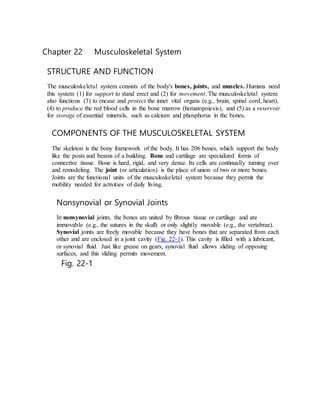
  • 1. Chapter 22 Musculoskeletal System STRUCTURE AND FUNCTION The musculoskeletal system consists of the body's bones, joints, and muscles. Humans need this system (1) for support to stand erect and (2) for movement. The musculoskeletal system also functions (3) to encase and protect the inner vital organs (e.g., brain, spinal cord, heart), (4) to produce the red blood cells in the bone marrow (hematopoiesis), and (5) as a reservoir for storage of essential minerals, such as calcium and phosphorus in the bones. COMPONENTS OF THE MUSCULOSKELETAL SYSTEM The skeleton is the bony framework of the body. It has 206 bones, which support the body like the posts and beams of a building. Bone and cartilage are specialized forms of connective tissue. Bone is hard, rigid, and very dense. Its cells are continually turning over and remodeling. The joint (or articulation) is the place of union of two or more bones. Joints are the functional units of the musculoskeletal system because they permit the mobility needed for activities of daily living. Nonsynovial or Synovial Joints In nonsynovial joints, the bones are united by fibrous tissue or cartilage and are immovable (e.g., the sutures in the skull) or only slightly movable (e.g., the vertebrae). Synovial joints are freely movable because they have bones that are separated from each other and are enclosed in a joint cavity (Fig. 22-1). This cavity is filled with a lubricant, or synovial fluid. Just like grease on gears, synovial fluid allows sliding of opposing surfaces, and this sliding permits movement. Fig. 22-1
  • 2. In synovial joints, a layer of resilient cartilage covers the surface of opposing bones. Cartilage is avascular; it receives nourishment from synovial fluid that circulates during joint movement. It is a very stable connective tissue with a slow cell turnover. It has a tough, firm consistency, yet is flexible. This cartilage cushions the bones and gives a smooth surface to facilitate movement. The joint is surrounded by a fibrous capsule and is supported by ligaments. Ligaments are fibrous bands running directly from one bone to another that strengthen the joint and help prevent movement in undesirable directions. A bursa is an enclosed sac filled with viscous synovial fluid, much like a joint. Bursae are located in areas of potential friction (e.g., subacromial bursa of the shoulder, prepatellar bursa of the knee) and help muscles and tendons glide smoothly over bone. Muscles Muscles account for 40% to 50% of the body's weight. When they contract, they produce movement. Muscles are of three types: skeletal, smooth, and cardiac. This chapter is concerned with skeletal, or voluntary, muscles, those under conscious control. Fig. 22-2
  • 3. Each skeletal muscle is composed of bundles of muscle fibers, or fasciculi. The skeletal muscle is attached to bone by a tendon-a strong fibrous cord. Skeletal muscles produce the following movements (Fig. 22-2): 1. Flexion—bending a limb at a joint 2. Extension—straightening a limb at a joint 3. Abduction—moving a limb away from the midline of the body 4. Adduction—moving a limb toward the midline of the body 5. Pronation—turning the forearm so that the palm is down 6. Supination—turning the forearm so that the palm is up 7. Circumduction—moving the arm in a circle around the shoulder 8. Inversion—moving the sole of the foot inward at the ankle 9. Eversion—moving the sole of the foot outward at the ankle
  • 4. 10. Rotation—moving the head around a central axis 11. Protraction—moving a body part forward and parallel to the ground 12. Retraction—moving a body part backward and parallel to the ground 13. Elevation—raising a body part 14. Depression—lowering a body part Fig. 22-3 Temporomandibular Joint The temporomandibular joint (TMJ) is the articulation of the mandible and the temporal bone (Fig. 22-3). You can feel it in the depression anterior to the tragus of the ear. The TMJ permits jaw function for speaking and chewing. The joint allows three motions: (1) hinge action to open and close the jaws, (2) gliding action for protrusion and retraction, and (3) gliding for side-to-side movement of the lower jaw. Spine The vertebrae are 33 connecting bones stacked in a vertical column (Fig. 22-4). You can feel their spinous processes in a furrow down the midline of the back. The furrow has paravertebral muscles mounded on either side down to the sacrum, where it flattens. Humans have 7 cervical, 12 thoracic, 5 lumbar, 5 sacral, and 3 to 4 coccygeal vertebrae. The following surface landmarks will orient you to their levels: • The spinous processes of C7 and T1 are prominent at the base of the neck.
  • 5. • The inferior angle of the scapula normally is at the level of the interspace between T7 and T8. • An imaginary line connecting the highest point on each iliac crest crosses L4. • An imaginary line joining the two symmetric dimples that overlie the posterior superior iliac spines crosses the sacrum. Fig. 22-4
  • 7.
  • 8. Fig. 22-6 A lateral view shows that the vertebral column has four curves (a double-S shape) (Fig. 22-5). The cervical and lumbar curves are concave (inward or anterior), and the thoracic and sacrococcygeal curves are convex. The balanced or compensatory nature of these curves, together with the resilient intervertebral disks, allows the spine to absorb a great deal of shock. The intervertebral disks are elastic fibrocartilaginous plates that constitute one fourth of the length of the column (Fig. 22-6). Each disk center has a nucleus pulposus, made of soft, semifluid, mucoid material that has the consistency of toothpaste in the young adult. The disks cushion the spine like a shock absorber and help it move. As the spine moves, the elasticity of the disks allows compression on one side, with compensatory expansion on the other. Sometimes compression can be too great. The disk then can rupture and the nucleus pulposus can herniate out of the vertebral column, compressing on the spinal nerves and causing pain. The unique structure of the spine enables both upright posture and flexibility for motion. The motions of the vertebral column are flexion (bending forward), extension (bending back), abduction (to either side), and rotation. Fig. 22-7
  • 9. Shoulder The glenohumeral joint is the articulation of the humerus with the glenoid fossa of the scapula (Fig. 22-7). Its ball-and-socket action allows great mobility of the arm on many axes. The joint is enclosed by a group of four powerful muscles and tendons that support and stabilize it. Together these are called the rotator cuff of the shoulder. The large subacromial bursa helps during abduction of the arm, so that the greater tubercle of the humerus moves easily under the acromion process of the scapula. Fig. 22-8
  • 10. The bones of the shoulder have palpable landmarks to guide your examination (Fig. 22- 8). The scapula and the clavicle connect to form the shoulder girdle. You can feel the bump of the scapula's acromion process at the very top of the shoulder. Move your fingers in a small circle outward, down, and around. The next bump is the greater tubercle of the humerus a few centimeters down and laterally, and from that the coracoid process of the scapula is a few centimeters medially. These surround the deeply situated joint. Fig. 22-9
  • 12. Elbow The elbow joint contains the three bony articulations of the humerus, radius, and ulna of the forearm (Fig. 22-9). Its hinge action moves the forearm (radius and ulna) on one plane, allowing flexion and extension. The olecranon bursa lies between the olecranon process and the skin. Palpable landmarks are the medial and lateral epicondyles of the humerus and the large olecranon process of the ulna in between them. The sensitive ulnar nerve runs between the olecranon process and the medial epicondyle. The radius and ulna articulate with each other at two radioulnar joints, one at the elbow and one at the wrist. These move together to permit pronation and supination of the hand and forearm.
  • 13. Wrist and Carpals Of the body's 206 bones, over half are in the hands and feet. The wrist or radiocarpal joint is the articulation of the radius (on the thumb side) and a row of carpal bones (Fig. 22-10). Its condyloid action permits movement in two planes at right angles: flexion and extension, and side-to-side deviation. You can feel the groove of this joint on the dorsum of the wrist. The midcarpal joint is the articulation between the two parallel rows of carpal bones. It allows flexion, extension, and some rotation. The metacarpophalangeal and the interphalangeal joints permit finger flexion and extension. The flexor tendons of the wrist and hand are enclosed in synovial sheaths. Fig. 22-11 Hip The hip joint is the articulation between the acetabulum and the head of the femur (Fig. 22-11). As in the shoulder, ball-and-socket action permits a wide range of motion on many axes. The hip has somewhat less range of motion (ROM) than the shoulder, but it has more stability as befits its weight-bearing function. Hip stability is due to powerful muscles that spread over the joint, a strong fibrous articular capsule, and the very deep insertion of the head of the femur. Three bursae facilitate movement. Fig. 22-12
  • 14. Palpation of these bony landmarks will guide your examination. You can feel the entire iliac crest, from the anterior superior iliac spine to the posterior. The ischial tuberosity lies under the gluteus maximus muscle and is palpable when the hip is flexed. The greater trochanter of the femur is normally the width of the person's palm below the iliac crest and halfway between the anterior superior iliac spine and the ischial tuberosity. Feel it when the person is standing, in a flat depression on the upper lateral side of the thigh. Knee The knee joint is the articulation of three bones—the femur, the tibia, and the patella (kneecap)—in one common articular cavity (Fig. 22-12). It is the largest joint in the body and is complex. It is a hinge joint, permitting flexion and extension of the lower leg on a single plane. The knee's synovial membrane is the largest in the body. It forms a sac at the superior border of the patella, called the suprapatellar pouch, which extends up as much as 6 cm behind the quadriceps muscle. Two wedge-shaped cartilages, called the medial and lateral menisci, cushion the tibia and femur. The joint is stabilized by two sets of ligaments. The cruciate ligaments (not shown) crisscross within the knee; they give anterior and posterior stability and help control rotation. The collateral ligaments connect the joint at both sides; they give medial and lateral stability and prevent dislocation. Numerous bursae prevent friction. One, the prepatellar bursa, lies between the patella and the skin. The infrapatellar fat pad is a small, triangular fat pad below the patella behind the patellar ligament. Landmarks of the knee joint start with the large quadriceps muscle, which you can feel on your anterior and lateral thigh (Fig. 22-13). The muscle's four heads merge into a common tendon that continues down to enclose the round bony patella. Then the tendon inserts down on the tibial tuberosity, which you can feel as a bony prominence in the midline. Move to the sides and a bit superiorly and note the lateral and medial condyles
  • 15. of the tibia. Superior to these on either side of the patella are the medial and lateral epicondyles of the femur. Fig. 22-13 Fig. 22-14
  • 16. Ankle and Foot The ankle or tibiotalar joint is the articulation of the tibia, fibula, and talus (Fig. 22-14). It is a hinge joint, limited to flexion (dorsiflexion) and extension (plantar flexion) on one plane. Landmarks are two bony prominences on either side—the medial malleolus and the lateral malleolus. Strong, tight medial and lateral ligaments extend from each malleolus onto the foot. These help the lateral stability of the ankle joint, although they may be torn in eversion or inversion sprains of the ankle. Joints distal to the ankle give additional mobility to the foot. The subtalar joint permits inversion and eversion of the foot. The foot has a longitudinal arch, with weight-bearing distributed between the parts that touch the ground—the heads of the metatarsals and the calcaneus (heel). DEVELOPMENTAL CARE Infants and Children By 3 months' gestation, the fetus has formed a “scale model” of the skeleton that is made up of cartilage. During succeeding months in utero, the cartilage ossifies into true bone and starts to grow. Bone growth continues after birth—rapidly during infancy and then steadily during childhood—until adolescence, when both boys and girls undergo a rapid growth spurt. Long bones grow in two dimensions. They increase in width or diameter by deposition of new bony tissue around the shafts. Lengthening occurs at the epiphyses, or growth plates. These specialized growth centers are transverse disks located at the ends of long bone. Any trauma or infection at this location puts the growing child at risk for bone deformity. This longitudinal growth continues until closure of the epiphyses; the last closure occurs at about age 20 years. Skeletal contour changes are apparent at the vertebral column. At birth the spine has a single C-shaped curve. At 3 to 4 months, raising the baby's head from prone position develops the anterior curve in the cervical neck region. From 1 year to 18 months, standing erect develops the anterior curve in the lumbar region. Although the skeleton contributes to linear growth, muscles and fat are significant for weight increase. Individual muscle fibers grow through childhood, but growth is marked during the adolescent growth spurt. Then muscles respond to increased secretion of growth hormone, to adrenal androgens, and in boys, to further stimulation by testosterone. Muscles vary in size and strength in different people. This is due to genetic programming, nutrition, and exercise. All through life, muscles increase with use and atrophy with disuse. The Pregnant Female
  • 17. Increased levels of circulating hormones (estrogen, relaxin from the corpus luteum, and corticosteroids) cause increased mobility in the joints. Increased mobility in the sacroiliac, sacrococcygeal, and symphysis pubis joints in the pelvis contributes to the noticeable changes in maternal posture. The most characteristic change is progressive lordosis, which compensates for the enlarging fetus; otherwise, the center of balance would shift forward. Lordosis compensates by shifting the weight farther back on the lower extremities. This shift in balance in turn creates strain on the low back muscles, which in some women is felt as low back pain during late pregnancy. Anterior flexion of the neck and slumping of the shoulder girdle are other postural changes that compensate for the lordosis. These upper back changes may put pressure on the ulnar and median nerves during the third trimester. Nerve pressure creates aching, numbness, and weakness in the upper extremities in some women. The Aging Adult With aging, loss of bone matrix (resorption) occurs more rapidly than new bone growth (deposition). The net effect is a loss of bone density, or osteoporosis. Although some degree of osteoporosis is nearly universal, females have it more than males, and whites more than blacks. Postural changes are evident with aging, and decreased height is the most noticeable. Long bones do not shorten with age. Decreased height is due to shortening of the vertebral column. This is caused by loss of water content and thinning of the intervertebral disks, which occurs more in the middle years. Also, decreased height is caused by a decrease in height of individual vertebrae, which occurs in later years from osteoporosis. Both men and women can expect a progressive decrease in height beginning at age 40 years in males and age 43 years in females, although this is not significant until age 60 years (Cline et al, 1989). A greater decrease occurs in the 70s and 80s as a result of osteoporotic collapse of the vertebrae. The result is a shortening of the trunk and comparatively long extremities. Other postural changes are kyphosis, and a backward head tilt to compensate for the kyphosis, and a slight flexion of hips and knees. The distribution of subcutaneous fat changes through life. Usually, men and women gain weight in their 40s and 50s. The contour is different, even if the weight is the same as when younger. They begin to lose fat in the face and deposit it in abdomen and hips. In the 80s and 90s, fat further decreases in the periphery, which is especially noticeable in the forearms and apparent over the abdomen and hips. Loss of subcutaneous fat leaves bony prominences more marked (e.g., tips of vertebrae, ribs, iliac crests), and body hollows deeper (e.g., cheeks, axillae). An absolute loss in muscle mass occurs; some muscles decrease in size, and some atrophy, producing weakness. The contour of muscles becomes more prominent, and muscle bundles and tendons feel more distinct.
  • 18. It has become more apparent that lifestyle affects musculoskeletal changes. A sedentary lifestyle hastens musculoskeletal changes of aging. However, physical exercise increases skeletal mass. This helps prevent or delay osteoporosis. Physical activity delays or prevents bone loss in postmenopausal and older women (Ebrahim et al, 1997; Gregg et al, 1998). CROSS-CULTURAL CARE The long bones of blacks are significantly longer, narrower, and denser than those of whites (Overfield, 1995). Measurement of bone density by race and gender reveals that black males have the densest bones, thus accounting for the relatively low incidence of osteoporosis in this population. Bone density in the Chinese, Japanese, and Inuits is below that of white Americans (Overfield, 1995). Curvature of the long bones varies widely among culturally diverse groups. American Indians have anteriorly convex femurs, blacks have markedly straight femurs, and in whites the femoral curvature is intermediate. This characteristic is related to both genetics and body weight. Thin blacks and whites have less curvature than average, whereas obese blacks and whites display increased curvatures. It is possible that the heavier density of the bones of blacks helps to protect them from increased curvature from obesity. TABLE 22-1 Biocultural Variations in the Musculoskeletal System Bone Variations Frontal Thicker in black males than in white males. Parietal occiput Thicker in white males than in black males. Palate Tori (protuberances) along the suture line of the hard palate; problematic for denture wearers. Incidence: blacks— 20%; whites—24%; Asians—up to 50%; American Indians—up to 50%. Mandible
  • 19. Tori (protuberances) on the lingual surface of the mandible near the canine and premolar teeth; problematic for denture wearers; most common in Asians and American Indians; incidence exceeds 50% in some Inuit groups. Humerus Torsion or rotation of proximal end with muscle pull; whites have a greater incidence than blacks; torsion in blacks is symmetric; torsion in whites greater on the right side than on the left side. Radius Length at the wrist variable. Ulna Ulna or radius may be longer. Equal length in Swedes—61%; in Chinese— 16%. Ulna longer than radius in Swedes—16%; in Chinese—48%. Radius longer than ulna in Swedes—23%; in Chinese—10%. Vertebrae Twenty-four vertebrae (cervical, thoracic, lumbar) are found in 85% to 93% of all people. Racial and gender differences reveal 23 or 25 vertebrae in select groups; 11% of black females have 23, and 12% of Inuit and American Indian males have 25. Increased number is related to lower back pain and lordosis. Pelvis Hip width is 1.6 cm (0.6 in) smaller in black women than in white women; Asian women have significantly smaller pelvises. Femur The femur has a convex anterior curve in American Indians, is straight in blacks, and has an intermediate curve in whites. Second tarsal Second toe longer than the great toe—incidence: whites—8%-34%; blacks— 8%-12%; Vietnamese—31%; Melanesians—21%—57%. Clinical significance for joggers and athletes, who reported increased foot problems. Height White males are 1.27 cm (0.5 in) taller than black males and 7.6 cm (2.9 in) taller than Asian males. White females have the same height as black females. Asian females are 4.14 cm (1.6 in) shorter than white or black females.
  • 20. Composition of long bones Longer, narrower, and denser in blacks than whites; bone density in whites is greater than in Chinese, Japanese, and Inuits. Osteoporosis incidence is lowest in black males; highest in white females. Muscle Variations Peroneus tertius Responsible for dorsiflexion of foot—muscle absent in 3%-10% of Asians, American Indians, and whites; in 10%-15% of blacks; and in 24% of Berbers (Sahara desert). No clinical significance because the tibialis anterior also dorsiflexes the foot. Palmaris longus Responsible for wrist flexion—muscle absent in 12%-20% of whites; in 2%- 12% of American Indians; in 5% of blacks; and in 3% of Asians. No clinical significance because three other muscles are also responsible for flexion. Data from Overfield T: Biologic variation in health and illness: Race, age, and sex differences, ed 2, New York, 1995, CRC Press; Andrews MM, Boyle JS, editors: Transcultural concepts in nursing care, ed 4, Philadelphia, 2003, Lippincott Williams & Wilkins. Table 22-1 summarizes reported biocultural variations occurring in the musculoskeletal system. SUBJECTIVE DATA 1. Joints Pain Stiffness Swelling, heat, redness Limitation of movement 2. Muscles Pain (cramps) Weakness
  • 21. 3. Bones Pain Deformity Trauma (fractures, sprains, dislocations) 4. Functional assessment (activities of daily living [ADLs]) 5. Self-care behaviors Examiner Asks 1. Joints. • Any problems with your joints? Any pain? • Location: Which joints? On one side or both sides? • Quality: What does the pain feel like: aching, stiff, sharp or dull, shooting? Severity: How strong is the pain? • Onset: When did this pain start? • Timing: What time of day does the pain occur? How long does it last? How often does it occur? • Is the pain aggravated by movement, rest, position, weather? Is the pain relieved by rest, medications, application of heat or ice? • Is the pain associated with chills, fever, recent sore throat, trauma, repetitive activity? • Any stiffness in your joints? • Any swelling, heat, redness in the joints? • Any limitation of movement in any joint? Which joint? • Which activities give you problems? (See Functional Assessment, p. 640.) Rationale Joint pain and loss of function are the most common musculoskeletal concerns that prompt a person to seek care. Rheumatoid arthritis (RA) involves symmetric joints; other musculoskeletal illnesses involve isolated or unilateral joints.
  • 22. Exquisitely tender with acute inflammation. RA pain is worse in morning when arising; osteoarthritis is worse later in the day; tendinitis is worse in morning, improves during the day. Movement increases most joint pain except in RA, in which movement decreases pain. Joint pain 10 to 14 days after an untreated strep throat suggests rheumatic fever. Joint injury occurs from trauma, repetitive motion. RA stiffness occurs in morning and after rest periods. Suggests acute inflammation. Decreased ROM may be due to joint injury to cartilage or capsule, or to muscle contracture. 2. Muscles. • Any problems in the muscles, such as any pain or cramping? Which muscles? • If in calf muscles: Is the pain with walking? Does it go away with rest? • Are your muscle aches associated with fever, chills, the “flu”? • Any weakness in muscles? • Location: Where is the weakness? How long have you noticed weakness? • Do the muscles look smaller there? Rationale Myalgia is usually felt as cramping or aching. Suggests intermittent claudication (see Chapter 20). Viral illness often includes myalgia. Weakness may involve musculoskeletal or neurologic systems (see Chapters 20 and 23). Atrophy. 3. Bones. • Any bone pain? Is the pain affected by movement? • Any deformity of any bone or joint? Is the deformity due to injury or trauma? Does the deformity affect ROM?
  • 23. • Any accidents or trauma ever affected the bones or joints: fractures, joint strain, sprain, dislocation? Which ones? • When did this occur? What treatment was given? Any problems or limitations now as a result? • Any back pain? In which part of your back? Is pain felt anywhere else, like shooting down leg? • Any numbness and tingling? Any limping? Rationale Fracture causes sharp pain that increases with movement. Other bone pain usually feels “dull” and “deep” and is unrelated to movement. 4. Functional assessment (ADL). Do your joint (muscle, bone) problems create any limits on your usual activities of daily living (ADLs)? Which ones? (Note: Ask about each category; if the person answers “yes,” ask specifically about each activity in category.) • Bathing—getting in and out of the tub, turning faucets? • Toileting—urinating, moving bowels, able to get self on/off toilet, wipe self? • Dressing—doing buttons, zipper, fasten opening behind neck, pulling dress or sweater over head, pulling up pants, tying shoes, getting shoes that fit? • Grooming—shaving, brushing teeth, brushing or fixing hair, applying makeup? • Eating—preparing meals, pouring liquids, cutting up foods, bringing food to mouth, drinking? • Mobility—walking, walking up or down stairs, getting in/out of bed, getting out of house? • Communicating—talking, using phone, writing? Rationale Functional assessment screens the safety of independent living, the need for home health services, and quality of life (see Chapter 30). Assess any self-care deficit. Impaired physical mobility. Impaired verbal communication.
  • 24. 5. Self-care behaviors. Any occupational hazards that could affect the muscles and joints? Does your work involve heavy lifting? Or any repetitive motion or chronic stress to joints? Any efforts to alleviate these? • Tell me about your exercise program. Describe the type of exercise, frequency, the warm-up program. • Any pain during exercise? How do you treat it? • Have you had any recent weight gain? Please describe your usual daily diet. (Note the person's usual caloric intake, all four food groups, daily amount of protein, calcium.) • Are you taking any medications for musculoskeletal system: aspirin, anti- inflammatory, muscle relaxant, pain reliever? • If person has chronic disability or crippling illness: How has your illness affected Your interaction with family Your interaction with friends The way you view yourself Rationale Assess risk for back pain or carpal tunnel syndrome. Self-care behaviors. Assess for • Self-esteem disturbance • Loss of independence • Body image disturbance • Role performance disturbance • Social isolation Additional History for Infants and Children 1. Were you told about any trauma to infant during labor and delivery? Did the baby come head first? Was there a need for forceps? 2. Did the baby need resuscitation?
  • 25. 3. Were the baby's motor milestones achieved at about the same time as siblings or age-mates? 4. Has your child ever broken any bones? Any dislocations? How were these treated? 5. Have you ever noticed any bone deformity? Spinal curvature? Unusual shape of toes or feet? At what age? Have you ever sought treatment for any of these? Rationale Traumatic delivery increases risk for fractures, (e.g., humerus, clavicle). Period of anoxia may result in hypotonia of muscles. Additional History for Adolescents 1. Involved in any sports at school or after school? How frequently (times per week)? 2. Do you use any special equipment? Does any training program exist for your sport? 3. What is the nature of your daily warm-up? 4. What do you do if you get hurt? 5. How does your sport fit in with other school demands and other activities? Rationale Assess safety of sport for child. Note if child's height and weight are adequate for the particular sport (e.g., football). Use of safety equipment and presence of adult supervision decrease risk of sports injuries. Lack of adequate warm-up increases risk of sports injury. Students may not report injury or pain for fear of limiting participation in sport. Additional History for the Aging Adult Use the functional assessment history questions in Chapter 5 (pp. 82 to 84) to elicit any loss of function, self-care deficit, or safety risk that may occur as a process of aging or musculoskeletal illness. (Review the complete functional assessment in Chapter 30.) 1. Any change in weakness over the past months or years?
  • 26. 2. Any increase in falls or stumbling over the past months or years? 3. Do you use any mobility aids to help you get around: cane, walker? OBJECTIVE DATA PREPARATION The purpose of the musculoskeletal examination is to assess function for ADL and to screen for any abnormalities. You already will have considerable data regarding ADL through the history. Note additional ADL data as the person goes through the motions necessary for an examination: gait, posture, how the person sits in a chair, raises from chair, takes off jacket, manipulates small object such as a pen, raises from supine. EQUIPMENT NEEDED Tape measure Goniometer, to measure joint angles Skin marking pen A screening musculoskeletal examination suffices for most people: • Inspection and palpation of joints integrated with each body region • Observation of ROM as person proceeds through motions described earlier • Age-specific screening measures, such as Ortolani's sign for infants or scoliosis screening for adolescents A complete musculoskeletal examination, as described in this chapter, is appropriate for persons with articular disease, a history of musculoskeletal symptoms, or any problems with ADLs. Make the person comfortable before and throughout the examination. Drape for full visualization of the body part you are examining without needlessly exposing the person. Take an orderly approach—head to toe, proximal to distal. Support each joint at rest. Muscles must be soft and relaxed to assess the joints under them accurately. Take care when examining any inflamed area where rough manipulation could cause pain and muscle spasm. To avoid this, use firm support, gentle movement, and gentle return to a relaxed state. Compare corresponding paired joints. Expect symmetry of structure and function and normal parameters for that joint.
  • 27. Normal Range of Findings ORDER OF THE EXAMINATION Inspection Note the size and contour of the joint. Inspect the skin and tissues over the joints for color, swelling, and any masses or deformity. Presence of swelling is significant and signals joint irritation. Abnormal Findings Swelling may be excess joint fluid (effusion), thickening of the synovial lining, inflammation of surrounding soft tissue (bursae, tendons) or bony enlargement. Deformities include dislocation (one or more bones in a joint being out of position), subluxation (partial dislocation of a joint), contracture (shortening of a muscle leading to limited ROM of joint), or ankylosis (stiffness or fixation of a joint). Palpation Palpate each joint, including its skin for temperature, its muscles, bony articulations, and area of joint capsule. Notice any heat, tenderness, swelling, or masses. Joints normally are not tender to palpation. If any tenderness does occur, try to localize it to specific anatomic structures (e.g., skin, muscles, bursae, ligaments, tendons, fat pads, or joint capsule). The synovial membrane normally is not palpable. When thickened, it feels “doughy” or “boggy.” A small amount of fluid is present in the normal joint, but it is not palpable. Abnormal Findings Palpable fluid is abnormal. Because fluid is contained in an enclosed sac, if you push on one side of the sac, the fluid will shift and cause a visible bulging on another side. Range of Motion (ROM) Ask for active ROM while stabilizing the body area proximal to that being moved. Familiarize yourself with the type of each joint and its normal ROM so that you can recognize limitations. If you see a limitation, gently attempt passive motion. Anchor the joint with one hand while your other hand slowly moves it to its limit. The normal ranges of active and passive motion should be the same.
  • 28. If any limitation or any increase in ROM occurs, use a goniometer to measure the angles precisely (Fig. 22-15). First extend the joint to neutral or 0 degrees. Center the 0 point of the goniometer on the joint. Keep the fixed arm of the goniometer on the 0 line and use the movable arm to measure; then flex the joint and measure through the goniometer to determine the angle of greatest flexion. Fig. 22-15 Joint motion normally causes no tenderness, pain, or crepitation. Do not confuse crepitation with the normal discrete “crack” heard as a tendon or ligament slips over bone during motion, such as when you do a knee bend. Abnormal Findings
  • 29. Crepitation is an audible and palpable crunching or grating that accompanies movement. It occurs when the articular surfaces in the joints are roughened, as with rheumatoid arthritis (see Table 22-2, p. 643). Muscle Testing Test the strength of the prime mover muscle groups for each joint. Repeat the motions you elicited for active ROM. Now ask the person to flex and hold as you apply opposing force. Muscle strength should be equal bilaterally and should fully resist your opposing force. (Note: Muscle status and joint status are interdependent and should be interpreted together. Chapter 23 discusses the examination of muscles for size and development, tone, and presence of tenderness.) A wide variability of strength exists among people. You may wish to use a grading system from no voluntary movement to full strength, as shown. Grade Description % Normal Assessment 5 Full ROM against gravity, full resistance 100 Normal 4 Full ROM against gravity, some resistance 75 Good 3 Full ROM with gravity 50 Fair
  • 30. 2 Full ROM with gravity eliminated (passive motion) 25 Poor 1 Slight contraction 10 Trace 0 No contraction 0 Zero TEMPOROMANDIBULAR JOINT With the person seated, inspect the area just anterior to the ear. Place the tips of your first two fingers in front of each ear and ask the person to open and close the mouth. Drop your fingers into the depressed area over the joint, and note smooth motion of the mandible. An audible and palpable snap or click occurs in many healthy people as the mouth opens (Fig. 22-16). Then ask the person to: Abnormal Findings Swelling looks like a round bulge over the joint, although it must be moderate or marked to be visible. Crepitus and pain occur with temporomandibular joint dysfunction. Fig. 22-16
  • 31. INSTRUCTIONS TO PERSON MOTION AND EXPECTED RANGE • Open mouth maximally. • Partially open mouth, protrude lower jaw, and move it side to side.
  • 32. • Stick out lower jaw. Vertical motion. You can measure the space between the upper and lower incisors. Normal is 3 to 6 cm, or three fingers inserted sideways. Lateral motion. Normal extent is 1 to 2 cm (Fig. 22-17). Protrude without deviation. Fig. 22-17
  • 33. Abnormal Findings Lateral motion may be lost earlier and more significantly than vertical. Palpate the contracted temporalis and masseter muscles as the person clenches the teeth. Compare right and left sides for size, firmness, and strength. Ask the person to move the jaw forward and laterally against your resistance, and to open mouth against your resistance. This also tests the integrity of cranial nerve V (trigeminal).
  • 34. CERVICAL SPINE Inspect the alignment of head and neck. The spine should be straight and the head erect. Palpate the spinous processes and the sternomastoid, trapezius, and paravertebral muscles. They should feel firm, with no muscle spasm or tenderness. Abnormal Findings Head tilted to one side. Asymmetry of muscles. Tenderness and hard muscles with muscle spasm. Ask the person to follow these motions (Fig. 22-18):* INSTRUCTIONS TO PERSON MOTION AND EXPECTED RANGE • Touch chin to chest. • Lift the chin toward the ceiling. • Touch each ear toward the corresponding shoulder. Do not lift up the shoulder. • Turn the chin toward each shoulder. Flexion of 45 degrees (Fig. 22-18, A). Hyperextension of 55 degrees. Lateral bending of 40 degrees (Fig. 22-18, B). Rotation of 70 degrees (Fig. 22-18, C). Abnormal Findings Limited ROM. Pain with movement. Fig. 22-18
  • 35. Repeat the motions while applying opposing force. The person normally can maintain flexion against your full resistance. This also tests integrity of cranial nerve XI (spinal). Abnormal Findings The person cannot hold flexion. * Do not attempt if you suspect neck trauma. UPPER EXTREMITY Shoulders Inspect and compare both shoulders posteriorly and anteriorly. Check the size and contour of the joint and compare shoulders for equality of bony landmarks. Normally, no redness, muscular atrophy, deformity, or swelling is present. Check the anterior aspect of the joint capsule and the subacromial bursa for abnormal swelling. Abnormal Findings Redness. Inequality of bony landmarks. Atrophy, shows as lack of fullness. Dislocated shoulder loses the normal rounded shape and looks flattened laterally.
  • 36. Swelling from excess fluid is best seen anteriorly. Considerable fluid must be present to cause a visible distention because the capsule normally is so loose (see Table 22-3). If the person reports any shoulder pain, ask that he or she point to the spot with the hand of the unaffected side. Be aware that shoulder pain may be from local causes or it may be referred pain from a hiatal hernia or a cardiac or pleural condition, which could be potentially serious. Pain from a local cause is reproducible during the examination by palpation or motion. Abnormal Findings Swelling of subacromial bursa is localized under deltoid muscle and may be accentuated when the person tries to abduct the arm. While standing in front of the person, palpate both shoulders, noting any muscular spasm or atrophy, swelling, heat, or tenderness. Start at the clavicle and methodically explore the acromioclavicular joint, scapula, greater tubercle of the humerus, area of the subacromial bursa, the biceps groove, and the anterior aspect of the glenohumeral joint. Palpate the pyramid-shaped axilla; no adenopathy or masses should be present. Abnormal Findings Swelling. Hard muscles with muscle spasm. Tenderness or pain. Test ROM by asking the person to perform four motions (Fig. 22-19). Cup one hand over the shoulder during ROM to note any crepitation; normally none is present. INSTRUCTIONS TO PERSON MOTION AND EXPECTED RANGE • With arms at sides and elbows extended, move both arms forward and up in wide vertical arcs, then move them back. • Rotate arms internally behind back, place back of hands as high as possible toward the scapulae. Forward flexion of 180 degrees. Hyperextension up to 50 degrees (Fig. 22-19, A). Internal rotation of 90 degrees (Fig. 22-19, B). Abnormal Findings Limited ROM.
  • 37. Asymmetry. Pain with motion. Crepitus with motion. Rotator cuff lesions may cause limited ROM, pain, and muscle spasm during abduction, whereas forward flexion stays fairly normal. Fig. 22-19
  • 38. Test the strength of the shoulder muscles by asking the person to shrug the shoulders, flex forward and up, and abduct against your resistance. The shoulder shrug also tests the integrity of cranial nerve XI, the spinal accessory.
  • 39. Elbow Inspect the size and contour of the elbow in both flexed and extended positions. Look for any deformity, redness, or swelling. Check the olecranon bursa and the normally present hollows on either side of the olecranon process for abnormal swelling. Abnormal Findings Subluxation of the elbow shows the forearm dislocated posteriorly. Swelling and redness of olecranon bursa are localized and easy to observe because of the close proximity of the bursa to skin. Effusion or synovial thickening shows first as a bulge or fullness in groove on either side of the olecranon process, and it occurs with gouty arthritis. Palpate with the elbow flexed about 70 degrees and as relaxed as possible (Fig. 22-20). Use your left hand to support the person's left forearm and palpate the extensor surface of the elbow—the olecranon process and the medial and lateral epicondyles of humerus— with your right thumb and fingers. Abnormal Findings Epicondyles, head of radius, and tendons are common sites of inflammation and local tenderness, or “tennis elbow.” Fig. 22-20
  • 40. With your thumb in the lateral groove and your index and middle fingers in the medial groove, palpate either side of the olecranon process using varying pressure. Normally, present tissues and fat pads feel fairly solid. Check for any synovial thickening, swelling, nodules, or tenderness. Abnormal Findings Soft, boggy, or fluctuant swelling in both grooves occurs with synovial thickening or effusion. Local heat or redness (signs of inflammation) can extend beyond synovial membrane. Palpate the area of the olecranon bursa for heat, swelling, tenderness, consistency, or nodules. Abnormal Findings Subcutaneous nodules are raised, firm, and nontender, and overlying skin moves freely. Common sites are in the olecranon bursa and along extensor surface of the ulna. These nodules occur with RA (see Table 22-4). Test ROM by asking the person to: INSTRUCTIONS TO PERSON
  • 41. MOTION AND EXPECTED RANGE • Bend and straighten the elbow (Fig. 22-21). • Movement of 90 degrees in pronation and supination (Fig. 22-22). Flexion of 150 to 160 degrees; extension at 0. Some healthy people lack 5 to 10 degrees of full extension, and others have 5 to 10 degrees of hyperextension. Hold the hand midway; then touch front and back sides of hand to table. Fig. 22-21
  • 43. While testing muscle strength, stabilize the person's arm with one hand (Fig. 22-23). Have the person flex the elbow against your resistance applied just proximal to the wrist. Then ask the person to extend the elbow against your resistance.
  • 44. Fig. 22-23 Wrist and Hand Inspect the hands and wrists on the dorsal and palmar sides, noting position, contour, and shape. The normal functional position of the hand shows the wrist in slight extension. This way the fingers can flex efficiently, and the thumb can oppose them for grip and
  • 45. manipulation. The fingers lie straight in the same axis as the forearm. Normally, no swelling or redness, deformity, or nodules are present. Abnormal Findings Subluxation of wrist. Ulnar deviation; fingers list to ulnar side. Ankylosis; wrist in extreme flexion. Dupuytren's contracture; flexion contracture of finger(s). The skin looks smooth with knuckle wrinkles present and no swelling or lesions. Muscles are full, with the palm showing a rounded mound proximal to the thumb (the thenar eminence) and a smaller rounded mound proximal to the little finger. Abnormal Findings Swan-neck or boutonnière deformity in fingers. Atrophy of the thenar eminence (see Table 22-5). Palpate each joint in the wrist and hands. Facing the person, support the hand with your fingers under it and palpate the wrist firmly with both your thumbs on its dorsum (Fig. 22-24). Make sure the person's wrist is relaxed and in straight alignment. Move your palpating thumbs side to side to identify the normal depressed areas that overlie the joint space. Use gentle but firm pressure. Normally the joint surfaces feel smooth, with no swelling, bogginess, nodules, or tenderness. Abnormal Findings Ganglion in wrist. Synovial swelling on dorsum. Generalized swelling. Tenderness. Fig. 22-24
  • 46. Palpate the metacarpophalangeal joints with your thumbs, just distal to and on either side of the knuckle (Fig. 22-25). Fig. 22-25
  • 47. Use your thumb and index finger in a pinching motion to palpate the sides of the interphalangeal joints (Fig. 22-26). Normally, no synovial thickening, tenderness, warmth, or nodules are present. Abnormal Findings Heberden's and Bouchard's nodules are hard and nontender and occur with osteoarthritis (see Table 22-5). Fig. 22-26
  • 48. Test ROM (Fig. 22-27) by asking the person to: INSTRUCTIONS TO PERSON MOTION AND EXPECTED RANGE
  • 49. • Bend the hand up at the wrist. • Bend hand down at the wrist. • Bend the fingers up and down at metacarpophalangeal joints. • With palms flat on table, turn them outward and in. • Spread fingers apart; make a fist. • Touch the thumb to each finger and to the base of little finger. Hyperextension of 70 degrees (Fig. 22-27, A). Palmar flexion of 90 degrees. Flexion of 90 degrees. Hyperextension of 30 degrees (Fig. 22-27, B). Ulnar deviation of 50-60 degrees, and radial deviation of 20 degrees (Fig. 22- 27, C). Abduction of 20 degrees; fist tight. The responses should be equal bilaterally (Fig. 22-27, D, E). The person is able to perform, and the responses are equal bilaterally (Fig. 22- 27, F). Abnormal Findings Loss of ROM here is the most common and most significant functional loss of the wrist. Limited motion. Pain on movement. Fig. 22-27
  • 50. For muscle testing, position the person's forearm supinated (palm up) and resting on a table (Fig. 22-28). Stabilize by holding your hand at the person's midforearm. Ask the person to flex the wrist against your resistance at the palm. Fig. 22-28
  • 51. Phalen's Test. Ask the person to hold both hands back to back while flexing the wrists 90 degrees. Acute flexion of the wrist for 60 seconds produces no symptoms in the normal hand (Fig. 22-29). Abnormal Findings Phalen's test reproduces numbness and burning in a person with carpal tunnel syndrome (see Table 22-5). Tinel's Sign. Direct percussion of the location of the median nerve at the wrist produces no symptoms in the normal hand (Fig. 22-30). Abnormal Findings In carpal tunnel syndrome, percussion of the median nerve produces burning and tingling along its distribution, which is a positive Tinel's sign. Fig. 22-29
  • 53. LOWER EXTREMITY Hip Wait to inspect the hip joint together with the spine a bit later in the examination as the person stands. At that time, note symmetric levels of iliac crests, gluteal folds, and equally sized buttocks. A smooth, even gait reflects equal leg lengths and functional hip motion. Help the person into a supine position and palpate the hip joints. The joints should feel stable and symmetric, with no tenderness or crepitance. Fig. 22-31
  • 54. Abnormal Findings Pain with palpation. Crepitation. Assess ROM (Fig. 22-31) by asking the person to: INSTRUCTIONS TO PERSON MOTION AND EXPECTED RANGE • Raise each leg with knee extended. • Bend each knee up to the chest while keeping the other leg straight. • Flex knee and hip to 90 degrees. Stabilize by holding the thigh with one hand and the ankle with the other hand. Swing the foot outward. Swing the foot inward. (Foot and thigh move in opposite directions.) • Swing leg laterally, then medially, with knee straight. Stabilize pelvis by pushing down on the opposite anterior superior iliac spine. • When standing (later in examination), swing straight leg back behind body. Stabilize pelvis to eliminate exaggerated lumbar lordosis. The most efficient
  • 55. way is to ask person to bend over the table and to support the trunk on the table. Or the person can lie prone on the table. Hip flexion of 90 degrees (Fig. 22-31, A). Hip flexion of 120 degrees. The opposite thigh should remain on the table (Fig. 22-31, B). Internal rotation of 40 degrees. External rotation of 45 degrees (Fig. 22-31, C). Abduction of 40 to 45 degrees. Adduction of 20 to 30 degrees (Fig. 22-31, D). Hyperextension of 15 degrees when stabilized. Abnormal Findings Limited motion. Pain with motion. Flexion flattens the lumbar spine; if this reveals a flexion deformity in the opposite hip, it represents a positive Thomas test. Limited internal rotation of hip is an early and reliable sign of hip disease. Limitation of abduction of the hip while supine is the most common motion dysfunction found in hip disease. Knee The person should remain supine with legs extended, although some examiners prefer the knees to be flexed and dangling for inspection. The skin normally looks smooth, with even coloring and no lesions. Abnormal Findings Shiny and atrophic skin. Swelling or inflammation (see Table 22-6). Lesions (e.g., psoriasis). Inspect lower leg alignment. The lower leg should extend in the same axis as the thigh. Abnormal Findings Angulation deformity: • Genu varum (bowlegs) (see p. 636)
  • 56. • Genu valgum (knock knees) • Flexion contracture Inspect the knee's shape and contour. Normally, distinct concavities, or hollows, are present on either side of the patella. Check them for any sign of fullness or swelling. Note other locations, such as the prepatellar bursa and the suprapatellar pouch, for any abnormal swelling. Abnormal Findings Hollows disappear; then they may bulge with synovial thickening or effusion. Check the quadriceps muscle in the anterior thigh for any atrophy. Because it is the prime mover of knee extension, this muscle is important for joint stability during weight bearing. Abnormal Findings Atrophy occurs with disuse or chronic disorders. First, it appears in the medial part of the muscle, although it is difficult to note because the vastus medialis is relatively small. Enhance palpation with the knee in the supine position with complete relaxation of the quadriceps muscle. Start high on the anterior thigh, about 10 cm above the patella. Palpate with your left thumb and fingers in a grasping fashion (Fig. 22-32). Proceed down toward the knee, exploring the region of the suprapatellar pouch. Note the consistency of the tissues. The muscles and soft tissues should feel solid, and the joint should feel smooth, with no warmth, tenderness, thickening, or nodularity. Abnormal Findings Feels fluctuant or boggy with synovitis of suprapatellar pouch. When swelling occurs, you need to distinguish whether it is due to soft tissue swelling or increased fluid in the joint. The tests for the bulge sign and ballottement of the patella aid this assessment. Fig. 22-32
  • 57. Bulge Sign. For swelling in the suprapatellar pouch, the bulge sign confirms the presence of small amounts of fluid as you try to move the fluid from one side of the joint to the other. Firmly stroke up on the medial aspect of the knee two or three times to displace any fluid (Fig. 22-33, A). Tap the lateral aspect (Fig. 22-33, B). Watch the medial side in the hollow for a distinct bulge from a fluid wave. Normally, none is present. Fig. 22-33 Bulge sign.
  • 58. Abnormal Findings The bulge sign occurs with very small amounts of effusion, 4 to 8 ml, from fluid flowing across the joint (Fig. 22-33, C). Ballottement of the Patella. This test is reliable when larger amounts of fluid are present. Use your left hand to compress the suprapatellar pouch to move any fluid into the knee joint. With your right hand, push the patella sharply against the femur. If no fluid is present, the patella is already snug against the femur (Fig. 22-34, A). Fig. 22-34 Ballottement. Abnormal Findings If fluid has collected, your tap on the patella moves it through the fluid, and you will hear a tap as the patella bumps up on the femoral condyles (Fig. 22-34, B). Continue palpation and explore the tibiofemoral joint (Fig. 22-35). Note smooth joint margins and absence of pain. Palpate the infrapatellar fat pad and the patella. Check for
  • 59. crepitus by holding your hand on the patella as the knee is flexed and extended. Some crepitus in an otherwise asymptomatic knee is not uncommon. Fig. 22-35 Abnormal Findings Irregular bony margins occur with osteoarthritis. Pain at joint line.
  • 60. Pronounced crepitus is significant and it occurs with degenerative diseases of the knee. Check ROM (Fig. 22-36) by asking the person to: INSTRUCTIONS TO PERSON MOTION AND EXPECTED RANGE • Bend each knee. • Extend each knee. • Check knee ROM during ambulation. Flexion of 130 to 150 degrees. A straight line of 0 degrees in some persons; a hyperextension of 15 degrees in others. Fig. 22-36
  • 62. Sudden locking—the person is unable to extend the knee fully. This usually occurs with a painful and audible “pop” or “click.” Sudden buckling, or “giving way,” occurs with ligament injury, which causes weakness and instability. Check muscle strength by asking the person to maintain knee flexion while you oppose by trying to pull the leg forward. Muscle extension is demonstrated by the person's success in rising from a seated position in a low chair or by rising from a squat without using the hands for support. Special Test for Meniscal Tears McMurray's Test. Perform this test when the person has reported a history of trauma followed by locking, giving way, or local pain in the knee. Position the person supine as you stand on the affected side. Hold the heel and flex the knee and hip. Place your other hand on the knee with fingers on the medial side. Rotate the leg in and out to loosen the joint. Externally rotate the leg and push a valgus (inward) stress on the knee. Then slowly extend the knee. Normally the leg extends smoothly with no pain (Fig. 22-37). Fig. 22-37 McMurray's test.
  • 63. Abnormal Findings If you hear or feel a “click,” McMurray's test is positive for a torn meniscus. Ankle and Foot Inspect while the person is in a sitting, non–weight-bearing position, as well as when standing and walking. Compare both feet, noting position of feet and toes, contour of joints, and skin characteristics. The foot should align with the long axis of the lower leg; an imaginary line would fall from midpatella to between the first and second toes. Weight-bearing should fall on the middle of the foot, from the heel, along the midfoot, to between the second and third toes. Most feet have a longitudinal arch, although that can vary normally from “flat feet” to a high instep. The toes point straight forward and lie flat. The ankles (malleoli) are smooth bony prominences. Normally the skin is smooth, with even coloring and no lesions. Note the
  • 64. locations of any calluses or bursal reactions because they reveal areas of abnormal friction. Examining well-worn shoes helps assess areas of wear and accommodation. Abnormal Findings Hallux valgus (see Table 22-7). Hammertoes. Claw toes. Swelling or inflammation. Calluses. Ulcers. Support the ankle by grasping the heel with your fingers while palpating with your thumbs (Fig. 22-38). Explore the joint spaces. They should feel smooth and depressed, with no fullness, swelling, or tenderness. Fig. 22-38 Abnormal Findings Swelling or inflammation. Tenderness. Palpate the metatarsophalangeal joints between your thumb on the dorsum and your fingers on the plantar surface (Fig. 22-39).
  • 65. Fig. 22-39 Abnormal Findings Swelling or inflammation; tenderness.
  • 66. Using a pinching motion of your thumb and forefinger, palpate the interphalangeal joints on the medial and lateral sides of the toes. Test ROM (Fig. 22-40) by asking the person to: INSTRUCTIONS TO PERSON MOTION AND EXPECTED RANGE • Point toes toward the floor. • Point toes toward your nose. • Turn soles of feet out, then in. (Stabilize the ankle with one hand, hold heel with the other to test the subtalar joint.) • Flex and straighten toes. Plantar flexion of 45 degrees. Dorsiflexion of 20 degrees (Fig. 22-40, A). Eversion of 20 degrees. Inversion of 30 degrees (Fig. 22-40, B). Abnormal Findings Limited ROM. Pain with motion. Assess muscle strength by asking the person to maintain dorsiflexion and plantar flexion against your resistance. Fig. 22-40
  • 67. Abnormal Findings Unable to hold flexion. SPINE The person should be standing, draped in a gown open at the back. Place yourself far enough back so that you can see the entire back. Inspect and note whether the spine is straight by following an imaginary vertical line from the head through the spinous processes and down through the gluteal cleft, and by noting equal horizontal positions for the shoulders, scapulae, iliac crests, and gluteal folds, and equal spaces between arm and lateral thorax on the two sides (Fig. 22-41, A). The person's knees and feet should be aligned with the trunk and should be pointing forward. Fig. 22-41
  • 68. Abnormal Findings A difference in shoulder elevation and in level of scapulae and iliac crests occur with scoliosis (see Table 22-8). From the side, note the normal convex thoracic curve and concave lumbar curve (Fig. 22- 41, B). An enhanced thoracic curve, or kyphosis, is common in aging people. A pronounced lumbar curve, or lordosis, is common in obese people. Abnormal Findings Lateral tilting and forward bending occur with a herniated nucleus pulposus (see Table 22-8).
  • 69. Palpate the spinous processes. Normally they are straight and not tender. Palpate the paravertebral muscles; they should feel firm with no tenderness or spasm. Abnormal Findings Spinal curvature. Tenderness. Spasm of paravertebral muscles. Check ROM of the spine by asking the person to bend forward and touch the toes (Fig. 22- 42). Look for flexion of 75 to 90 degrees and smoothness and symmetry of movement. Note that the concave lumbar curve should disappear with this motion, and the back should have a single convex C-shaped curve. Fig. 22-42
  • 70. If you suspect a spinal curvature during inspection, this may be more clearly seen when the person touches the toes. While the person is bending over, mark a dot on each spinous process. When the person resumes standing, the dots should form a straight vertical line. Abnormal Findings
  • 71. If the dots form a slight S-shape when the person stands, a spinal curve is present. Stabilize the pelvis with your hands. Check ROM (Fig. 22-43) by asking the person to: INSTRUCTIONS TO PERSON MOTION AND EXPECTED RANGE • Bend sideways. • Bend backward. • Twist shoulders to one side, then the other. Lateral bending of 35 degrees (Fig. 22-43, A). Hyperextension of 30 degrees. Rotation of 30 degrees, bilaterally (Fig. 22-43, B). Fig. 22-43
  • 72. Abnormal Findings Limited ROM. Pain with motion. These maneuvers reveal only gross restriction. Movement is still possible even if some spinal fusion has occurred. Finally, ask the person to walk on his or her toes for a few steps; then return walking on the heels. Straight Leg Raising or LaSegue's Test. These maneuvers reproduce back and leg pain and help confirm the presence of a herniated nucleus pulposus. Straight leg raising while keeping the knee extended normally produces no pain. Raise the affected leg just short of the point where it produces pain. Then dorsiflex the foot (Fig. 22-44). Fig. 22-44 Abnormal Findings LaSegue's test is positive if it reproduces sciatic pain. If lifting the affected leg reproduces sciatic pain, it confirms the presence of a herniated nucleus pulposus. Raise the unaffected leg while leaving the other leg flat. Inquire about the involved side. Abnormal Findings
  • 73. If lifting the unaffected leg reproduces sciatic pain, it strongly suggests a herniated nucleus pulposus. Measure Leg Length Discrepancy. Perform this measurement if you need to determine whether one leg is shorter than the other. For true leg length, measure between fixed points, from the anterior iliac spine to the medial malleolus, crossing the medial side of the knee (Fig. 22-45). Normally these measurements are equal or within 1 cm, indicating no true bone discrepancy. Fig. 22-45 Abnormal Findings Unequal leg lengths. Sometimes the true leg length is equal, but the legs still look unequal. For apparent leg length, measure from a nonfixed point (the umbilicus) to a fixed point (medial malleolus) on each leg. Abnormal Findings True leg lengths are equal, but apparent leg lengths unequal—this condition occurs with pelvic obliquity or adduction or flexion deformity in the hip. DEVELOPMENTAL CARE Review the developmental milestones discussed in Chapter 2. Keep handy a concise chart of the usual sequence of motor development so that you can refer to expected findings for the age of each child you are examining. Use the Denver II test to screen the fine and gross motor skills for the child's age. Because some overlap exists between the musculoskeletal and neurologic examinations, assessment of muscle tone, resting posture, and motor activity are discussed in the next chapter.
  • 74. Infants Examine the infant fully undressed and lying on the back. Take care to place the newborn on a warming table to maintain body temperature. Feet and Legs. Start with the feet and work your way up the extremities. Note any positional deformities, a residual of fetal positioning. Often the newborn's feet are not held straight but in a varus (apart) or valgus (together) position. It is important to distinguish whether this position is flexible (and thus usually self-correctable) or fixed. Scratch the outside of the bottom of the foot. If the deformity is self- correctable, the foot assumes a normal right angle to the lower leg. Or immobilize the heel with one hand and gently push the forefoot to the neutral position with the other hand. If you can move it to neutral position, it is flexible. Abnormal Findings A true deformity is fixed and assumes a right angle only with forced manipulation or not at all. Note the relationship of the forefoot to the hindfoot. Commonly, the hindfoot is in alignment with the lower leg and just the forefoot angles inward. This forefoot adduction is metatarsus adductus. It is usually present at birth and usually resolves spontaneously by age 3 years. Abnormal Findings Metatarsus varus—adduction and inversion of forefoot. Talipes equinovarus (see Table 22-9). Check for tibial torsion, a twisting of the tibia. Place both feet flat on the table, and push to flex up the knees. With the patella and the tibial tubercle in a straight line, place your fingers on the malleoli. In an infant, note that a line connecting the four malleoli is parallel to the table. Abnormal Findings More than 20 degrees of deviation; or if lateral malleolus is anterior to medial malleolus, it indicates tibial torsion. Tibial torsion may originate from intrauterine positioning and then may be exacerbated at a later age by continuous sitting in a reverse tailor position, the “TV squat.” This is sitting with the buttocks on the floor and the lower legs splayed back and out on either side.
  • 75. Hips. Check the hips for congenital dislocation. The most reliable method is Ortolani's maneuver, which should be done at every professional visit until the infant is 1 year old. With the infant supine, flex the knees holding your thumbs on the inner mid- thighs, and your fingers outside on the hips touching the greater trochanters. Adduct the legs until your thumbs touch (Fig. 22-46, A). Then gently lift and abduct, moving the knees apart and down so their lateral aspects touch the table (Fig. 22-46, B). This normally feels smooth and has no sound. Fig. 22-46 Ortolani's maneuver. Abnormal Findings With a dislocated hip, the head of the femur is not cupped in the acetabulum but rests posterior to it. Hip instability feels like a clunk as the head of the femur pops back into place. This is a positive Ortolani's sign and warrants referral. The Allis test also is used to check for hip dislocation by comparing leg lengths (Fig. 22-47). Place the baby's feet flat on the table and flex the knees up. Scan the tops of the knees; normally they are at the same elevation. Fig. 22-47 Allis test.
  • 76. Abnormal Findings Finding one knee significantly lower than the other is a positive indication of Allis' sign and suggests hip dislocation. Note the gluteal folds. Normally they are equal on both sides. However, some asymmetry may occur in healthy children. Abnormal Findings Unequal gluteal folds may accompany hip dislocation after 2 to 3 months of age. Hands and Arms. Inspect the hands, noting shape, number, and position of fingers and palmar creases. Abnormal Findings Polydactyly is the presence of extra fingers or toes. Syndactyly is webbing between adjacent fingers or toes (see Table 22-5). A simian crease is a single palmar crease that occurs with Down syndrome, accompanied by short broad fingers, incurving of little fingers, and low-set thumbs. Palpate the length of the clavicles because the clavicle is the bone most frequently fractured during birth. The clavicles should feel smooth, regular, and without crepitus. Also note equal ROM of arms during Moro's reflex. Abnormal Findings
  • 77. Fractured clavicle: Note irregularity at the fracture site, crepitus, and angulation. The site has rapid callus formation with a palpable lump within a few weeks. Observe limited arm ROM and unilateral response to Moro's reflex. Back. Lift up the infant and examine the back. Note the normal single C curve of the newborn's spine (Fig. 22-48). By 2 months of age, the infant can lift the head while prone. This builds the concave cervical spinal curve and indicates normal forearm strength. Inspect the length of the spine for any tuft of hair, dimple in midline, cyst, or mass. Normally, none are present. Fig. 22-48 Abnormal Findings A tuft of hair over a dimple in the midline may indicate spina bifida. A small dimple in the midline-anywhere from the head to the coccyx-suggests dermoid sinus. Mass, such as meningocele. Observe ROM through spontaneous movement of extremities. Fig. 22-49
  • 78. Test muscle strength by lifting up the infant with your hands under the axillae (Fig. 22-49). A baby with normal muscle strength wedges securely between your hands. Abnormal Findings A baby who starts to “slip” between your hands shows weakness of the shoulder muscles. Preschool- and School-Age Children Once the infant learns to crawl and then to walk, the waking hours show perpetual motion. This is convenient for your musculoskeletal assessment; you can observe the muscles and joints during spontaneous play before a table-top examination. Most young children enjoy showing off their physical accomplishments. For specific motions, coax the toddler: “Show me how you can walk to Mom,” “Climb the step stool.” Ask the preschooler to hop on one foot or to jump. Back.
  • 79. While the child is standing, note the posture. From behind, you should note a “plumb line” from the back of the head, along the spine, to the middle of the sacrum. Shoulders are level within 1 cm, and scapulae are symmetric. From the side, lordosis is common throughout childhood, appearing more pronounced in children with a protuberant abdomen. Abnormal Findings Lordosis is marked with muscular dystrophy and rickets. Legs and Feet. Anteriorly, note the leg position. A “bowlegged” stance (genu varum) is a lateral bowing of the legs (Fig. 22-50, A). It is present when you measure a persistent space of more than 2.5 cm between the knees when the medial malleoli are together. Genu varum is normal for 1 year after the child begins walking. The child may walk with a waddling gait. This resolves with growth; no treatment is indicated. Abnormal Findings Genu varum also occurs with rickets. “Knock knees” (genu valgum) are present when there is more than 2.5 cm between the medial malleoli when the knees are together (Fig. 22-50, B). It occurs normally between 2 and 3½ years of age. Also, treatment is not indicated. (Note: To remember the two conditions, remember to link the r's and g's: genu varum—knees apart; genu valgum—knees together.) Fig. 22-50 A, Genu varum. B, Genu valgum.
  • 80. Abnormal Findings Genu valgum also occurs with rickets, poliomyelitis, and syphilis. Often, parents tell you they are concerned about the child's foot development. The most common questions are about “flatfeet” and “pigeon toes.” Flatfoot (pes planus) is pronation, or turning in, of the medial side of the foot. The young child may look flatfooted because the normal longitudinal arch is concealed by a fat pad until age 3 years. When standing begins, the child takes a broad-based stance, which causes pronation. Thus pronation is common between 12 and 30 months. You can see it best from behind the child, where the medial side of the foot drops down and in. Abnormal Findings Pronation beyond 30 months. Pigeon toes, or toeing in, are demonstrated when the child tends to walk on the lateral side of the foot, and the longitudinal arch looks higher than normal. It often starts as a forefoot adduction, which usually corrects spontaneously by age 3 years, as long as the foot is flexible. Abnormal Findings Toeing in from forefoot adduction that is fixed, or lasts beyond age 3 years. Toeing in from tibial torsion. Check the child's gait while walking away from and returning to you. Let the child wear socks, because a cold tile floor will distort the usual gait. From 1 to 2 years of age, expect a broad-based gait, with arms out for balance. Weight-bearing falls on the inside of the foot. From 3 years of age, the base narrows and the arms are closer to the sides. Inspect the shoes for spots of greatest wear to aid your judgment of the gait. Normally the shoes wear more on the outside of the heel and the inside of the toe. Abnormal Findings Limp, usually caused by trauma, fatigue, or hip disease. Abnormal gait patterns (see Chapter 23). Check the Trendelenburg's sign to screen progressive subluxation of the hip (Fig. 22-51). Watching from behind, ask the child to stand on one leg, then the other. Watch the iliac crests; they should stay level when weight is shifted. Fig. 22-51
  • 81. Abnormal Findings The sign occurs with severe subluxation of one hip. When the child stands on the good leg, the pelvis looks level. When the child stands on the affected leg, the pelvis drops toward the “good” side. The child may sit for the remainder of the examination. Start with the feet and hands of the child from 2 to 6 years of age because the child is happy to show these off, and proceed through the examination described earlier. Particularly, check the arm for full ROM and presence of pain. Look for subluxation of the elbow (head of the radius). This occurs most often between 2 and 4 years of age as a result of forceful removal of clothing or dangling while adults suspend the child by the hands. Abnormal Findings Inability to supinate the hand while the arm is flexed, together with pain in elbow, indicates subluxation of the head of the radius.
  • 82. Palpate the bones, joints, and muscles of the extremities as described in the adult examination. Abnormal Findings Pain or tenderness in extremities is usually caused by trauma or infection. Fractures are usually due to trauma and are exhibited as an inability to use the area, a deformity, or an excess motion in the involved bone with pain and crepitation. Enlargement of the tibial tubercles with tenderness suggests Osgood-Schlatter disease (see Table 22-6). Adolescents Proceed with the musculoskeletal examination you provide for the adult, except pay special note to spinal posture. Kyphosis is common during adolescence because of chronic poor posture. Be aware of the risk of sports-related injuries with the adolescent, because sports participation and competition often peak with this age-group. The U.S. Preventive Services Task Force (2005) no longer recommends the routine screening of asymptomatic adolescents for idiopathic scoliosis. They found that most cases detected through screening did not progress to clinically significant scoliosis. Also, the harms of unnecessary follow-up visits and psychologic adverse effects of brace wear did not offset any potential benefits of screening. However, when idiopathic scoliosis is discovered incidentally or when the teen or parent expresses concern, clinicians should be prepared to evaluate. Screen for scoliosis with the forward bend test (Fig. 22-52). Seat yourself behind the standing child, and ask the child to stand with the feet shoulder width apart and bend forward slowly to touch the toes. Expect a straight vertical spine while standing and also while bending forward. Posterior ribs should be symmetric, with equal elevation of shoulders, scapulae, and iliac crests. You may wish to mark each spinous process with a felt marker. The lineup of ink dots highlights even a subtle curve. Fig. 22-52
  • 83. Abnormal Findings Scoliosis is most apparent during the preadolescent growth spurt. Asymmetry suggests scoliosis—ribs hump up on one side as child bends forward, and with unequal landmark elevation (see Table 22-8). The Pregnant Female Proceed through the examination described in the adult section. Expected postural changes in pregnancy include progressive lordosis and, toward the third trimester, anterior cervical flexion, kyphosis, and slumped shoulders (Fig. 22-53, A). When the pregnancy is at term, the protuberant abdomen and the relaxed mobility in the joints create the characteristic “waddling” gait (Fig. 22-53, B). Fig. 22-53
  • 84. The Aging Adult Postural changes include a decrease in height, more apparent in the eighth and ninth decades (Fig. 22-54). “Lengthening of the arm-trunk axis” describes this shortening of the trunk with comparatively long extremities. Kyphosis is common, with a backward head tilt to compensate. This creates the outline of a figure 3 when you view this older adult from the left side. Slight flexion of hips and knees is also common. Fig. 22-54
  • 85.
  • 86. Contour changes include a decrease of fat in the body periphery and fat deposition over the abdomen and hips. The bony prominences become more marked. For most older adults, ROM testing proceeds as described earlier. ROM and muscle strength are much the same as with the younger adult, provided no musculoskeletal illnesses or arthritic changes are present. Functional Assessment. For those with advanced aging changes, arthritic changes, or musculoskeletal disability, perform a functional assessment for ADLs. This applies the ROM and muscle strength assessments to the accomplishment of specific activities. You need to determine adequate and safe performance of functions essential for independent home life. See Chapter 30 for further assessments. INSTRUCTIONS TO PERSON COMMON ADAPTATION FOR AGING CHANGES 1. Walk (with shoes on). 2. Climb up stairs. 3. Walk down stairs. 4. Pick up object from floor. 5. Rise up from sitting in chair. 6. Rise up from lying in bed. Shuffling pattern; swaying; arms out to help balance; broader base of support; person may watch feet. Person holds hand rail; may haul body up with it; may lead with favored (stronger) leg. Holds hand rail, sometimes with both hands. If the person is weak, he or she may descend sideways, lowering the weaker leg first. If the person is unsteady, he or she may watch feet. Person often bends at waist instead of bending knees; holds furniture to support while bending and straightening. Person uses arms to push off chair arms, upper trunk leans forward before body straightens, feet planted wide in broad base of support.
  • 87. May roll to one side, push with arms to lift up torso, grab bedside table to increase leverage. Click here for Audio Examination Summary Summary Checklist: Musculoskeletal Exam For each joint to be examined: 1. Inspection Size and contour of joint Skin color and characteristics 2. Palpation of joint area Skin Muscles Bony articulations Joint capsule 3. ROM Active Passive (if limitation in active ROM is present) Measure with goniometer (if abnormality in ROM is present) 4. Muscle testing For a PDA-downloadable version, go to http://evolve.elsevier.com/Jarvis/. Promoting a Healthy Lifestyle: Preventing Osteoporosis Don't Overlook Osteoporosis? Bone is a living tissue that is continually growing and changing. Every day old bone dissolves and is replaced with new, stronger bone. Up until a person is 30 years old, new bone appears at a faster rate than the old bone disappears. But as we age, the opposite begins to occur. When this happens, bones can become “spongy,” weak, and more likely to break with even the slightest of twists or bumps. This condition is called osteoporosis. The bones of the wrist, hip, and spine are most often affected. Osteoporosis is more common in women than in men and more common in individuals of European descent. Postmenopausal women are especially vulnerable because of hormone
  • 88. changes. Further, osteoporosis occurs more often in slender people. It tends to run in families, but lifestyle choices can also increase the risk of osteoporosis in individuals without a family history. A person whose calcium intake is low, lacks exercise, smokes, or consumes alcohol is at higher risk for development of osteoporosis. Prevention of osteoporosis is important because, although there is available treatment for osteoporosis, there is no cure. The gold standard for treatment of osteoporosis has traditionally been hormone replacement therapy (HRT) which has been shown to prevent bone loss and to improve bone mineralization. However, new research indicates that HRT also carries an increased risk for breast cancer and myocardial infarction in women. Alternative therapies that include exercise and diet provide similar, if not equal, benefits for women who are at risk for osteoporosis. The National Osteoporosis Foundation (NOF) shares five steps to prevent osteoporosis. No one step by itself is enough to prevent osteoporosis, but doing all five may. NOF's Five Steps to Bone Health and Osteoporosis Prevention 1. DIET. The bottom line is you need the daily recommended amounts of calcium and vitamin D. a. Drink milk. Low-fat and skim milk, nonfat yogurt, and reduced-fat cheese are healthy sources of calcium. Fortified milk products also contain vitamin D which is needed to absorb calcium. b. Go fish. Canned salmon and sardines, which are packed with their bones, are also rich in calcium. Further, oily fish such as mackerel are rich in vitamin D. c. Eat greens with gusto. Leafy green vegetables have a lot of calcium. Fill up on broccoli, kale, Swiss chard, turnip greens, and bok choy. In addition to the calcium, you will also get the benefits of potassium and vitamin K, which help block calcium loss from bones. d. Try soy. Soy contains calcium and plant estrogens. Try substituting soy flour for regular flour in recipes, nibbling on soybean “nuts,” or drinking soy milk. e. Limit caffeine. Caffeine causes the body to excrete calcium more readily. Caffeine is in coffee, tea, hot chocolate, and many sodas. f. Eat onions. Although onions are not known to have any nutritive value, they appear to reduce the bone breakdown process that can lead to osteoporosis. 2. EXERCISE. The key here is weight bearing. Your bones and muscles must work against gravity for a bone-building effect. A regular program of weight-bearing exercise for at least 30 minutes three times a week is recommended as the minimum. Try walking, low-impact aerobics, dancing, or stationary cycling. The bonus: by improving your posture, balance, and flexibility, it also reduces your risk of falls. Don't
  • 89. forget sunshine either. Sunshine helps the body produce vitamin D. About 15 minutes of sunshine a day is all that is needed to maintain a good vitamin D supply. 3. LIFESTYLE. Avoid smoking and excessive alcohol and seek help for depression. Smokers have twice the risk of spinal and hip fractures. Further, fractures heal slower in smokers and are more apt to heal improperly. Too much alcohol prevents your body from absorbing calcium. And don't let depression linger. Seek help. Research has shown that women with clinical depression have lower bone densities. 4. MEDICAL OPTIONS. Talk to your health care provider about bone health. Although many people consider osteoporosis prevention to be important, less than half discuss the topic with their health care providers or undergo bone density screening. Ask that your height be measured on an annual basis. A loss of 1-2 inches is an early sign of undiagnosed vertebral fractures and osteoporosis. Seek treatment for those conditions that can threaten bone density, including thyroid disease, certain intestinal and kidney diseases, and some cancers. Also, remember that other medications may contribute to bone loss. These medications include corticosteroids, anticoagulants, thyroid supplements, and certain anticonvulsants. Bone-density tests are the only way to predict your fracture risk and diagnose osteoporosis. They should be done on all women over 65 years old, postmenopausal women with one or more risk factors, and others as a baseline. 5. SUPPLEMENTS. When appropriate, take supplemental calcium. Adults need 1000 mg of elemental calcium a day during their middle age years. The need rises to 1500-2000 mg daily after menopause in women and after age 65 years in men. Most people do not get enough calcium in their diets, and supplements may help to make up the difference. Make sure the supplement contains vitamin D, which helps your body absorb the calcium. Benton MJ, White A: Osteoporosis: Recommendations for resistance exercise and supplementation with calcium and vitamin D to promote bone health, J Commun Health Nurs 23:201-211, 2006. National Institutes of Health: Osteoporosis and related bone diseases—National Resource Center: www.niams.nih.gov/bone. National Osteoporosis Foundation: www.nof.org. DOCUMENTATION AND CRITICAL THINKING Sample Charting SUBJECTIVE
  • 90. States no joint pain, stiffness, swelling, or limitation. No muscle pain or weakness. No history of bone trauma or deformity. Able to manage all usual daily activities with no physical limitations. Occupation involves no musculoskeletal risk factors. Exercise pattern is brisk walk 1 mile 53/week. OBJECTIVE Joints and muscles symmetric; no swelling, masses, deformity; normal spinal curvature. No tenderness to palpation of joints; no heat, swelling, or masses. Full ROM; movement smooth, no crepitance, no tenderness. Muscle strength—able to maintain flexion against resistance and without tenderness. ASSESSMENT Muscles and joints—healthy and functional Focused Assessment: Clinical Case Study M.T. is a 45-year-old white female salesperson with a diagnosis of rheumatoid arthritis 3 years PTA, who seeks care now for “swelling and burning pain in my hands” for 1 day. SUBJECTIVE M.T. was diagnosed as having rheumatoid arthritis at age 41 years by staff at this agency. Since that time, her “flare-ups” seem to come every 6 to 8 months. Acute episodes involve hand joints and are treated with aspirin, which gives relief. Typically experiences morning stiffness, lasting ½ to 1 hour. Joints feel warm, swollen, tender. Has had weight loss of 15 pounds over last 4 years and feels fatigued much of the time. States should rest more, but “I can't take the time.” Daily exercises have been prescribed but doesn't do them regularly. Takes aspirin for acute flare-ups, feels better in a few days, decreases dose by herself. OBJECTIVE Body joints within normal limits with exception of joints of wrist and hands. Radiocarpal, metacarpophalangeal, and proximal interphalangeal joints are red, swollen, tender to palpation. Spindle-shaped swelling of proximal interphalangeal joints of third digit right hand and second digit left hand; ulnar deviation of metacarpophalangeal joints. ASSESSMENT Acute pain R/T inflammation Impaired physical mobility R/T inflammation
  • 91. Deficient knowledge about aspirin treatment R/T lack of exposure Noncompliance with exercise program R/T lack of perceived benefits of treatment Noncompliance with advised rest periods R/T lack of perceived benefits of treatment Nursing Diagnoses Commonly Associated With Musculoskeletal Disorders All nursing diagnoses can be found on the Evolve website at http://evolve.elsevier.com/Jarvis/ ABNORMAL FINDINGS FOR ADVANCED PRACTICE TABLE 22-2 Abnormalities Affecting Multiple Joints INFLAMMATORY CONDITIONS DEGENERATIVE CONDITIONS
  • 92. Rheumatoid Arthritis (RA) This is a chronic, systemic inflammatory disease of joints and surrounding connective tissue. Inflammation of synovial membrane leads to thickening; then to fibrosis, which limits motion, and finally to bony ankylosis. The disorder is symmetrical and bilateral and is characterized by heat, redness, swelling, and painful motion of the affected joints. RA is associated with fatigue, weakness, anorexia, weight loss, lowgrade fever, and lymphadenopathy. Associated signs are described in the following tables, especially Table 22-5.
  • 93. Ankylosing Spondylitis (not illustrated) Chronic progressive inflammation of spine, sacroiliac, and larger joints of the extremities, leading to bony ankylosis and deformity. A form of RA, this affects primarily men by a 10:1 ratio, in late adolescence or early adulthood. Spasm of para-spinal muscles pulls spine into forward flexion, obliterating cervical and lumbar curves. Thoracic curve exaggerated into single kyphotic rounding. Also includes flexion deformities of hips and knees. Osteoarthritis (Degenerative Joint Disease) Noninflammatory, localized, progressive disorder involving deterioration of articular cartilages and subchondral bone and formation of new bone (osteophytes) at joint surfaces. Aging increases incidence; nearly all adults over 60 years old have some radiographic signs of osteoarthritis. Asymmetric joint involvement commonly affects hands, knees, hips, and lumbar and cervical segments of the spine. Affected joints have stiffness, swelling with hard, bony protuberances, pain with motion, and limitation of motion (see Table 22-5).
  • 94. Osteoporosis Decrease in skeletal bone mass occurring when rate of bone resorption is greater than that of bone formation. The weakened bone state increases risk for stress fractures, especially at wrist, hip, and vertebrae. Occurs primarily in postmenopausal white women. Osteoporosis risk also is associated with smaller height and weight, younger age at menopause, lack of physical activity, and lack of estrogen replacement therapy. TABLE 22-3 Abnormalities of the Shoulder
  • 95. Atrophy Loss of muscle mass is exhibited as a lack of fullness surrounding the deltoid muscle. In this case, atrophy is due to axillary nerve palsy. Atrophy also occurs from disuse, muscle tissue damage, or motor nerve damage.
  • 96. Joint Effusion Swelling from excess fluid in the joint capsule, here from rheumatoid arthritis. Best observed anteriorly. Fluctuant to palpation. Considerable fluid must be present to cause a visible distention because the capsule normally is so loose.
  • 97. Dislocated Shoulder Anterior dislocation (95%) is exhibited when hunching the shoulder forward and the tip of the clavicle dislocates. It occurs with trauma involving abduction, extension, and rotation (e.g., falling on an outstretched arm or diving into a pool). Tear of Rotator Cuff
  • 98. Characteristic “hunched” position and limited abduction of arm. Occurs from traumatic adduction while arm is held in abduction, or from fall on shoulder, throwing, or heavy lifting. Positive drop arm test: If the arm is passively abducted at the shoulder, the person is unable to sustain the position and the arm falls to the side. ◂Frozen Shoulder—Adhesive Capsulitis Fibrous tissues form in the joint capsule, causing stiffness, progressive limitation of motion, and pain. Motion limited in abduction and external rotation; unable to reach overhead. It may lead to atrophy of shoulder girdle muscles. Gradual onset; unknown cause. It is associated with prolonged bed rest or shoulder immobility. May resolve spontaneously. Subacromial Bursitis (not illustrated) Inflammation and swelling of subacromial bursa over the shoulder cause limited ROM and pain with motion. Localized swelling under deltoid muscle may increase by partial passive abduction of the arm. Caused by direct trauma, strain during sports, local or systemic inflammatory process, or repetitive motion with injury. TABLE 22-4 Abnormalities of the Elbow
  • 99. Olecranon Bursitis Large soft knob, or “goose egg,” and redness from inflammation of olecranon bursa. Localized and easy to see because bursa lies just under skin.
  • 100. Subcutaneous Nodules Raised, firm, nontender nodules that occur with rheumatoid arthritis. Common sites are in the olecranon bursa and along extensor surface of arm. The skin slides freely over the nodules.
  • 101. Gouty Arthritis Joint effusion or synovial thickening, seen first as bulge or fullness in grooves on either side of olecranon process. Redness and heat can extend beyond area of synovial membrane. Soft, boggy, or fluctuant fullness to palpation. Limited extension of elbow.
  • 102. Epicondylitis—Tennis Elbow Chronic disabling pain at lateral epicondyle of humerus, radiates down extensor surface of forearm. Pain can be located with one finger. Resisting extension of the hand will increase the pain. Occurs with activities combining excessive pronation and supination of forearm with an extended wrist (e.g., racquet sports or using a screwdriver). Medial epicondylitis is rarer and is due to activity of forced palmar flexion of wrist against resistance. TABLE 22-5 Abnormalities of the Wrist and Hand
  • 103. Ganglion Cyst Round, cystic, nontender nodule overlying a tendon sheath or joint capsule, usually on dorsum of wrist. Flexion makes it more prominent. A common benign tumor; it does not become malignant.
  • 104. Ankylosis Wrist in extreme flexion, due to severe rheumatoid arthritis. This is a functionally useless hand because when the wrist is palmar flexed, a good deal of power is lost from the fingers, and the thumb cannot oppose the fingers. Colles' Fracture (not illustrated) Nonarticular fracture of distal radius, with or without fracture of ulna at styloid process. Usually from a fall on an out-stretched hand; occurs more often in older
  • 105. women. Wrist looks puffy, with “silver fork” deformity, a characteristic hump when viewed from the side. Carpal Tunnel Syndrome with Atrophy of Thenar Eminence Atrophy occurs from interference with motor function from compression of the median nerve inside the carpal tunnel. Caused by chronic repetitive motion; occurs between 30 and 60 years of age and is five times more common in women than in men. Symptoms of carpal tunnel syndrome include pain, burning and numbness, positive findings on Phalen's test, positive indication of Tinel's sign, and often atrophy of thenar muscles.
  • 106. Dupuytren's Contracture Chronic hyperplasia of the palmar fascia causes flexion contractures of the digits, first in the fourth digit, then the fifth digit, and then the third digit. Note the bands that extend from the midpalm to the digits and the puckering of palmar skin. The condition occurs commonly in men past 40 years of age and is usually bilateral. It
  • 107. occurs with diabetes, epilepsy, and alcoholic liver disease and as an inherited trait. The contracture is painless but impairs hand function. Swan-Neck and Boutonnière Deformity Flexion contracture resembles curve of a swan's neck. Note flexion contracture of metacarpophalangeal joint, then hyperextension of the proximal interphalangeal joint, and flexion of the distal interphalangeal joint. It occurs with chronic rheumatoid arthritis and is often accompanied by ulnar drift of the fingers. In boutonnière deformity the knuckle looks as if it is being pushed through a buttonhole. It is a relatively common deformity and includes flexion of proximal interphalangeal joint with compensatory hyperextension of distal interphalangeal joint.
  • 108. Degenerative Joint Disease orOsteoarthritis Osteoarthritis is characterized by hard, nontender nodules, 2 to 3 mm or more. These osteophytes (bony overgrowths) of the distal interphalangeal joints are called Heberden's nodes, and those of the proximal interphalangeal joints are called Bouchard's nodes.
  • 109. Ulnar Deviation or Drift Fingers drift to the ulnar side because of stretching of the articular capsule and muscle imbalance. Also note subluxation and swelling in the joints and muscle atrophy on the dorsa of the hands. This is caused by chronic rheumatoid arthritis.
  • 110. Acute Rheumatoid Arthritis Painful swelling and stiffness of joints, with fusiform or spindle-shaped swelling of the soft tissue of proximal interphalangeal joints. Fusiform swelling is usually symmetric, the hands are warm, and the veins are engorged. The inflamed joints have a limited range of motion.
  • 111. Syndactyly Webbed fingers are a congenital deformity, usually requiring surgical separation. The metacarpals and phalanges of the webbed fingers are different lengths, and the joints do not line up. To leave the fingers fused would thus limit their flexion and extension.
  • 112. Polydactyly Extra digits are a congenital deformity, usually occurring at the fifth finger or the thumb. Surgical removal is considered for cosmetic appearance. The sixth finger shown here was not removed because it had full ROM and sensation and a normal appearance. Reprinted from the Clinical Slide Collection on the Rheumatic Diseases, © 1991, 1995, 1997. Used by permission of the American College of Rheumatology. TABLE 22-6 Abnormalities of the Knee
  • 113. Mild Synovitis Loss of normal hollows on either side of the patella, which are replaced by mild distention. Occurs with synovial thickening or effusion (excess fluid). Also note mild distention of the suprapatellar pouch.
  • 114. Swelling of Menisci Localized soft swelling from cyst in lateral meniscus shows at the midpoint of the anterolateral joint line. Semiflexion of the knee makes swelling more prominent. Chondromalacia Patellae (not illustrated) Degeneration of articular surface of patellae. The condition occurs most often in females, and its cause is unknown. May produce mild effusion. Joint motion is painless, but crepitus may be present. Pain starts with kneeling.
  • 115. Prepatellar Bursitis Localized swelling on anterior knee between patella and skin. A tender fluctuant mass indicates swelling; in some cases, infection spreads to surrounding soft tissue. The condition is limited to the bursa, and the knee joint itself is not involved. Overlying skin may be red, shiny, atrophic, or coarse and thickened.
  • 117. Painful swelling of the tibial tubercle just below the knee, probably from repeated stress on the patellar tendon. Occurs most in puberty during rapid growth and most often in males. Pain increases with kicking, running, bike riding, stair climbing, or kneeling. The condition is usually self-limited, and symptoms resolve with rest. TABLE 22-7 Abnormalities of the Ankle and Foot Achilles Tenosynovitis
  • 118. Inflammation of a tendon sheath near the ankle (here, the Achilles tendon) produces a superficial linear swelling and a localized tenderness along the route of the sheath. Movement of the involved tendon usually causes pain. Tophi With Chronic Gout Hard, painless nodule (tophi) over metatarsophalangeal joint of first toe. Tophi are collections of sodium urate crystals due to chronic gout in and around the joint that cause extreme swelling and joint deformity. They sometimes burst with a chalky discharge.
  • 119. Acute Gout ▸ Acute episode of gout usually involves first the metatarsophalangeal joint. Clinical findings consist of redness, swelling, heat, and extreme tenderness. Gout is a metabolic disorder of disturbed purine metabolism, associated with elevated serum uric acid. It occurs primarily in men over 40 years of age. Callus (not illustrated) Hypertrophy of the epithelium develops because of prolonged pressure, commonly on the plantar surface of the first metatarsal head in the hallux valgus deformity. The condition is not painful. Plantar Wart (not illustrated) Vascular papillomatous growth is probably due to a virus and occurs on the sole of the foot, commonly at the ball. The condition is extremely painful.
  • 120. ◂Hallux Valgus With Bunion and Hammertoes Hallux valgus is a common deformity from rheumatoid arthritis. It is a lateral or outward deviation of the great toe with medial prominence of the head of the first metatarsal. The bunion is the inflamed bursa that forms at the pressure point. The great toe loses power to push off while walking; this stresses the second and third metatarsal heads, and they develop calluses and pain. Chronic sequelae include corns, calluses, hammertoes, and joint subluxation. Note the hammertoe deformities in the second, third, fourth, and fifth toes. Often associated with hallux valgus, hammertoe includes hyperextension of the metatarsophalangeal joint and flexion of the proximal interphalangeal joint. Corns (thickening of soft tissue) develop on the dorsum over the bony prominence from prolonged pressure from shoes. Ingrown Toenail (not illustrated) A misnomer; the nail does not grow in, but the soft tissue grows over the nail and obliterates the groove. It occurs almost always on the great toe on the medial or lateral side. It is due to trimming the nail too short or toe-crowding in tight shoes. The area becomes infected when the nail grows and its corner penetrates the soft tissue. TABLE 22-8 Abnormalities of the Spine
  • 121.
  • 122. Herniated Nucleus Pulposus ▸ The nucleus pulposus (at the center of the intervertebral disk) ruptures into the spinal canal and puts pressure on the local spinal nerve root. Usually occurs from stress, such as lifting, twisting, continuous flexion with lifting, or fall on buttocks. Occurs mostly in men 20 to 45 years of age. Lumbar herniations occur mainly in interspaces L4 to L5 and L5 to S1. Note: sciatic pain, numbness, and paresthesia of involved dermatome; listing away from affected side; decreased mobility; low back tenderness; and decreased motor and sensory function in leg. Straight leg raising tests reproduce sciatic pain.