2. INTRODUCTION
kingdom : Protista
sub kingdom: protozoa
Phylum: Sarcomastigophora
Sub phylum: Sarcodina
Class: Lobosea
Sub class: Gymnamoebia
Order: Amoebida
Sub order: Tubulina
Genus: Entamoeba
Species: histolytica
3. INTRODUCTION
- Lambl (1859) first discovered the parasite.
- Losch (1875) prove its pathogenic nature.
- Schaudinn (1903) differentiated pathogenic and non
pathogenic types of amoebae.
The amoeba infecting man may be
classified according to their pathogenecity and habitat
as follows:
4. Contd…
A. PATHOGENIC:
Intestinal Amoeba : E. histolytica
B. Non Pathogenic ( Harmless commensals)
1. Mouth Amoeba: E. gingivalis
2. Intestinal Amoeba: E. coli, E. nana, I.
butschlii
5. Geographical Distribution
- World wide
- More common in tropics and sub tropics than in
temprate zone.
Habitat:
Trophozoites of E. histolytica live in the
mucous and submucous layers of the large intestine of
man.
7. MORPHOLOGY
The morphology of E. histolytica
shows three different stages.
1. Trophozoite (the growing
or feeding stage).
2. Pre- cystic stage.
3. Cystic stage.
8. Contd..
Trophozoite:
Shape: not fixed because of constantly changing
position.
Size: Average being 20-30 um.
Cytoplasm: divisible into two portion,
- a clear translucent ectoplasm.
- a granular endoplasm.
- Red blood cells, occasionally leucocytes and
tissue debris are found inside the endoplasm.
10. Contd…
Nucleus:
shape – spherical
size – 4-6um
In stained preparation, the nuclear structure shows, i)
karyosome- a small dot like, central in position and
surrounded by a clear halo ii) nuclear membrane.iii)
the space between the karyosome and the nuclear
membrane is traversed by a fine thread of linin
network.
11. Pre-cystic stage
Size – 10-20um
shape – round or slightly ovoid with a blunt
pseudopodium projecting from the periphery.
Endoplasm is free of red blood cells and other food
particles.
Nuclear structure is same as that of trophozoite.
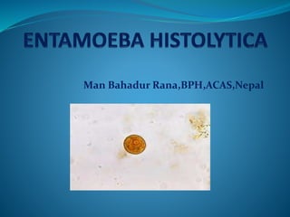
12. Cystic stage
The cyst varies greatly in size:
- the small race being 6to9um.
- the large race being 12-15um.
During encystment, the parasite becomes rounded and
is surrounded by a highly refractile little membrane,
called the cyst wall.
A mature cyst is a quqdrinucleate spherical body.
The cyst begins as a uninucleate body but soon
divides by binary fission and develops into binucleate and
quadrinucleate bodies.
14. Contd…
- The single nucleus multiplies by two successive
divisions into 2 and ultimately to 4 daughter nuclei.
During the process of division the nuclei undergo
gradual reduction in size becoming 2 um in diameter.
- In early stage of development, the cytoplasm of the
cyst shows the following.
15. Contd..
i) chromatoid or chromidial bars:
- These do not stain with iodine
- seen as refractile oblong bars with normal saline
preparation.
ii) glycogen vacuole: stains brown with iodine.
- As the cyst matures (passing from the
uninucleate to the quadrinucleate stage), both
the chromatoid bars and glycogen mass gradually
disappear .
18. Reproduction
The reproduction occurs in three
stages:
. Excystation
. Encystation
. Multiplication
Excystation: This is the process of
transformation of cyst to trophozoites. During
excystation a quadrinucleate cyst give rise to eight
amoebulae each one of which is being capable of
developing into a trophozoite.
19. Contd…
Encystation: This is the process of
transformation of trophozoite to cyst and occurs
inside the lumen of the intestine of an infected
individual.
Multiplication: This occurs only in the
trophozoite forms of Entamoeba histolytica,
growing and multiplication takes place inside the
tissue . Reproduction of trophozoites occurs by
simple binary fission.
21. Life Cycle
- E. histolytica passes its life cycle in only one host.
- The mature quadrinucleate cysts are the infective
forms. These infective cyst are passed in the
faeces of carriers.
22. Contd…- Man acquires the infection by ingestion of water
and food containing these cyst.
- When the cyst reaches the caecum or the lower
part of the ileum, the excystation occurs (due to
lysis of cyst wall by trypsin in the small intestine).
23. Contd…
- During this process, each mature cyst liberates a
single amoeba with four nuclei, a tetranucleate
amoeba which eventually produce eight
metacystic trophozoites by the division of nuclei
by binary fission.
- The metacystic trophozoite ultimately lodge in
the submucous tissue of the large intestine, their
normal habitat. Here they grow and multiply by
binary fission.
24. Contd…
- During growth, E. histolytica secretes a
proteolytic enzyme which cause destruction and
necrosis of tissue leading to flask shaped ulcer.
- Sometimes the trophozoite enter into deeper
layer and may gain entry into deep layers into the
radicals of portal vein to be carried away to liver
producing amoebic liver abscess.
25. Contd…
- When the effect of the parasite on the host is
toned down and there is increase in the tolerance
of the host, the lesion start healing . The
trophozoites, in the lumen of large intestine
occurs encystation.
- These quadrinucleate cyst again pass into the
faeces and continued their life cycle.
27. MODE OF TRANSMISSION
- Transmission of E. histolytica from man to
man is effected through its encysted stage
and infection occurs through the ingestion
of these cyst.
- Faecal contamination of drinking water,
vegetables, and food are the primary
causes.
- Eating of uncooked vegetables and fruits
which have been fertilised with infected
human faeces has often lead to occurrence
of disease.
28. Pathogenicity
- Incubation period: 4-5 days.
- Clinical features or Symptomatology: The term
amoebiasis is used to denote all those condition which
are produce in the human host by infection with E.
histolytica.
- Amoebic dysentry: is a condition in which the
infection is confined to the intestinal canal and is
characterised by the passage of blood and mucus in
the stool.
35. Contd…
Secondary or metastatic lesion:
Some individual with intestinal amoebiasis develop
hepatic amoebiasis.
Amoebic liver abscess is formed.
It is the most common form of hepatic amoebiasis and
accounts for upto 10% of all intestinal amoebiasis.
It may occur in any part of liver but is generally confined to
posteriosuperior surface of right lobe of liver.
36. Contd…
Clinical features of Amoebic Liver abscess:
- Onset is insidious
- Pain and tenderness in the right
hypochondrium.
- Shoulder pain due to the irritation of phrenic
nerve
- Fever.
- Jaundice is an unusual manifestation.
- The patient becomes emarciated ( excessively
thin)
39. Contd…
Metastatic lesions in other
organs:
The various extra hepatic amoebiasis include:
- Pulmonary Amoebiasis
- Cerebral Amoebiasis
- Cutaneous Amoebiasis
- Splenic Amoebiasis
- Vaginal and Penile Amoebiasis.
44. Contd…
Specimen:
The various sample collected for laboratory
investigation include stool, swabs,
aspirated pus, blood, CSF,
biopsied and autopsied
material.
45. Contd…
1.Examination of Stool:
a) Naked eye or Macroscopic appearance:
- An offensive dark brown semi-fluid stool
- Acid in reaction
- Admixed with blood and mucus.
48. Contd…
2. Examination of Blood:
- Shows moderate leucocytosis.
3. Serological Test:
- In early cases it is always negative.
ELISA, Indirect haemagglutination, Dot
ELISA, latex agglutination test are in use.
49. Contd…
Extra intestinal amoebiasis:
1. Parasitological:
wet (saline and iodine) and permanent
preparation of clinical material obtained from
lesion.
2. Serological:
CFT ( Complement Fixation Test)
ELISA, Dot ELISA, IFA test are in use.
50. Contd…
3. Biochemical Test:
Liver Function Test shows raised alkaline
phosphatase level and SGOT level.
4. Intradermal Skin Test:
In an infected individual injection of 0.1ml of
an antigen prepared from a culture of E.
histolytica produces at the site of injection, an
erythema which manifest after 3 hrs (9-10cm) in
diameter and disappearing in another 24-48hrs.
51. Contd…
5. Radiological Examination:
- USG of upper abdomen
- CT-Scan of liver.
- MRI scan of liver may be
found useful for detection of
Amoebic liver abscess.
53. Contd…
6. Histopathological:
Typical cellular architecture at the site of lesion
(liver abscess) is seen.
7. Detection of antibodies in
liver pus:
Gel diffusion and Counter Current Immuno
Electrophoresis (CCIEP) test are commonly used.
54. CULTURE
Cultures are not done routinely.
Boeck and Drbohlavs medium
modified by Laidlaw extensively used for
isolation and maintenance of E.
histolytica .
Diamonds axenic medium used in
studies on Pathogenecity, antigenic
characterization, and drug sensitivity
test.