Call Girl Lucknow Mallika 7001305949 Independent Escort Service Lucknow
Enterobius vermicularis
1. Enterobius vermicularis
The pinworms are one of the most common intestinal nematodes.
The adult worms inhabit the cecum and colon.
Right after mating, the male dies. Therefore, the male worms are rarely seen.
The female worms migrate out the anus depositing eggs on the perianal skin.
Humans get this infection by mouth and by autoinfection.
Morphology
Adults:
The adults look like a pin and are white in color.
The female worm measures about 8 to 13 mm in size and is fusiform in shape.
The male adult is only 2-5mm.
The tail of a male is curved.
They die right after mating, thus males are rarely seen.
The anterior end tapers and is flanked on each side by cuticular extensions called
“cephalic alae”.
The esophagus is slender, terminating in a prominent posterior bulb , which is called
esophageal bulb.
The cephalic alae and esophageal bulb are important in identification of the species.
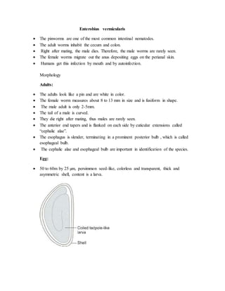
Egg:
50 to 60m by 25 µm, persimmon seed-like, colorless and transparent, thick and
asymmetric shell, content is a larva.
2. Life Cycle
Site of inhabitation: cecum and colon
Infective stage: embryonated egg
Infective route: by mouth
Without intermediate host and reservoir host
Life span of female adults: 1-2 months
3.
4. Pathogenesis
The eggs hatch in the large intestine.
Worms mature in 2-4 weeks and live for 2 months. Continuous reinfection is common.
approximately one-third of infections are asymptomatic.
The most common presentation is irritation and pruritus ani. sometimes itching is severe,
and secondary bacterial infection occurs.
Occasionally, necrosis of the mucosal surface produces pain when nerve endings are
exposed.
Worms often occur in the appendix and may be associated with appendicitis, but
causation has not been proved.
Rarely, worms may migrate to ectopic sites, mostly within the female genitourinary tract
Laboratory Diagnosis
Cellophane tape test.
E. vermicularis females lay their eggs on the perineum during the night.
Touching the perianal skin with the sticky side of the tape will pick up the eggs; the tape
is affixed to a microscope slide and examined.
Eggs are oval, approximately 55x25 µm in size, and flattened on one side, and they
contain a larva.
Specimens should be collected prior to bathing or using the toilet.
Four to six consecutive negative pinworm tape preparations are required to rule
out infection.
NIH swab
• Eggs are deposited in large number on the perianal and perineal skin at night can be
demonstrated by scraping this area by NIH swab in the morning before taking bath.
• Spread over glass slide and examined microscopically.
• This procedure should be repeated on three successive days
5. Stool samples.
Eggs are only rarely seen in stool, but in patients with heavy worm burdens, adult female
worm may be seen in stool samples.
TREATMENT :
Mebandazole and albendazole can be usesd .
These drugs are given in 1 dose at first, the second dose is repeated after 2 weeks.
Topical insecticide containing malathion can be applied on skin.