Nano-adjuvanted polio vaccine: Preparation and characterization of chitosan and trimethylchitosan (TMC) nanoparticles loaded with inactivated polio virus and coated with sodium alginate

Abstract Objective(s): It is proposed that particulate antigens could better interact with the antigen presenting cells (APCs). A fast, simple and scalable process for preparation of polymeric nanoparticles (NPs) is coating of charged antigenic particles, like viruses, with oppositely charged polymers. A second coating with a charged polymer could increase the stability and modify the immunomodulatory potentials of NPs. Materials and Methods: Negatively charged inactivated polio virus (IPV) was coated with cationic polymers, chitosan (CHT) and trimethylchitosan (TMC) by a simple incubation method. CHT: IPV and TMC: IPV NPs were coated by anionic polymer, sodium alginate (ALG). Physical characteristics and stability of NPs were studied. Cytocompatibility of NPs was checked with MTT assay. DC maturation study was used for evaluation of the NPs potential in interaction with DCs. Results: Among the various polymer to antigen ratios tested, the least size and PDI and the highest ZP was seen in TMC: IPV (2:1), CHT: IPV (2:1), ALG: TMC: IPV (2:2:1) and ALG: CHT: IPV (4:2:1). The physical stability of TMC: IPV and CHT: IPV was preserved until 15 days. After an early de-association of some part of coated alginate, ALG: CHT: IPV and ALG: TMC: IPC NPs were stable until the end of study (25th day). No one of the NPs formulations had a negative effect on cell viability. Compared with plain IPV, nanoparticulate IPV formulations failed to increase the expression of CD40 and CD86 markers of DCs. Conclusion: NPs prepared with simple and scalable method, had reasonable physical characteristics, stability and cytocompatibility and could be tested in vivo for their immunoadjuvant potential.

Recommended
Recommended
More Related Content
What's hot
What's hot (12)
Similar to Nano-adjuvanted polio vaccine: Preparation and characterization of chitosan and trimethylchitosan (TMC) nanoparticles loaded with inactivated polio virus and coated with sodium alginate
Similar to Nano-adjuvanted polio vaccine: Preparation and characterization of chitosan and trimethylchitosan (TMC) nanoparticles loaded with inactivated polio virus and coated with sodium alginate (20)
More from Nanomedicine Journal (NMJ)
More from Nanomedicine Journal (NMJ) (20)
Recently uploaded
Recently uploaded (20)
Nano-adjuvanted polio vaccine: Preparation and characterization of chitosan and trimethylchitosan (TMC) nanoparticles loaded with inactivated polio virus and coated with sodium alginate
- 1. 220 Nanomed J, Vol. 1, No. 4, Summer 2014 Original Research (font 12) Received: Feb. 25, 2014; Accepted: Apr. 12, 2014 Vol. 1, No. 4, Summer 2014, page 220-228 Received: Apr. 22, 2014; Accepted: Jul. 12, 2014 Vol. 1, No. 5, Autumn 2014, page 298-301 Online ISSN 2322-5904 http://nmj.mums.ac.ir Original Research Nano-adjuvanted polio vaccine: Preparation and characterization of chitosan and trimethylchitosan (TMC) nanoparticles loaded with inactivated polio virus and coated with sodium alginate Mohsen Tafaghodi1,2* , Gideon Kersten2,3 , Wim Jiskoot2 1 Nanotechnology Research Center, School of Pharmacy, Mashhad University of Medical Sciences, Mashhad, Iran 2 Division of Drug Delivery Technology, Leiden Academic Centre for Drug Research, Leiden University, Leiden, Netherlands 3 Institute for Translational Vaccinology, Bilthoven, Netherlands Abstract Objective(s): It is proposed that particulate antigens could better interact with the antigen presenting cells (APCs). A fast, simple and scalable process for preparation of polymeric nanoparticles (NPs) is coating of charged antigenic particles, like viruses, with oppositely charged polymers. A second coating with a charged polymer could increase the stability and modify the immunomodulatory potentials of NPs. Materials and Methods: Negatively charged inactivated polio virus (IPV) was coated with cationic polymers, chitosan (CHT) and trimethylchitosan (TMC) by a simple incubation method. CHT: IPV and TMC: IPV NPs were coated by anionic polymer, sodium alginate (ALG). Physical characteristics and stability of NPs were studied. Cytocompatibility of NPs was checked with MTT assay. DC maturation study was used for evaluation of the NPs potential in interaction with DCs. Results: Among the various polymer to antigen ratios tested, the least size and PDI and the highest ZP was seen in TMC: IPV (2:1), CHT: IPV (2:1), ALG: TMC: IPV (2:2:1) and ALG: CHT: IPV (4:2:1). The physical stability of TMC: IPV and CHT: IPV was preserved until 15 days. After an early de-association of some part of coated alginate, ALG: CHT: IPV and ALG: TMC: IPC NPs were stable until the end of study (25th day). No one of the NPs formulations had a negative effect on cell viability. Compared with plain IPV, nanoparticulate IPV formulations failed to increase the expression of CD40 and CD86 markers of DCs. Conclusion: NPs prepared with simple and scalable method, had reasonable physical characteristics, stability and cytocompatibility and could be tested in vivo for their immunoadjuvant potential. Keywords: Alginate, Chitosan, Inactivated Polio Virus, Nanoparticles, Trimethylchitosan (TMC) *Corresponding Author: Nanotechnology Research Center, School of Pharmacy, Mashhad University of Medical Sciences, Mashhad, Iran. Division of Drug Delivery Technology, Leiden Academic Centre for Drug Research, Leiden University, Leiden, Netherlands. Tel: +98 511 8823255, E-mail: tafaghodim@mums.ac.ir
- 2. Nanoadjuvanted polio vaccine Nanomed J, Vol. 1, No. 4, Summer 2014 221 Introduction Most of the available vaccines are currently designed for parenteral administration. Most of these vaccines can induce high systemic immune responses, but because of their disadvantages due to the use of needles, like pain and their inability to induce mucosal immunity, many studies are in progress to investigate mucosal routes of administration (1-7). Among the mucosal vaccines, the vaccines designed for nasal administration are very attractive because of their simple administration and absence of harsh environments like the gastro-intestinal tract (8). To induce systemic and mucosal immune responses after nasal administration, many challenges should be overcome. The mucociliary clearance limits the residence time of the antigen in the nasal cavity. Moreover, because of low permeability of the nasal epithelium and the large size of most antigens and their delivery systems, as well as the tolerogenic nature of the mucosal epithelium, induction of strong immune responses is difficult. Polymeric nano-particles as adjuvant/delivery system can be efficient mucosal adjuvants (8, 9). Mucoadhesive polymers like chitosan may prolong the residence time of the antigen, protect the antigen against the enzymes (10), and increase the permeability of intercellular tight junctions (11). The specialized antigen-sampling cells (microfold (M) cells) in the nasal associated lymphoid tissue (NALT) can efficiently uptake the particulate antigens and promote the mucosal and systemic immune responses (12). Multimerization of antigenic epitopes on the surface of the nanoparticles and the possibility of co- encapsulating antigen and adjuvants can also potentiate and modulate the immune responses (13). As a mucoadhesive, safe and biodegradable polymer, chitosan (CHT) has been frequently used for nasal vaccine delivery (4, 8, 14, 15). Because of its limited aqueous solubility at neutral or alkaline pH, CHT derivatives with improved solubility have been developed. N, N, N-trimethyl chitosan (TMC) is one of the most studied synthetic derivatives of chitosan used for nasal delivery of antigens. TMC is also a mucoadhesive and biodegradable polymer (11) and has successfully been tested pre-clinically for nasal, pulmonary and oral delivery of antigens (7, 16, 17). As both CHT and TMC NPs can be prepared by simple and scalable methods like polyelectrolyte complexation or ionotropic gelation (18, 19) and possess limited toxicity, these have great potential as delivery system/adjuvants for nasal vaccination. CHT and TMC NPs have been widely used as adjuvant/delivery system for nasal immunization (14, 16, 20- 23). In the ionotropic gelation method, the polycationic polymer is incubated with a polyanionic molecule like tripolyphosphate (TPP). For negatively charged nanoparticulate antigens like whole inactivated influenza virus, simple incubation with CHT or TMC has been shown to coat the antigen with polymer and form antigen:polymer nanoparticles (7, 21). Positively charged chitosan nanoparticles could be simply coated by negatively charged alginate to modify their immunomodulatory capacity and increase their stability (24, 25). Adjuvants/delivery systems can improve the immunogenicity of a vaccine and reduce the antigen dose required for mucosal vaccination. For eradication of poliomyelitis, use of inactivated poliomyelitis vaccine is very important. In the present study we prepared and characterized the CHT and TMC NPs, coated with sodium alginate, with encapsulated inactivated polio virus (IPV), by polyelectrolyte complexation.
- 3. Tafaghodi M, et al 222 Nanomed J, Vol. 1, No. 4, Summer 2014 Materials and Methods Materials IPV (protein content determined by BCA assay method: 50 µg/ml in PB 8 mM, pH 7.2, particle size: 93.9 nm, Zeta potential: - 21 mV) was provided by the National Institute for Public Health and the Environment (RIVM, Bilthoven, The Netherlands). TMC with a degree of quaternization of 23.8% was synthesized from 92% deacetylated (MW 120 KDa) chitosan (Primex, Avaldsnes, Norway) and charac- terized by NMR, as described by Bal et al. (26). All of cell culture reagents were bought from Invitrogen (Breda, The Nether-lands). Anti-CD86-APC and anti-CD83-PE were provided by Becton Dickinson (Breda, The Netherlands). BCA protein assay kit was purchased from Pierce (Thermo Scientific, Rockford, IL). Sodium alginate, Thiazolyl blue tetrazolium bromide (MTT), acetic acid, sodium hydroxide and hydrochloric acid were obtained from Sigma-Aldrich Chemical Co (Zwijndrecht, the Nether- lands). Preparation of TMC: IPV and CHT: IPV NPs The IPV Nps were first titrated (pH 4 to pH 10) for finding the best pH for the smallest size and PDI and the highest surface charge (Zetasizer Nano, Malvern, UK). The smallest particle size and PDI (34.8 nm, 0.114) was found at pH 7.2. At this pH the IPV NPs showed an acceptable zeta potential of -21 mV. The TMC:IPV and CHT:IPV NPs were prepared by adding equal volumes of TMC or CHT solution to IPV dispersion by a Gilson pipette and gently mixing for about 5 seconds (7, 8, 27). Formulations containing different w/w ratios of polymer/antigen (0.5:1 to 10:1) were prepared. Preparation of ALG: TMC: IPV and ALG: CHT: IPV NPs The TMC: IPV and CHT: IPV NPs were incubated with equal volume of ALG solution and gently mixed for about 5 seconds. Formulations containing different ratios of the ALG/antigen (0.5:1 to 6:1) were prepared. Characterization of NPs Particle size (z-average mean), polydispersity index (PDI) and zeta potential were measured by a Zetasizer Nano (Malvern Instruments, Malvern, UK). The fraction of associated CHT and TMC to the IPV NPs were measured by quantification of the free polymer in the supernatant of ultracentrifuged (40000 rpm, 90 min) TMC: IPV and CHT: IPV NPs. To quantify the free polymer in the supe-rnatants, a sensitive colorimetric method using a dye reagent, Cibacron Brilliant Red 3B-A (Aldrich, Taufkirchen, Germany) was used. The method was introduced by Muzzarelli (28) and was modified for more sensitivity (10-80 µg/ml) (29). According to the improved method, one part of the dye reagent was added to five parts of a chitosan solution (5-100 µg/ml) and vortexed. The dye reagent contained 0.9 mg/ml Cibacron Brilliant Red 3B-A in a 0.3 M glycine.HCl buffer with a pH of 3.2. The absorbance difference between 575 and 750 nm was measured immediately against a mixture of water and dye reagent in 96 well microplates (29). The sensitivity of the method to TMC was increased by decreasing the reagent concentration to 0.3 mg/ml. The percent of IPV-associated TMC and chitosan was calculated as (7): TMCbound(%) = 100 – (TMCfree (g) × 100) / total TMC (g) NPs prepared in different situations were kept in 4 o C for 20 days. Each five days, NPs size, PDI and zeta potential NPs were evaluated for their size, PDI and zeta potential. Cell viability test by MTT method The cell viability test by MTT method was performed according to Mosman with
- 4. Nanoadjuvanted polio vaccine Nanomed J, Vol. 1, No. 4, Summer 2014 223 some modifications (30). Briefly, Caco-2 cells were added to 96 well plates (12000 cells/well) and incubated at 37°C and 5% CO2 for 48 hours. For better simulation of the nasal administrations, TMC: IPV, CHT: IPV, ALG: TMC: IPV and ALG: CHT: IPV NPs were prepared in suitable concentration for in vivo studies. NPs were diluted 1:1 by culture medium and added to the cells and incubated for 0.5 hour. Plates were emptied and washed with PBS pH 7.4 (2×) and then 100 µl of 0.5 mg/ml MTT solution in DMEM was added to each well and incubated for 3 hours. After aspiration of the MTT solution, 100 µl of DMSO was added to dissolve the purple crystals. Absorbances were read at 570 nm (23, 31). Human monocytes derived dendritic cells culture Monocytes were isolated from buffy coat (obtained from blood bank, Sanquin, The Netherlands) by Ficoll and Percoll density centrifugation (32). The monocytes were separated from platelets by their adherence to 24 well plates (Corning, Schiphol, Netherlands) followed by washing. Monocytes (5×10⁵ cells/well) were cultured in RPMI 1640, supplemented with 10% v/v FBS, 1% glutamine, 1% v/v penicillin-/streptomycin, 250 U/ml GM- CSF and 100 U/ml IL-4 at 37 o C and 5% CO2 for 6 days. Medium was refreshed after 3 days (23, 33). Impact of nanoparticles on DC maturation DCs were incubated for 48 hours at 37 o C in RPMI 1640 and 500 U/ml GM-CSF with 45 µg/ml IPV, free or as TMC: IPV, CHT: IPV, ALG: TMC: IPV or ALG: CHT: IPV nanoparticles, 100 ng/ml LPS as positive control and medium as negative control. After 48 hours, cells were washed three times with PBS supplemented with 1% w/v BSA and 2% v/v FBS and incubated with 50 times diluted anti- CD40PE and anti-CD86APC in the dark at 4 o C. These markers will show the expression of CD 40 and CD 86 molecules on the surface of DCs. The cells were washed and expression of above molecules was quant-ified by flowcytometry. Living cells were gated based on forward and side scatter and the amount of CD 40 and CD 86. Positive cells were expressed as the MFI relative to the LPS control (13, 23, 33). Statistical analysis Unless otherwise stated, One-way analysis of variance (ANOVA) statistical test with Tukey’s post test was performed using GraphPad InStat version 3.05 for Windows (GraphPad Software, San Diego, USA) to determine the significance of the differences between various groups. Results and Discussion Characterization of TMC: IPV and CHT: IPV NPs The TMC: IPV and CHT: IPV NPs were prepared by adding the equal volume of TMC or CHT solution to IPV dispersion. Different w/w ratios of the polymer/antigen (0.5:1 to 10:1 w/w ratios) were prepared and characterized. Among the NPs tested, the lowest ratio in which NPs had an acceptable size, PDI and zeta potential was 2:1 w/w ratio. In other ratios, both CHT: IPV and TMC: IPV NPs showed larger sizes and PDI. In higher ratios of polymer: antigen, ZP of TMC:IPV and CHT:IPV NPs was nearly constant and indicated that coating of IPV NPs by polymers has reached to maximum in this ratio. NPs prepared with 2:1 ratio were used for further studies. Quaternization of NH2 groups of chitosan in the synthesis process of TMC provides more positive charge and TMC NPs have usually higher zeta potentials than CHT NPs (12, 18). Both CHT: IPV and TMC: IPV NPs prepared, had similar size and
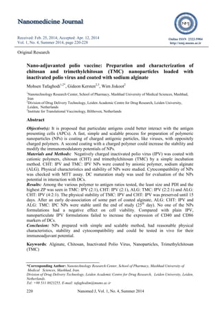
- 5. Tafaghodi M, et al 224 Nanomed J, Vol. 1, No. 4, Summer 2014 PDI (t test, P>0.05, Table I). Alginate coated CHT or TMC nanoparticles have been used in several studies as adjuvant/delivery systems for immun-izations (24, 25, 34-38). Coating of chitosan nanoparticles with sodium alginate, increase the stability of these particles. After coating, inversion of the particles' zeta potential from positive to negative values and substantial increase in size suggest the presence of an alginate coating layer. Additionally, based on in vitro release reported in other studies, the presence of the alginate layer around the particles could prevent a burst release of loaded antigen (34). These nanoparticles have great stability and both chitosan and alginate shows immunostimulatory properties (25). To determine the percent of TMC and CHT polymers associated with IPV NPs, an indirect method was used. After ultracen-trifugation, the amount of the free polymer in NPs supernatants was quantified by Cibacron Brilliant Red 3B-A dye reagent (0.3 mg/ml for TMC and 0.9 mg/ml for CHT) (Table II). The association of both TMC and CHT polymers with IPV was similar in 2:1 ratio of polymer:antigen. The association percent of CHT in CHT: IPV (4:1) NPs was significantly lower than CHT: IPV (2:1) NPs (P< 0.01). It confirms the saturability of coating process. Based on the results, in the NPs prepared with 2:1 ratio of polym- er:antigen, the amount of free polymer in supernatant is not high and most of the polymer used have been condensed on the surface of viral particles. TMC: IPV, CHT: IPV, ALG: TMC: IPV and ALG: CHT: IPV NPs were prepared and their size, PDI and ZP were evaluated for 20 days at 4o C (Figure 1). Based on the figures 1A and 1C, ALG: TMC: IPV and ALG: CHT: IPV NPs shows a dramatic change in size in the 5th day. Regards to the figure. 1C, in the same day, zeta potentials of these NPs changes from about -20 to -10. These changes could be attributed to the de-association of some of attached alginate. This led to decrease in negative charge and repulsion forces between NPs. This phenomenon could result in aggregated NPs and increase in size. The same phenomenon could be observed for TMC: IPV and CHT: IPV NPs. In day 15th , de-association of cationic polymers (TMC and CHT) has lowered the zeta potential from positive to negative (Figure. 1C). At the same day a dramatic change in NPs size could be seen in Figure. 1A. Disregards from the primary changes in the 5th day for alginate coated NPs; in the rest of the study these NPs were more stable. Table 1. Size and zeta potential of NPs prepared in PB 6.7(mean ± SD, n=3). NPs Size (nm) PDI Zeta potential (mV) TMC:IPV (2:1) 158.7 (±30.6) 0.305 (±0.050) 11.03 (±0.93) CHT:IPV (2:1) 88.8 (±32.3) 0.309 (±0.122) 2.57 (±1.22) ALG:TMC:IPV (2:2:1) 254.3 (±8.8) 0.292 (±0.002) -29.1 (±4.67) ALG:CHT:IPV (4:2:1) 184.4 (±8.9) 0.405 (±0.08) -27.1 (±2.55) Cell viability test by MTT method Caco-2 cells were incubated with TMC: IPV, CHT: IPV, ALG: TMC: IPV and ALG: CHT: IPV NPs. All formulations showed more that 90% cell viability (figure. 2). Table 2. Association percent of the polymers in TMC: IPV (2:1) and CHT: IPV (2:1) NPs (mean ± SD, n=4). NPs Association percent TMC:IPV (2:1) 87 ± 3.3 CHT:IPV (2:1) 85.5 ± 5.2 CHT:IPV (4:1) 68 ± 3.8
- 6. Nanoadjuvanted polio vaccine Nanomed J, Vol. 1, No. 4, Summer 2014 225 Figure 1. Stability of TMC: IPV, CHT: IPV, ALG: TMC: IPV and ALG: CHT: IPV NPs in 4 o C within 25 days. Each 5 days NPs were checked for size (A), PDI (B) and ZP (C). Error bars represents mean ± SD (n=3). There was no significant differences amongst buffers and NPs tested (P>0.05). Therefore the cytotoxicity of the NPs is negligible. Impact of nanoparticles on DC maturation The effect of IPV dispersion, TMC: IPV, CHT: IPV, ALG: TMC: IPV and ALG: CHT: IPV NPs dispersed in isotonic medium on maturation of DCs was evaluated by measuring the expression of CD 40 and CD 86 surface markers appeared on the surface of matured DCs. Culture medium was used as negative and LPS as positive control. Based on the results presented in Figure 3, no one of the 4 NPs formulation could significantly increase the expression of CD40 and CD86 markers, as compared with plain IPV antigen. Figure 2. TMC: IPV, CHT: IPV, ALG: TMC: IPV and ALG: CHT: IPV NPs in different media were diluted 1:1 by culture medium and added to the caco-2 cells and incubated for 0.5 hour. Plates were washed and MTT solution was added and incubated for 3 hours. After aspiration of the MTT solution, 100 µl of DMSO was added to dissolve the purple crystals. Absorbances were read at 570 nm. Error bars represent mean ± SD (n=6). In our previous study, CHT and TMC NPs were loaded with Hepatitis B surface antigen (HBsAg) with the same method. TMC:HBsAg NPs could significantly increase the expression of MHCII and CD86 markers, as compared with plain HBsAg, however the expression induced with CHT:HBsAg NPs was similar to plain antigen (8). More complementary studies are required to better explain the inefficiency of these 4 NPs formulation in this test. Conclusion The CHT:IPV, TMC:IPV, ALG:CHT:IPV and ALG:TMC:IPC NPs prepared with a Cell Viability test- Calu-3 cells 0 20 40 60 80 100 120 140 IPV sol PB 6.7 PB 7.0 M edium CH T:IPV TM C:IPV ALG :CHT:IPV ALG :TM C :IPV ALG :CHT ALG :TM C Cellviabilitypercent
- 7. Tafaghodi M, et al 226 Nanomed J, Vol. 1, No. 4, Summer 2014 simple and scalable method, had reasonable size, PDI and zeta potential. The CHT:IPV and TMC:IPV NPs showed physical stability for 2 weeks, and ALG:CHT:IPV and ALG:TMC:IPC NPs kept their stability for 4 weeks. Figure 3. DCs were incubated with IPV, TMC: IPV, CHT: IPV, ALG: TMC: IPV and ALG: CHT: IPV nanoparticles, LPS as positive control and medium as negative control. After 48 hours, cells were washed and incubated with anti-CD 40FITC and anti-CD86APC and expression of above molecules was quantified by flowcytometry. Values are expressed as mean fluorescence intensity (MFI) relative to the LPS. Error bars represent mean ± SD (n=5, 3 separate measurements). No one of the NPs formulations had negative effect on cell viability, as tested by MTT assay. Compared with plain IPV, nanoparticulate IPV formulations failed to increase the expression of CD40 and CD86 markers of DCs. These NPs could be tested in vivo for their immunoadjuvant potential. Acknowledgements The authors acknowledge Mashhad University of Medical Sciences, Mashhad, Iran, for providing a Research Fellowship to MT for carrying out the research work. The authors thank Christophe Barnier and Jean-Pierre Amorij for their kind assistance. References 1. Amorij JP, Hinrichs W, Frijlink HW, Wilschut JC, Huckriede A. Needle- free influenza vaccination. Lancet Infect Dis. 2010; 10: 699-711. 2. Saluja V, Amorij JP, van Roosmalen ML, et al. Intranasal delivery of influenza subunit vaccine formulated with GEM particles as an adjuvant. AAPS J. 2010; 12: 109-16. 3. Tafaghodi M, Rastegar S. Preparation and in vivo study of dry powder microspheres for nasal immunization. J Drug Target. 2010; 18: 235-42. 4. Amin M, Jaafari MR, Tafaghodi M. Impact of chitosan coating of anionic liposomes on clearance rate, mucosal and systemic immune responses following nasal administration in rabbits. Colloids Surf B Biointerfaces. 2009; 74: 225-9. 5. Slutter B, Hagenaars N, Jiskoot W. Rational design of nasal vaccines. J Drug Target. 2008; 16: 1-17. 6. Hirschberg HJHB, de Wijdeven GGPv, Kraan H, Amorij J-P, Kersten GFA. Bioneedles as alternative delivery system for hepatitis B vaccine. J Control Release. 2010; 147: 211-7. 7. Hagenaars N, Mastrobattista E, Verheul RJ, et al. Physicochemical and immunological characterization of N,N,N-trimethyl chitosan-coated whole inactivated influenza virus vaccine for intranasal administration. Pharm Res. 2009; 26: 1353-64. 8. Tafaghodi M, Saluja V, Kersten GF, et al. Hepatitis B surface antigen nanoparticles coated with chitosan and trimethyl chitosan: Impact of formulation on physicochemical and immunological characteristics. Vaccine. 2012; 30: 5341-8. 9. Koping-Hoggard M, Sanchez A, Alonso MJ. Nanoparticles as carriers for nasal vaccine delivery. Expert Rev Vaccines. 2005; 4: 185-96. 10. Almeida AJ, Alpar HO. Mucosal immunization with antigen-containing CD 40 0 20 40 60 80 100 120 LPS Blank IPV Sol. TM C :IPV C H T:IPV ALG :TM C :IPV ALG :C H T:IPV %Maturation[MFI]
- 8. Nanoadjuvanted polio vaccine Nanomed J, Vol. 1, No. 4, Summer 2014 227 microparticles. In: Gander B, Merkle HP, (eds) Antigen delivery systems. New York: Harwood academic publishers, 1997. 207-26. 11. Verheul RJ, Amidi M, van der Wal S, van Riet E, Jiskoot W, Hennink WE. Synthesis, characterization and in vitro biological properties of O-methyl free N,N,N-trimethylated chitosan. Biomaterials. 2008; 29: 3642-9. 12. Mangal S, Pawar D, Garg NK, et al. Pharmaceutical and immunological evaluation of mucoadhesive nanoparticles based delivery system(s) administered intranasally. Vaccine. 2011; 29: 4953-62. 13. Bal SM, Ding Z, van Riet E, Jiskoot W, Bouwstra JA. Advances in transcutaneous vaccine delivery: do all ways lead to Rome? J Control Release. 2010; 148: 266-82. 14. Illum L, Jabbal-Gill I, Hinchcliffe M, Fisher AN, Davis SS. Chitosan as a novel nasal delivery system for vaccines. Adv Drug Deliv Rev. 2001; 51: 81-96. 15. van der Lubben IM, Kersten G, Fretz MM, Beuvery C, Coos Verhoef J, Junginger HE. Chitosan microparticles for mucosal vaccination against diphtheria: oral and nasal efficacy studies in mice. Vaccine. 2003; 21: 1400-8. 16. Amidi M, Mastrobattista E, Jiskoot W, Hennink WE. Chitosan-based delivery systems for protein therapeutics and antigens. Adv Drug Deliv Rev. 2010; 62: 59-82. 17. Boonyo W, Junginger HE, Waranuch N, Polnok A, Pitaksuteepong T. Chitosan and trimethyl chitosan chloride (TMC) as adjuvants for inducing immune responses to ovalbumin in mice following nasal administration. J Control Release. 2007; 121: 168-75. 18. Sadeghi AM, Dorkoosh FA, Avadi MR, Saadat P, Rafiee-Tehrani M, Junginger HE. Preparation, characterization and antibacterial activities of chitosan, N-trimethyl chitosan (TMC) and N-diethylmethyl chitosan (DEMC) nanoparticles loaded with insulin using both the ionotropic gelation and polyelectrolyte complexation methods. Int J Pharm. 2008; 355: 299-306. 19. Amidi M, Romeijn SG, Borchard G, Junginger HE, Hennink WE, Jiskoot W. Preparation and characterization of protein-loaded N-trimethyl chitosan nanoparticles as nasal delivery system. J Control Release. 2006; 111: 107-16. 20. Amidi M, Romeijn SG, Verhoef JC, et al. N-Trimethyl chitosan (TMC) nanoparticles loaded with influenza subunit antigen for intranasal vaccination: Biological properties and immunogenicity in a mouse model. Vaccine. 2007; 25: 144-53. 21. Hagenaars N, Verheul RJ, Mooren I, et al. Relationship between structure and adjuvanticity of N,N,N-trimethyl chitosan (TMC) structural variants in a nasal influenza vaccine. J Control Release. 2009; 140: 126-33. 22. Slutter B, Bal S, Keijzer C, et al. Nasal vaccination with N-trimethyl chitosan and PLGA based nanoparticles: nanoparticle characteristics determine quality and strength of the antibody response in mice against the encapsulated antigen. Vaccine. 2010; 28: 6282-91. 23. Slutter B, Plapied L, Fievez V, et al. Mechanistic study of the adjuvant effect of biodegradable nanoparticles in mucosal vaccination. J Control Release. 2009; 138: 113-21. 24. Demoulins T, Bassi I, Thomann- Harwood L, et al. Alginate-coated chitosan nanogel capacity to modulate the effect of TLR ligands on blood dendritic cells. Nanomedicine. 2013. 25. Oliveira CR, Rezende CM, Silva MR, Pego AP, Borges O, Goes AM. A new strategy based on SmRho protein loaded chitosan nanoparticles as a candidate oral vaccine against schistosomiasis. PLoS Negl Trop Dis. 2012; 6: e1894. 26. Bal SM, Slutter B, van Riet E, et al. Efficient induction of immune responses through intradermal vaccination with N-trimethyl chitosan containing antigen formulations. J Control Release. 2009; 142: 374-83. 27. Hagenaars N, Mania M, de Jong P, et al. Role of trimethylated chitosan (TMC) in nasal residence time, local
- 9. Tafaghodi M, et al 228 Nanomed J, Vol. 1, No. 4, Summer 2014 distribution and toxicity of an intranasal influenza vaccine. J Control Release. 2010; 144: 17-24. 28. (28) Muzzarelli RA. Colorimetric determination of chitosan. Anal Biochem. 1998; 260: 255-7. 29. (29) Wischke C, Borchert HH. Increased sensitivity of chitosan determination by a dye binding method. Carbohydr Res. 2006; 341: 2978-9. 30. Mosmann T. Rapid colorimetric assay for cellular growth and survival: application to proliferation and cytotoxicity assays. J Immunol Methods. 1983; 65: 55-63. 31. Jafari T, Simchi A, Khakpash N. Synthesis and cytotoxicity assessment of superparamagnetic iron-gold core- shell nanoparticles coated with polyglycerol. J Colloid Interface Sci. 2010; 345: 64-71. 32. de Jong EC, Vieira PL, Kalinski P, et al. Microbial compounds selectively induce Th1 cell-promoting or Th2 cell-promoting dendritic cells in vitro with diverse th cell-polarizing signals. J Immunol. 2002; 168: 1704-9. 33. Bal SM, Slutter B, van Riet E, et al. Efficient induction of immune responses through intradermal vaccination with N-trimethyl chitosan containing antigen formulations. J Control Release. 2010; 142: 374-83. 34. Borges O, Borchard G, Verhoef JC, de Sousa A, Junginger HE. Preparation of coated nanoparticles for a new mucosal vaccine delivery system. Int J Pharm. 2005; 299: 155-66. 35. Borges O, Cordeiro-da-Silva A, Tavares J, et al. Immune response by nasal delivery of hepatitis B surface antigen and codelivery of a CpG ODN in alginate coated chitosan nanoparticles. Eur J Pharm Biopharm. 2008; 69: 405-16. 36. Borges O, Silva M, de Sousa A, Borchard G, Junginger HE, Cordeiro- da-Silva A. Alginate coated chitosan nanoparticles are an effective subcutaneous adjuvant for hepatitis B surface antigen. Int Immunopharmacol. 2008; 8: 1773-80. 37. Chen F, Zhang ZR, Huang Y. Evaluation and modification of N- trimethyl chitosan chloride nanoparticles as protein carriers. Int J Pharm. 2007; 336: 166-73. 38. Zhou J, Romero G, Rojas E, Ma L, Moya S, Gao C. Layer by layer chitosan/alginate coatings on poly(lactide-co-glycolide) anoparticles for antifouling protection and Folic acid binding to achieve selective cell targeting. J Colloid Interface Sci. 2010; 345: 241-7