New technology for fish oral vaccine - A review of a published article
•Download as PPTX, PDF•
0 likes•174 views

A published article was reviewed for it content on developing a novel technology for effective fish oral vaccine against vibriosis.

Recommended
More Related Content
What's hot
What's hot (20)
Similar to New technology for fish oral vaccine - A review of a published article
Similar to New technology for fish oral vaccine - A review of a published article (20)
Recently uploaded
Recently uploaded (20)
New technology for fish oral vaccine - A review of a published article
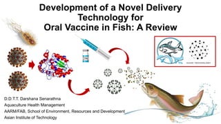
- 1. Development of a Novel Delivery Technology for Oral Vaccine in Fish: A Review D.D.T.T. Darshana Senarathna Aquaculture Health Management AARM/FAB, School of Environment, Resources and Development Asian Institute of Technology
- 2. Outline • Approach • Introduction • Objectives • Materials and methods • Results & Discussion • Conclusion • Recommendations and suggestions • Acknowledgement • References • Supplementary details
- 3. Approach Aquaculture Economic losses Antibiotics Environmental issues Diagnosis, Treatment, Biosecurity • Inactivated vaccine • Live attenuated vaccine • Toxoids (inactivated toxins) • Subunit vaccine • DNA vaccine or mRNA vaccine Immersion, Injection, Oral ORAL VACCINE 1
- 4. pH-Controlled Release of Antigens Using Mesoporous Silica Nanoparticles Delivery System for Developing a Fish Oral Vaccine Zhang W, Zhu C, Xiao F, Liu X, Xie A, Chen F, Dong P, Lin P, Zheng C, Zhang H, Gong H and Wu Y (2021) pH-Controlled Release of Antigens Using Mesoporous Silica Nanoparticles Delivery System for Developing a Fish Oral Vaccine. Front. Immunol. 12:644396. doi: 10.3389/fimmu.2021.644396 Present as a review by: D.D.T.T. Darshana Senarathna, as an assignment for Aquaculture Health Management at Asian Institute of Technology, 2021.11. 20 2
- 5. 1. Introduction Vibriosis Vibrio parahaemolyticus Vibrio alginolyticus Vibrio harveyi largest mariculture fish in China (2017): 177,640 tons Convenient, safe, efficient vaccine and delivery system High-performance Cost-effective Stable vaccines Better release kinetics Whole-pathogen vaccines Single proteins and peptides as antigens Antigens have a greatly reduced immunogenicity Not achieve the desired level of immune protection Necessary to develop adjuvants and effective delivery systems Larimichthys polyactis 3 Xu and Lu, 2018 Mahboubeh et al., 2017
- 6. Adjuvant Activate antigen-presenting cells Strong immune response Less toxicity and side effects Long-term protection Carrier systems Low antigen degradation Controlled antigen release Enhance bioavailability Suitable size Absorb by endocytosis Promote antigen absorption Enhance Antigen presentation Nanoliposomes Macromolecule Inorganic nanoparticles Immunostimulatory Complexes Mesoporous silica nanoparticles (MSN) Humoral and Cell-mediated immune responses 4 Smith et al., 2015 Gregory et al., 2013 Shaalan et al., 2016
- 7. Hypothesis Objectives Methylcellulose phthalate (HP55) coated Mesoporous silica nanoparticles (MSN) might act as an effective delivery system for an oral vaccine developed from purified antigen, Dihydrolipoamide dehydrogenase (DLDH) of Vibrio alginolyticus. • To develop DLDH loaded MSN coated with HP55. • To analyze the antigen releasing behavior of developed nanoparticles. • To analyze the toxicity of nanoparticles. • To analyze the effectiveness of nanoparticle based oral vaccine against vibrio. 5
- 8. Conceptual Framework 6 Gene Clone and Protein Expression Characterization of Materials Acid-Base Release Characteristics In Vitro Cytotoxicity Analysis of Vaccine Relative Percent Survival Assays Analysis of Serum Antibody Levels Expression Levels of Cytokines Vibrio alginolyticus DLDH DLDH loaded MSN DLDH loaded MSN mixed feed Processing and antigen presenting cells T cells IFNγ B cells Antibodies
- 9. 2. Materials • Large yellow croaker • Kidney cells of large yellow croaker • Vibrio alginolyticus • Monoclonal antibody 2H5F4 • HP55 and span 80 • N, N, N-trimethylhexadecan-1-aminium 4- methylbenzenesulfonate (CTATos) • Horseradish peroxidase (HRP) conjugated goat anti-mouse IgG • Triethanolamine (TEAH3), tetraethyl orthosilicate (TEOS), polyvinyl alcohol (PVA) and triethyl citrate • Luminescent cell viability assay kit 7
- 10. 3. Method 3.1 Gene Cloning and Protein Expression DLDH Centrifugation (6,720 g for 10 min) + ultrasonic cell disruptor Remove cell fragments (centrifugation at 98,900 g) nickel-nitrilotriacetic acid (Ni2+-NTA) pET-28a Recombination 3h in LB + IPTG 0.3mM at 16°C,12h PCR E.coli Tag removal 1 2 3 4 5 6 7 8 9 10 8
- 11. Swiss-model on line server used for homology modeling to build the three-dimensional structure of DLDH. The DLDH protein from Colwellia psychrerythraea 34H (PDB ID 3IC9, 60.17% identity) has used as a template for modeling, and the three-dimensional structure diagram was performed using PyMOL software 9 Rigsby and Parker, 2016
- 12. CTATos (4.80 g) TEAH3 (0.7 mL) H2O (250 mL) Stir 1h, 80⁰C 3.2 Preparation of Nanoparticles & Feed Stir 2h, 80⁰C Extract using NH4NO3 methanol solution, 60⁰C Buffer solution 1:1.28 (MSN/DLDH) DLDH MSN DLDH load(%) = DLDHload× 100%. (DLDHload + MSNmass) MSN-DLDH HP55 enteric coatings Double emulsion Chitosan solution (3s) + H2O (5s) + Dry Commercial feed + binder 1 2 3 4 5 MSN-DLDH@HP55 10
- 13. 3.3 Acid-Base Release Characteristics 11
- 14. 3.4 In Vitro Cell Cytotoxicity Assay Cell Titer-Lumi™ plus luminescent cell viability assay kit 96-well plates (104/well) in M199 media supplemented with 10% fetal bovine serum for 24 h at 27°C. Yellow croaker kidney cells Culture DLDH proteins & MSN-DLDH@HP55 (0, 4, 8, 10, 20, 40, 60, 80, 100, 120, 140, 160, 180, 200 μg/ml) Incubate for 24 h 100 ml Cell Titer- Lumi™ plus solution shake at room temperature for 2 min and incubate at 25°C for 10 min microplate reader Cell viability (%) = (Sample/Control) × 100%. 12
- 15. 3.4 Relative Percent Survival Assay One week after the second stimulation intraperitoneal inoculated with 0.2 ml PBS 10-fold median lethal dose (1.1 × 10⁷ CFU/ml) of Vibrio alginolyticus RPS = [1 - (mortality in immunized group / mortality in control group)] x 100% Kole et al., 2019 13
- 16. 3.5 Analysis of Serum Antibody Levels Coated with 20 mg/ml of purified DLDH protein Primary antibody: Antiserum of large yellow croaker (1:50) Secondary antibody: Monoclonal antibody 2H5F4 against large yellow croaker (1:500) Third antibody: Goat anti-mouse IgG conjugated to HRP (1:5,000) PI (%) = (1-OD490 of sample serum/OD490 of control serum) ×100%. Absolute value of the PI >18.4% = POSITIVE (OD) value of each well was determined by microplate spectrophotometer Gaps 0,7,14,21 days Blocking ELISA 14
- 17. 3.5 Analysis of the Expression Levels of Cytokines Total RNA Trizol reagent cDNA Reverse transcription: PrimeScript™ RT reagent kit with gDNA Eraser expression of IFNγ, IL-1β, IL-2, IL-4, and IL-13 genes RT-qPCR TB GreenTM Premix Relative expression 2−ΔΔCt primers used for RT-qPCR assay. A B 1 2 3 4 15
- 18. 4.Results and Discussion 4.1 Gene Clone and Protein Expression (A) PCR amplification of the DLDH gene. Lane M: 2 KB DNA marker; Lane 1: 1500 bp DLDH gene product. (B) SDS-PAGE analysis of DLDH purification by Ni2+-NTA column. 16 Lane 1: After removal of His-tag Lane M: Protein marker Lane 2: Precipitation Lane 3: Supernatant Lane 4: Flowthrough Lanes 5–9: Fractions eluted with 0, 10, 20, 30, and 40 mM imidazole Lane 10: Target protein eluted with 300 mM imidazole.
- 19. 4.2 Characterization of Materials Transmission electron microscopy (TEM) Energy dispersive X-ray spectroscopy (EDX) A B C A B hydrodynamic particle size (81.6 nm) A. MSN 71.39 ± 8.00 nm B. MSN-DLDH 72.49 ± 8.07 nm C. MSN-DLDH coated with HP55 A. MSN B. MSN-DLDH 15
- 20. Dynamic light scattering analysis of MSN and MSN-DLDH. Particle size analysis of (A) MSN and (B) MSN-DLDH. Zeta Potential analysis of (C) MSN and (D) MSN-DLDH. (ddH2O at 25°C) 16
- 21. Thermo gravimetric analysis (TGA) Nitrogen adsorption-desorption analysis 11.26% 53.41% Loading degree of DLDH protein in MSN = 42.15% MSN MSN-DLDH Specific surface area 679.90 m2/g 29.38 m2/g Pore volume 1.13 cm3/g 0.06 cm3/g Pore diameter 7.83 nm 6.67 nm 17
- 22. 4.3 Acid-Base Release Characteristics In vitro assay of nanoparticles: Acid-base triggered release of MSN-DLDH@HP55. Enteric-coated MSN-DLDH@HP55 nanoparticles are stable in acidic conditions and release the loaded DLDH protein in weak alkaline conditions 18
- 23. 4.4 In Vitro Cytotoxicity Analysis of Vaccine Analysis of cytotoxicity of MSN, DLDH and MSN-DLDH@HP55 in vitro by CellTiter-Lumi™ plus luminescent cell viability assay kit 71% 79% 19
- 24. 4.5 Relative Percent Survival Assays 76.92% 15.39% 20
- 25. 4.6 Analysis of Serum Antibody Levels Serum antibody level of large yellow croaker was assessed at 7, 14, and 21 days after immunization with MSN-DLDH@HP55 vaccine 65.05% 89.61% <6% 21
- 26. 4.7 Analysis of the Expression Levels of Cytokines 22 IFNγ IL-1β Fredriksen et al., 2011
- 28. 5. Conclusion Theoretical foundation for industrialization of oral vaccine against Vibrio species. New directions for developing vaccines with good stability and biocompatibility under a gastrointestinal environment. 24
- 29. 6. Recommendation and suggestions Further optimization is required to refine the vaccine formula ratio, the most suitable does of oral administration feeding, and the best immunization periods prior to clinical application. DLDH is the common protective antigen with cross-protection effect for the pathogen of Vibrio alginolyticus, Vibrio parahaemolyticus, and Vibrio harveyi. The antigenic epitopes were similar, thus providing the theoretical basis for the future development Additional clinical tests are required for vaccine biosafety evaluation. Systematic exploration of the effects of various factors on the MSN nanomaterials and evaluation of its long-term in vivo mechanism will be the focus of future work. 25
- 30. Acknowledgement All the authors of the original research article: Zhang W, Zhu C, Xiao F, Liu X, Xie A, Chen F, Dong P, Lin P, Zheng C, Zhang H, Gong H and Wu Y and institutions where the research was carried out. All the journal reviewers and editors of the original research article. 26
- 31. Reference Xu JL, Lu YH. China fishery statistics yearbook. Beijing: China Agriculture Press (2018). Mahboubeh E, Maryam H, Mohsen M, Gholamreza H, Khalil A, Mohammad R, et al. Co-delivery of dual toll-Like receptor aagonists and antigen in poly(lactic-co-glycolic) acid polyethylenimine cationic hybrid nanoparticles promote efficient in vivo immune responses. Front Immunol (2017) 8:1077. doi: 10.3389/fimmu.2017.01077 Smith JD, Morton LD, Ulery BD. Nanoparticles as synthetic vaccines. Curr Opin Biotech (2015) 34:217–24. doi: 10.1016/j.copbio.2015.03.014 Gregory AE, Titball R, Williamson D. Vaccine delivery using nanoparticles. Front Cell Infect Mi (2013) 3:13. doi: 10.3389/fcimb.2013.00013 Shaalan M, Saleh M, El-Mahdy M, El-Matbouli M. Recent progress in applications of nanoparticles in fish medicine: a review. Nanomed-Nanotechnol (2016) 12:701–10. doi: 10.1016/j.nano.2015.11.005 Mahony D, Cavallaro AS, Mody KT, Xiong L, Mahony TJ, Qiao SZ, et al. In vivo delivery of bovine viral diahorrea virus, E2 protein using hollow mesoporous silica nanoparticles. Nanoscale (2014) 6:6617–26. doi: 10.1039/c4nr01202j
- 32. Rigsby RE, Parker AB. Using the PyMOL application to reinforce visual understanding of protein structure. Biochem Mol Biol Edu (2016) 44:433–7. doi: 10.1002/bmb.20966 Kole S, Qadiri SSN, Shin SM, Kim WS, Lee J, Jung SJ. Nanoencapsulation of inactivated-viral vaccine using chitosan nanoparticles: evaluation of its protective efficacy and immune modulatory effects in olive flounder (Paralichthys olivaceus) against viral haemorrhagic septicaemia virus (VHSV) infection. Fish Shellfish Immun (2019) 91:136–47. doi: 10.1016/j.fsi.2019.05.017 A B RNA extraction using trizol/tri, https://openwetware.org/wiki/RNA_extraction_using_trizol/tri https://www.takarabio.com/learning-centers/real-time-pcr/overview/one-step-rt-qpcr-kits
- 33. Thank you!
- 34. Supplementary Details ELISA plate are coated with antigen Antigen-coated plates are washed with PBS Nonspecific binding sites are blocked with 100 µL of blocking buffer (PBS containing 5% skim milk) Diluted serum were added to each well and incubated washed 3 times with PBST Incubate with Monoclonal antibody washed 3 times with PBST goat anti-mouse IgG (Sigma, USA) conjugated to HRP is added and incubate washed 3 times with PBST 3,3′,5,5′-tetramethyl benzidine was added and cells were incubated reaction was then stopped by adding 0.1 N sulfuric acid The optical density (OD) was measured at 450 nm Blocking ELISA Xuesong et al., 2012
- 35. Transmission electron microscopy (TEM), Energy dispersive Xray spectroscopy (EDX) Dynamic light scattering Thermo gravimetric analysis (TGA)