SlideShare a Scribd company logo
1 of 157
PowerPoint Template JurnalDivisiPenyakitInfeksidanMikrobiologi I Rapid detection of Pseudomonas aeruginosa from positive blood cultures by  quantitative PCR Annals of Clinical Microbiology and Antimicrobials 2010, 9:21 Vincent Cattoir, Audrey Gilibert, Jeanne-Marie Le Glaunec, Nathalie Launay,  Lilia Bait-Mérabet,Patrick Legrand YUNI SETYOWATININGSIH /ARYATI Jumat, 1 April 2011
2 Pseudomonas  aeruginosa 6,8 % Bakteremiakarenabakteribatang Gram negatif PENDAHULUAN Patogenoportunisutamainfeksinosokomial, terutamadi unit perawatanintensif Di Amerika patogenterbanyakke 7 darialirandarah,
3 Pseudomonas  aeruginosa Lanjutan (PENDAHULUAN) Bakteremiasecaraklinistidakdapatdibedakandariinfeksikarenabakteri Gram negatiflainnya Terapiantibiotika awal yang  tidaktepatmengakibatkandampak negatifhebat.
Metodeidentifikasifenotipmembutuhkanwaktu lama dankebanyakanpunyapembatasaninheren Beberapametodeberbasis PCR telahditelitiuntukmengidentifikasiP.aeruginosa, terutamapadasampel  yang diambildarisistimrespiratoriuspasien fibrosis kistik. Teknikmolekulertelahditelitisebagaimetode yang cepatdandapatdiandalkanuntukidentifikasipatogenbakterial. Lanjutan (PENDAHULUAN) 4
5 Lanjutan (PENDAHULUAN) Target  MolekulerqPCR ecfX 16 S rRNA algD gyrB Pseudomonas  aeruginosa oprI toxA oprL Adahasilpositifpalsu Adahasilnegatifpalsu
Kelemahanmetode PCR  masihharusditeliti target gen yang sesuaikarenatelahdilaporkansebelumnya target gen oprIdanoprLtidak 100% spesifikuntukP.aeruginosa. Metode PCR dari BC positifcepat, akuratsampai level spesiesdilanjutkanpengobatanempiris yang sesuai 6 Lanjutan (PENDAHULUAN) KelemahanteknikidentifikasiP.aeruginosakonvensionaldari BC positifbutuhwaktu minimal 24 jam
7
Spesimen 1 IdentifikasiFenotip 2 Ekstraksi DNA 3 PCR kuantitatif 4 Batas deteksi 5 METODE 8
9 Diambildari 100 hasil BC positifdari 100 pasienrawatinap METODE Waktupenelitian :  Agustus 2008  sampaiJuni 2009 SPESIMEN  DarahbotolBacT/ALERT aerobdananaerob DiinkubasidalamalatBacT/ALERT Pengecatan Gram : Batang Gram Negatif 87 aerob + 13 anaerob + Konsentrasibakteri KulturKuantitatif Identifikasifenotipdenganmorfologikoloni, tesoksidasedanhasil API 20 E Diinokulasisecaraaerobdananaerob,  37oC, 24  -  48  jam pada TSA, Drigalski Agar, Blood Agar, Chocolate Agar Diinokulasi SecaraAnaerob Diinokulasi SecaraAerob
Ekstraksi DNA denganRapid Boiling Procedure 10 0,5 ml  darah Sentrifus 850 g 2’ Supernatandiambil 1% Triton X-100 0,5% Tween 20 10 mMTris-HCl 1 mM EDTA Inkubasi pada 100oC 10 ‘ Hasil : Template DNA Waktu : 20 menit Sentrifus 11500 g 5’                Pellet  diambil, diresuspen 200 µL buferpelisis Supernatandiambil Sentrifus 850 g 2’
11 PCR  KUANTITATIF CAMPURAN  AMPLIFIKASI : 2 µl 10x LightCyclerFastStart DNA Master Hybridization (Roche) 2 mM MgCl2 0,5 µM masing-masing primer 0,2 µM masing-masingprobe 5 µl template DNA  volume akhir 20 µl.  Primeroligonukleotida Amplifikasi Kontrolnegatif: SuspensiTris-EDTA  KontrolPositif: Ekstrak DNA P. aeruginosaATCC 27853  Gen ecfX fragmen152 bp Probe berlabelfluoresen Deteksi
12 Tabel 1 PrimeroligonukleotidadanProbehibridisasiLightCycler              yang digunakandalampemeriksaanPCR a [FAM], fluorescein; [LC705], LightCycler™-Red 705; Ph, 3’-phosphate.  b Extracytoplasmic function gene(GenBank accession no. DQ996551).
13 Lanjutan (PENDAHULUAN) LOKASI  ecfX ecfX-HP2 154-172 ecfX-F 46-62 ecfX-HP1 135-151 ecfX-R 179-197
Tahap1 Tahap 2 Tahap3 PCR  KUANTITATIF SatuSiklus PCR  Denaturasiawalpada suhu 95 °C selama 10 menit SEQUENCING/ ELONGATION 72 °C, 20” DENATURASI 95 °C, 10” ANNEALING 50 °C, 10” 14
PCR  KUANTITATIF 15 DNA teramplifikasidiukurdengandeteksifluoresensipada705 nm WaktuProsesqPCR  : 1,5 jam Padasaat yang bersamaan, dilakukankontrolinhibisi PCR untuksemuasampeldenganmenggunakantabungreaksikedua yang berisi 100 ngekstrak DNA darikontrolpositif.
BATAS DETEKSI Ligasi ecfX Plasmid pRT-ecfX Media berisi 30 µg/mL Kanamycin Escherichia Coli Plasmid DNA diekstraksi,dimurnikan, ditambahelution buffer dandi-sequencing untukmenentukanadanyaecfX 16
BATAS DETEKSI Konsentrasi DNA  Spektrofotometer Bahan Plasmid DNA rujukandiencerkan serial   plasmid genome equivalen10 sampai 1010 AnalisisKurvaStandar. AnalisiskuantitatifdilakukandenganLightCycler software v 3.5 (Roche) Rasio signal diukurpada 705 nm/ signal pada 530 nm untukmenghitungnilaiCrossing Point 17
KurvaamplifikasiqPCRdariplasmid DNA reference material dengan 11 konsentrasi DNA Eksternal(dari 1011sampai 10 copies/ml) 18
KurvakalibrasiqPCRmenggunakanpengenceran serial dariplasmid DNA reference material 19
HASIL 20 33 strain P. aeruginosa 53 strain Enterobacteriaceae 100 BC 9 strain Stenotrophomonasmaltophilia 2 spesies gram negatif lain 3 BC adalahpolimikrobial Dijumpaiinhibisi PCR padadua  (2 %)  preparasi DNA
Tabel 2 HasiltesqPCRecfX Pseudomonas aeruginosadari 98 botol BC positif KulturdarahPolimikrobial .  Didapatkan P. aeruginosasebanyak 20 CFU/ml dan K. pneumoniae 108 CFU/ml darikulturkuantitatif 21
Lanjutan HASIL 22 qPCRternyatatidakdapatmendeteksiadanyaP.aeruginosadalamkulturPolimikrobial PenelitimelakukantesqPCRlangsungdariisolatP.aeruginosa ecfXberhasildiamplifikasi KulturkuantitatifditemukanbahwakonsentrasiP.aeruginosadi BC inisangatrendah (20 CFU/ml). Batas deteksi  PCR Kuantitatifpenelitianiniadalah 102 CFU/ml.
Pada Dari 98 BC yang dapat ditafsirkan (98 %), assay qPCR menunjukkan sensitivitas 97%, spesifisitas 100 %, PPV 98,5 % dan NPV 100%. 23
Lanjutan HASIL Sensitivitas 97% Spesifisitas 100 % 98 BC NPV 98.5 % 24 PPV 100 %
DISKUSI DenganteknikFISH (disetujuioleh FDA) yang secaralangsungdigunakanpada BC positif, metodeqPCRterbarumenunjukkanperforma yang serupanamunlebihcepat (1,5 jam vs 2,5 jam)  Inhibitor teknikqPCRyang potensialdarispesimen BC positifadalahcharcoal dan hemoglobin 25
Lanjutan DISKUSI Informasiakuratdantepatwaktu yang diberikanolehassayqPCRiniakanmembantudoktermengidentifikasibakteremiaP. aeruginosadanmemberiterapilebihcepat 18-24 jam. SulituntukmemprediksikepekaanantibiotikapadaP. aeruginosadenganteknikmolekuler. P. aeruginosaMultidrug resistance  terapiantibiotikadikombinasi 26
27 PenelitimengembangkanteknikqPCR  identifikasicepat (< 1,5 jam) dengansensitivitasdanspesifisitastinggiuntukidentifikasiPseudomonas aeruginosa dari BC positif.  Penelitianlanjutanmengevaluasi dampakklinisdarimetodeqPCRbaruinidibandingkandenganmetode konvensional. 2 1 KESIMPULAN
www.themegallery.com Thank You ! www.themegallery.com
KurvaamplifikasiqPCRdariplasmid DNA reference material dengan 11 konsentrasi DNA Eksternal(dari 1011sampai 10 copies/ml) 29 Treshold CT CT CT
KurvakalibrasiqPCRmenggunakanpengenceran serial dariplasmid DNA reference material 30
Inhibitor  PCR  Inhibitor PCR: Darah, jaringan, fabrics, soil Sumber lain : material danreagen yang kontakdengansampelselamaproses PCR misal : KelebihanKCl, NaCl, Ion detergen (Sodium Deoxycholate), Isopropanolol, Phenol Cara mengatasi : 	- inhibitor PCR daridarahdanjaringantidakbisadihindari 	- inhibitor PCR misal saliva dengan media transfer swab pemurnian DNA dengancaraekstraksi  memurnikanTemplate DNA 31
32
PCR CONTAMINATION Contamination is defined as the unwanted presence of DNA (or RNA) amplicons At present, no experimental means 	exist which can directly detect the ultra-trace amounts of this template (e.g., picogram amounts or less).  The best way to monitor is to use reagent-only blanks  The presence of contamination is then signalled by the appearance of the test amplicon in the reagent-only blank tubes. 33
Triton X-100 	Triton X-100 (C14H22O(C2H4O)n) is a nonionicsurfactant which has a hydrophilicpolyethylene oxide group (on average it has 9.5 ethylene oxide units) and a hydrocarbonlipophilic or hydrophobic group.  34
Tween 20 Polysorbate 20 (a common commercial brand name is Tween 20) is a polysorbatesurfactant whose stability and relative non-toxicity allows it to be used as a detergent and emulsifier in a number of domestic, scientific, and pharmacological applications.  It is a polyoxyethylene derivative of sorbitanmonolaurate, and is distinguished from the other members in the polysorbate range by the length of the polyoxyethylene chain and the fatty acid ester moiety.  35
Tris-HCl TE buffer is a commonly used buffer solution in molecular biology, especially in procedures involving DNA or RNA.  "TE" is derived from its components: Tris, a common pH buffer, and EDTA, a molecule that chelatescations like Mg2+.  The purpose of TE buffer is to solubilize DNA or RNA, while protecting it from degradation. 36
TBE TBE or Tris/Borate/EDTA, is a buffer solution containing a mixture of Tris base, boric acid and EDTA. In molecular biology, TBE and TAE buffers are often used in procedures involving nucleic acids, the most common being electrophoresis.  Tris-acid solutions are effective buffers for slightly basic conditions, which keep DNA deprotonated and soluble in water.  EDTA is a chelator of divalent cations, particularly of magnesium (Mg2+). As these ions are necessary co-factors for many enzymes, including contaminant nucleases, the role of the EDTA is to protect the nucleic acids against enzymatic degradation.  37
KANAMYCIN Kanamycin is used in molecular biology as a selective agent most commonly to isolate bacteria (e.g., E. coli) which have taken up genes (e.g., of plasmids) coupled to a gene coding for kanamycin resistance (primarily Neomycin phosphotransferase II [NPT II/Neo]).  Bacteria that have been transformed with a plasmid containing the kanamycin resistance gene are plated on kanamycin (50-100ug/ml) containing agar plates or are grown in media containing kanamycin (50-100ug/ml).  Only the bacteria that have successfully taken up the kanamycin resistance gene become resistant and will grow under these conditions.  As a powder kanamycin is white to off-white and is soluble in water (50mg/ml). 38
Dampaknegatif Pasientidaksembuh Waktuopnamelebih lama Memicuresistensiantibiotika lain 39
Kelemahan PCR Tidakbisamenunjukkanbakteriinihidupataumati.  KulturtetapdipercayasebagaiGold Standard  karenabakteritumbuhdanhidup Tetapidarisampeldarahbiasanyahanya 20-30% yang positif  menunjukkansulitnyamencapaiGold Standard  butuhmetodealternatifuntukidentifikasi. 40
Antimicrobial Resistance in P. aeruginosa Intrinsic resistance to most antibiotics is attributed to: Efflux pumps: Chromosomally-encoded genes (e.g. mexAB-oprM, mexXY, etc) and  Low permeability of the bacterial cellular envelope Acquired resistance with development of multi-drug resistant strains by: Mutations in chromosomally-encoded genes, or  Horizontal gene transfer of antibiotic resistance determinants   41
Metodefenotip MetodekonvensionaluntukidentifikasiPseudomonas aeruginosabutuhwaktu lama Tesgula-gula : Tesoksidasepositif Mamputumbuhpada 42oC TSI slant  tidakadareaksi MH agar : Warnahijaukebiruan, merahataucoklat Produksinitrat, Gas + 42
Metodefenotip LanjutanTesGula-gula : Arginindehidrogenase + Gelatin Liquified : variabel Lysine decarboxylase – Urea Hydrolysis : variabel Mengoksidaseglukosa & Xylosa (Lactose -, mannitolvariabel) 43
Pseudomonas aeruginosa Scientific Classification : Kingdom	: Bacteria Phylum	: Proteobacteria Class		: Gamma Proteobacteria Order		: Pseudomonadales Family	: Pseudomonadaceae Genus	: Pseudomonas Species	: Pseudomonas aeruginosa 44
Synonims Bacterium aeruginosumSchroeter 1872 Bacterium aeruginosum Cohn 1872 Micrococcus pyocyaneusZopf 1884 Bacillus aeruginosus (Schroeter 1872) Trevisan 1885 Bacillus pyocyaneus (Zopf 1884) Flügge 1886 Pseudomonas pyocyanea (Zopf 1884) Migula 1895 Bacterium pyocyaneum (Zopf 1884) Lehmann and Neumann 1896 Pseudomonas polycolor Clara 1930 Pseudomonas vendrellinomennudum 1938 45
[object Object]
This organism can achieve anaerobic growth with nitrate as a terminal electron acceptor, and, in its absence, it is also able to ferment arginine by substrate-level phosphorylation.46
47
Biofilms and treatment resistance Biofilms of Pseudomonas aeruginosa can cause chronic opportunistic infections.  They often cannot be treated effectively with traditional antibiotic therapy.  Biofilms seem to protect these bacteria from adverse environmental factors.  Pseudomonas aeruginosa can cause nosocomial infections and is considered a model organism for the study of antibiotic-resistant bacteria.  48
DIAGNOSIS First, a Gram stain is performed, which should show Gram-negative rods with no particular arrangement. Then, if the specimen is pure, the organism is grown on MacConkey agar plate to produce colorless colonies (as it does not ferment lactose) Cetrimide agarP. aeruginosa may express the exopigmentpyocyanin, which is blue-green in color, and the colonies will appear flat, large, and oval. It also has a characteristic fruity smell.  P. aeruginosa is catalase+, oxidase+, nitrase+, and lipase+. When grown on TSI medium, it has a K/K profile, meaning that the medium will not change color.  Finally, serology could help, which is based on H and Oantigens. 49
TREATMENT aminoglycosides (gentamicin, amikacin, tobramycin) quinolones (ciprofloxacin, levofloxacin, and moxifloxacin) cephalosporins (ceftazidime, cefepime, cefoperazone, cefpirome, but notcefuroxime, ceftriaxone, cefotaxime) antipseudomonalpenicillins: ureidopenicillins and carboxypenicillins (piperacillin, ticarcillin: P. aeruginosa is intrinsically resistant to all other penicillins) carbapenems (meropenem, imipenem, doripenem, but notertapenem) polymyxins (polymyxin B and colistin)[31] monobactams (aztreonam) In the rare occasions where infection is superficial and limited (for example, ear infections or nail infections), topicalgentamicin or colistin may be used. 50
51
PENGGUNAAN INKUBATOR SESUAI KEBUTUHAN LINGKUNGAN UNTUK PERTUMBUHAN BAKTERI 52
TOPLES LILIN Komponen Toplesterbuatdarikaca Tutuptoples Lilinputih yang kering 53
ANAEROBIC JAR DENGAN GAS PACK KomponenAlat: Toplesterbuatdarimika yang dilengkapikaretmelingkardiatasnya Tutuptoplesdengan 3 lubangsaluranuntuk gas CO2, O2 dan N2 Penjepitantaratutupdantoples yang diperkuatsekrupdiatasnya Gas pack Indikatoranaerob Katalis 54
INKUBATOR CO2 55
The basics Aerobic, opportunistic pathogen Gram-negative bacillus Flagella 56
Pathogenesis Virulence Factors (structural components) “Alginate” Adherence proteins and “pili” Lipopolysaccharide (LPS) Procyanin 57
Structure 58
Structural Components Adherence to host cells mediated by pili and nonpilusadhesins.   LPS (lipopolysaccharide) inhibiting antibiotic killing and suppress neutrophil and lymphocyte activity Alginate – mucoidexopolysaccharide that forms a shiny biofilm protecting from antibodies, complement, phagocytosis, and antibiotics Procyanin – impairs ciliary function, mediates tissue damage through production of oxgen radicals 59
Pathogenesis Virulence Factors (toxins and enzymes): Exotoxin A Exoenzyme S Endotoxins Phospholipase C Elastase and Alkaline Protease 60
Exotoxin A Similar in structure to Diptheria toxin  Inhibits protein synthesis by ADP-ribosylating EF-2 (G-protein) Causes Dermatonecrosis in burn wounds, corneal damage in ocular infections, and tissue damage in chronic pulmonary infections. Also this toxin is immunosuppressive 61
Exoenzyme S & T ADP-ribosylates G-proteins including p21 RAS interfering with host cell growth 62
Phospholipase C Heat labile hemolysin Breaks down lipids and lecithin causing tissue destruction Stimulates inflammatory response 63
Elastase and Alkaline Protease Destruction of elastin-containing tissues (blood vessels, lung tissue, skin), collagen, immunoglobulins, and complement factors Can produce hemorrhagic lesions (ecthyma gangrenosum) associated with disseminated infection Inactivation of interferon and TNF-Alpha 64
Epidemiology Ubiquitous in moist environmental sites in the hospital as well as nature No seasonal incidence Can transiently colonize the respiratory and GI tract of hospitalized patients Minimal nutritional requirements and can tolerate broad temperature spectrum 65
Clinical Disease Pulmonary Infections Burn Wound Infections and other skin and soft tissue infections (life threatening) UTI’s (especially catheterized) External Otitis (malignant OE, swimmer’s ear) Eye Infections and corneal ulceration via contaminated contact lens cleaning fluids Pseudomonal Endocarditis 66
Pulmonary Infections Can range from asymptomatic colonization to severe necrotizing bronchopneumonia Colonization is seen in patients with cystic fibrosis, chronic lung disease, and neutropenia Mucoid strains are commonly isolated from chronic pulmonary patients and are more difficult to eradicate Predisposing conditions include previous therapy with broad spectrum abx (disrupts normal protective bacteria population and use of respiratory therapy equipment (can introduce the organism to lower airways) Mortality rate can be as high as 70% for invasive bronchopneumonia 67
Ecthyma Gangrenosum Ecthyma gangrenosum is a well recognized cutaneous manifestation of severe, invasive infection by Pseudomonas aeruginosa that is usually seen in immunocompromised, burn patients, and other critically ill patients  68
Antibiotic Resistance Inherently resistant to many abx Can mutate to more resistant strains during therapy Penetration of abx highly dependent on outer membrane pores which can be altered Production of B-lactamases Combination of active abx generally required for successful therapy 69
Drigalski Agar Drigalski agar is a selective isolation and differentiation medium used to identify Enterobacteriaceae and other Gram-negative bacteria. Microorganisms that ferment lactose form yellow or yellowish-green colonies; the others produce blue, green, or bluish-green colonies. The presence of sodium deoxycholate and crystal violet inhibits the growth of Gram-positive. 70
Chocolate Agar A non-selective, enriched growth medium.  A variant of the blood agar plate.  Contains red blood cells, which have been lysed by heating very slowly to 56 °C.  Chocolate agar is used for growing fastidious (fussy) respiratory bacteria, such as Haemophilusinfluenzae. These bacteria need growth factors, like NAD and hematin, which are inside red blood cells; thus, a prerequisite to growth is lysis of the red blood cells.  71
Trypticase Soy Agar The medium contains enzymatic digests of casein and soybean meal which provides amino acids and other nitrogenous substances. Dextrose is the energy source. Sodium chloride maintains the osmotic equilibrium, while dipotassium phosphate acts as buffer to maintain pH. The medium may be supplemented with blood to facilitate the growth of more fastidious bacteria or antimicrobial agents to permit the selection of various microbial groups from pure flora. 72
Microbiology Family Pseudomonadaceae Aerobic, non-spore forming Gram negative straight or slightly curved rod (1 to 3 um in length), polar flagella Non-fermenters Catalase and oxidase positive Morphologic characteristics on lab media: Production of pigments: Soluble blue-coloured phenazine pigment called pyocyanin) Some strains produce red or black colonies due to pigments termed pyorubin and pyomelanin, respectively P. aerugnosa produces pyoverdin (diffusible yellow-green to yellow-brown pigment) which, when produced with pyocyanin gives rise to green-blue colonies on solid media Term ‘aeruginosa” stems from green-blue hue  73
Microbiology Term Pseudo = “false”; monas = “single unit” Term ‘aeruginosa” stems from green-blue hue Pseudomonas are classified as strict aerobes but some exceptions: May use nitrate Biofilm formation 74
Microbiology Pseudomonads classified into five rRNA homology groups: Pseudomonas (sensu stricto) Burkholderia species Comamonas, Acidovorax, and Hydrogenophaga genera Brevundimonas species Stenotrophomonas and Xanthomonas genera Genus Pseudomonas contains over 160 species but only 12 are clinically relevant 75
Microbiology P. aeruginosa is the type species and may have highly varied morphology Typical colonies may appear to spread over the plate, lie flat with a metallic sheen and frequently produce a gelatinous or “slimy” appearance Most strains produce characteristic ‘grapelike’ or ‘corn taco-like’ odor 76
P. aeruginosa on blood agar 77
Non-Lactose Fermenter Lactose Fermenter MacConkey Agar 78
P. aeruginosa Mucoid P. aeruginosa MacConkey Agar 79
Epidemiology and Transmission Natural habitat: Temperature between 4 to 36oC (can survive up to 42oC) Found throughout nature in moist environment (hydrophilic) (e.g. sink drains, vegetables, river water, antiseptic solutions, mineral water, etc.) P. aeruginosa rarely colonizes healthy humans Normal skin does not support P. aeruginosa colonization (unlike burned skin) Acquisition is from the environment, but occasionally can be from patient-to-patient spread 80
Range of clinical infections caused by P. aeruginosa P. aeruginosa is an opportunistic infection: Individuals with normal host defenses are not at risk for serious infection with P. aeruginosa Those at risk for serious infections include: Profoundly depressed circulating neutrophil count (e.g. cancer chemotherapy) Thermal burns Patients on mechanical ventilation Cystic fibrosis patients 81
Range of clinical infections caused by P. aeruginosa Immunocompetent Host: Most common cause of osteochondritis of dorsum of foot following puncture wounds (running shoes) Hot tub folliculitis Swimmer’s ear Conjunctivitis in contact lens users (poor hygiene or if lenses are worn for extended periods) Other Hosts: Malignant otitis externa in diabetics Meningitis post trauma or surgery Sepsis and meningitis in newborns Endocarditis or osteomyelitis in IVDUs Community-acquired pneumonia in pts with bronchiectasis UTI in patients with urinary tract abnormalitis 82
PLASMID A plasmid is a DNA molecule that is separate from, and can replicate independently of, the chromosomal DNA.  They are double stranded and, in many cases, circular.  Plasmids usually occur naturally in bacteria. 83
PLASMID Plasmids can be considered to be part of the mobilome, since they are often associated with conjugation, a mechanism of horizontal gene transfer. Plasmids are considered transferable genetic elements, or "replicons", capable of autonomous replication within a suitable host. plasmids are "naked" DNA and do not encode genes necessary to encase the genetic material for transfer to a new host, though some classes of plasmids encode the sex pilus necessary for their own transfer. 84
TesOksidase Digunakanuntukmembantuidentifikasispesies yang memproduksienzimoksidase Prinsip Sepotongkertassaringdibasahidenganbeberapatetesreagenoksidase. Jikaorganismememproduksioksidase, phenylenediaminepadareagenakanteroksidasimenjadiwarna deep purple. Reagen yang dibutuhkan: ReagenOksidase yang berisilarutan 10 g/l  tetramethyl-p- phenylenediaminedihydrochloride Ctt: reageninimudahteroksidasi. Jikareagentelahberwarnabiru, jangangunakan. 85
Metode ,[object Object]
Denganbatangkaca, pindahkankoloniorganismetesdanoleskanpadakertassaring
Lihatperubahanwarnamenjadi blue-purple dalambeberapadetikHasil Warna blue purple (dalam 10 detik) positifmemproduksienzimoksidase Warnabukan blue purple (dlam 10 detik) negatif Ctt:abaikanwarna blue purple yang terjadisetelah 10 detik Kontrolpositif: Pseudomonas aeruginosa Kontrolnegatif: Eschericia Coli 86
87
Pseudomonas aeruginosa Cultural features 	blood agar: large, flat, haemolitic colonies. Strict aerob. Most strain produce pyocyanin (yellow green in medium) McConkey:non lactose fermenting colonies with yellow green in medium 	KIA: Pink-red slope and butt, gas (-), H2S (-) identifikasi: gram negatif, oksidasepositif, citrate positif MIU: Motility positif, indolnegatif, tesureasetergantung strain  88
Citrate utilization test Untukmembantuidentifikasienterobactericiaea. Tesiniberdasarkankemampuanorganismemenggunakansitratsebagaisumberkarbondan ammonia sebagaisumber nitrogen Prinsip: Organismedikulturpada media yang mengandung sodium citrate, garam ammonium, danindikatorbromothymol blue. Pertumbuhanpada media ditandaiolehkekeruhandanperubahanwarnaindikatordarihijauterangmenjadibiru, olehkarenareaksialkali,mengikutipemakaiancitrat 89
Bahan yang dibutuhkan: 	Media Koser’s citrate (Simon citrate dapatdigunakantapilebihmahal) Dengansterile straight wire, inokulasiorganismedi media kultur broth ke 3-4 ml media Koser’s Citrate 	(harushati-hati  agar tidakmengkontaminasi media denganpartikelkarbon, sepertikawat yang terbakar Inkubasipada 35-37oC, hingga 4 hari, periksatiaphariadanyapertumbuhan 90
Hasil ,[object Object]
Takadapertumbuhan, tesnegatif, citrate tidakterpakai
Kontrolpositif : Klebsiellapneumoniae
Kontrolnegatif : Eschericia coli91
CITRATE UTILIZATION TEST 92 Inkubasi Positifnegatif
93
Urease Test  Tesureasepentinguntukmembedakanenterobacteriaceaeberdasarkankemampuanmemproduksiurease. Prinsip Organismetesdikulturpada media yang mengandung urea danindikator phenol warnamerah. Jika strain memproduksiurease, enziminiakanmemecah urea menghasilkan ammonia dankarbondioksida. Denganmenghasilkan ammonia, media menjadi alkali yang ditunjukkanperubahanwarnamenjadimerahmuda 94
Tesurease Bahan yang dibutuhkan 	Media Motility Indole urea (MIU) Metode menggunakankawatsterillurus, inokulasi media pada media MIU Tempatkanindole paper strip dilehertabung MIU diatas media. Tutuptabungdaninkubasipada 35-37oC semalam Periksaproduksiureasedenganmelihatwarnamerahmudapada media 95
Hasil 	Media merahmuda, positifmenghasilkanurease 	Media tidakmerahmudanegatifmenghasilkanurease Kontrol kontrolpositifurease : Proteus vulgaris kontrolnegatifurease : Eschericia Coli Motilitas ditunjukkandenganadanyapenyebarankekeruhandarigaristusukan 96
97
TesIndol Tesproduksiindolpentinguntukidentifikasienterobacteriaceae. Kebanyakan strain dariE.Colimemecahasam amino tryptophan  denganmenghasilkanindole. Organismetesdikulturpada media yang mengandung tryptophan. ProduksiindoldideteksiolehreagenKovac’satau Ehrlich yang berisi 4 (p)-dimethylaminobenzaldehyde. Bahaninibereaksidenganindolsehinggamemproduksiwarnamerah 98
Bahan 	media MIU Kovac’s reagent strips Hasil 	strip kemerahan, positifmenghasilkanindol bukanwarnamerah, negatifmenghasilkanindol Jikabelumjelas, tambahkan 1 ml reagent Kovacdanlihatadanyawarnamerahdalam 10 menit 99
100
gyrB DNA gyrase subunit B Location : sitoplasma Genomic location : 4275 - 6695 (+)  Function : DNA replication, recombination and repair 101
gyrB 102 Dari gyrB dicari  primer  misalnya 20 bpdengan urutansbb : Diambildari  www.pseudomonas.com
Exotoxin A 103
algD 104 Gene name : algD Genomic Location : 3962825 - 3964135 (+)  Function :   Cell wall / LPS / capsule		Adaptation, Protection		Secreted Factors (toxins, enzymes, alginate)
oprI Gene Name :oprI Location : 3206915 – 3207166 PseudoCAP Function Class : Membrane Protein Product name : Outer membrane lipoprotein OprI precursor 105
oprL Gene Name :oprL Location : 1057400 - 1057906 PseudoCAP Function Class :Membrane proteins,Transport of small molecules Product name : Peptidoglycan associated lipoprotein OprL precursor 106
toxA Gene Name : toxA Location : 1242500 - 1240584 PseudoCAP Function Class : Secreted Factors (toxins, enzymes, alginate) Product name : Exotoxin A precursor 107
PCR PCR is a technique used to amplify segments of DNA in vitro PCR can generate millions of copies of DNA fragments 108
109
BacT/ALERT Fungsinya sama dengan BACTEC sebagai media pemupuk kuman. Macam Media : 110 FAN ANAEROB PEDIATRIC FAN  FAN AEROB   SA   SN  FA   FN  PF MP  Lytic
Media BacT/ALERT 111
Prinsip dan Cara Kerja BacT/ALERT Menggunakan sensor kolorimetri dan sinar refleksi untuk memonitor  CO2 yang larut pada botol media. CO2  dihasilkan oleh mikroorganisme   berdifusi melewati membran dan melepas ion H+perubahan pH      ditangkap oleh sensor warna pada dasar botol BacT/ALERT perubahan warna dari abu-abu menjadi kuning. 112
113 Perubahan warna botol Prinsip kerja kolorimetri
114
115
API 20 E API 20 E is an identification system for Enterobacteriaceae and other non-fastidious Gram-negative rods which uses 23 standardized and miniaturized biochemical tests and a database.  The API 20 E strip consists of 20 microtubes containing dehydrated substrates. These tests are inoculated with a bacterial suspension which reconstitutes the media. During incubation, metabolism produces color changes that are either spontaneous or revealed by the addition of reagents. Incubate at 35-37°C for 18-24 hours. 116
Preparation of the strip Prepare an incubation box (tray and lid) and distribute about 5 ml of distilled water or demineralized water [or any water without additives or chemicals which may release gases (e.g., Cl2, CO2, etc.)] into the honeycombed wells of the tray to create a humid atmosphere. Record the strain reference on the elongated flap of the tray. Remove the strip from its packaging. Place the strip in the tray. Perform the oxidase test on a colony identical to the colony which will be tested. Refer to the Oxidase Test Kit Package Insert. This reaction should be recorded on the result sheet as it constitutes the 21st test. 117
Preparation of the inoculum Open an ampule of NaCl 0.85 % Medium (5 ml) or an ampule of Suspension Medium (5ml) as indicated in the paragraph "Warnings and Precautions" or use any tube containing 5 ml of sterile NaCl 0.85% Medium, pH 5.5 - 7.0, or sterile distilled water, without additives. With the aid of a pipette, remove a single well-isolated colony from an isolation plate. Carefully emulsify to achieve a homogeneous bacterial suspension. 118
Inoculation of the strip With the same pipette, fill both tube and cupule of test CIT , VP and GEL with the bacterial suspension. Fill only the tubes (and not the cupules) of the other tests. Create anaerobiosis in the tests _A_D_H_, _L_D_C_, _O_D_C_, _H_2_S_ and U__R_E_ by overlaying with mineral oil. Close the incubation box. Incubate at 35-37°C for 18-24 hours. 119
PCR(Polymerase Chain Reaction)
Pendahuluan ,[object Object]
PCR  suatutekniksintesisdanamplifikasifragmen DN A secara in vitro
Proses PCR miripdenganprosesreplikasi DNA in vivo
PCR membutuhkan template untaiganda yang mengandung DNA target yang akandiamplifikasi, enzim DNA polimerase, nukleotidatrifosfat, dansepasang primer oligonukleotida.121
PRIMER Primer oligonukleotidadisintesismenggunakanalatDNA Synthesizer Untukmerancangurutan primer, perludiketahuiurutannukleotidapadaawaldanakhir DNA target. Padakondisitertentu, kedua primer menempelpadauntai DNA komplemennya yang terletakpadaawaldanakhir DNA target.  122
Lanjutan (PRIMER) Primer haruspunyasuhuTm(Melting Temperature) yang samadengansuhuprosesanneling menghindarimishybridizedan agar tidakmenempelkesegmen DNA lain Kedua primer menempelpadaawaldanakhir DNA target  berfungsimenyediakangugushidroksilbebaspadakarbon 3’. SetelahituEnzim DNA Polimerasemengkatalisisprosespemanjangankedua primer tersebutdenganmenambahkannukleotida yang sesuai 123
Primer dipilihdaridaerah yang khususuntukmenghindarimishybridizationdari sequence yang sama.  Method BLASTseringdigunakanuntukmencari primer disemua area tersebut.  Basic Local Alignment Search Tool 124
Komponen PCR 125 DNA Template Enzim DNA Polymerase Primer dNTP (deoxynucleosidetriphosphate) Buffer Ion Logam
16 S rRNA 126 16S ribosomal RNA (or 16SrRNA) is a component of the 30S subunit of prokaryoticribosomes. It is 1,542 nucleotides in length. Multiple sequences of 16S rRNA can exist within a single bacterium.[2] In addition to highly conserved primer binding sites, 16S rRNA gene sequences contain hypervariable regions that can provide species-specific signature sequences useful for bacterial identification. As a result, 16S rRNA gene sequencing has become prevalent in medical microbiology as a rapid, accurate alternative to phenotypic methods of bacterial identification[9].
16 S rRNA 127 It has several functions: Like the large (23S) ribosomal RNA, it has a structural role, acting as a scaffold defining the positions of the ribosomal protein The 3' end contains the anti-Shine-Dalgarno sequence, which binds upstream to the AUG start codon on the mRNA Interacts with 23S, aiding in the binding of the two ribosomal subunits (50S+30S) Stabilizes correct codon-anticodon pairing in the A site, via a hydrogen bond formation between the N1 atom of Adenine (see image of Purine chemical structure) residues 1492 and 1493 and the 2'OH group of the mRNA backbone
128 16 S rRNA
Enzim DNA Polimerase 129 Enzim DNA Polimerasebersifattermostabil, tahansampaisuhu 95oC BakteriThermusAquaticus(enzimdisebutTaqPolymerase) Meningkatkanspesifisitas PCR karenasintesis DNA dilakukanpadasuhu 72oC
dNTP (deoxynucleosidetriphosphate) 130 Untuksintesis DNA : Deoksiadenosintrifosfat (dATP) Deoksitidintrifosfat (dCTP) Deoksiguadintrifosfat (dGTP) Deoksitimidintrifosfat (dTTP) dNTPinimenempelpadagugus 3’ hidroksilbebas primer membentukrantaiuntaibarudenganrantai DNA templat
Buffer dan Magnesium klorida 131 Buffer yang biasanyaterdiriatasbahan-bahankimiauntukmengkondisikanreaksi agar berjalan optimum danmenstabilkanenzim DNA polymerase. Ion Magnesium membentukkomplekslarutantaradNTP. Ion Magnesium menstimulasiaktivitaspolimerase, meningkatkan Tm DNA untaigandadaninteraksidengan template
Primer 132 Panjangoligonukleotida :15 – 30 bp Temperatur melting ™ : 55 – 65oC Komposisinukleotida Lokasipada DNA target Panjang target yang diamplifikasi
Pradenaturasi 133 Dilakukanselama 1-10 menitdiawalreaksiuntukmemastikankesempurnaandenaturasidanmengaktifasi DNA Polymerase (jenis hot-start alias baruaktifkalaudipanaskanterlebihdahulu).
DNA 134 DNA adalahasamnukleat yang mengandungmaterigenetikdanberfungsiuntukmengaturperkembanganbiologisseluruhbentukkehidupansecaraseluler.  DNA terdapatpadanukleus, mitokondriadankloroplas.  Perbedaandiantaraketiganyaadalah: DNA nukleusberbentuk linear danberasosiasisangateratdengan protein histon, sedangkan DNA mitokondriadankloroplasberbentuksirkulardantidakberasosiasidengan protein histon.  Struktur DNA prokariotberbedadenganstruktur DNA eukariot. DNA prokariottidakmemiliki protein histondanberbentuksirkular, sedangkan DNA eukariotberbentuk linear danmemiliki protein histon (Klug & Cummings 1994: 315--316; Raven & Johnson 2002: 94).
Bentuk DNA 135 ContohGambar  Double helix
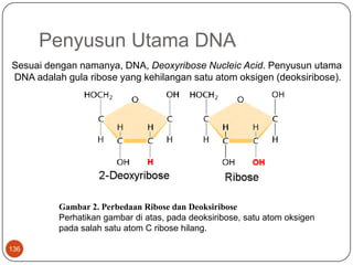
PenyusunUtama DNA 136 Sesuaidengannamanya, DNA, Deoxyribose Nucleic Acid. Penyusunutama  DNA adalahgula ribose yang kehilangansatu atom oksigen (deoksiribose). Gambar 2. Perbedaan Ribose danDeoksiribose Perhatikangambardiatas, padadeoksiribose, satu atom oksigen padasalahsatu atom C ribose hilang.
Nukleotida 137 Tiap pita/rantai double helix terbuatdari unit-unit berulang yang disebutnukleotida. Satunukleotidaterdiridaritigagugusfungsi; satugula ribose, triphosphate, dansatubasa nitrogen.
138 Satuhal yang perludiingatadalahposisitriphosphatedanbasa nitrogen yang terikatpadaribosa. Gugustriphosphatterikatpada atom C no 5′ dariribosa (Lihatgambardiatas). Gugustriphosphateinihanyadimilikiolehnukleotidabebas. Sedangkannukleotida yang terikatpadarantai DNA kehilanganduadarigugus phosphate ini, sehinggahanyasatu phosphate yang masihtertinggal. Ketikanukleotidabergabungmenjadi DNA, nukleotida-nukleotidatersebutdihubungkanolehikatanphosphodiester. Ikatankovalen yang terjadiantaragugus phosphate padasatunukleotida, dengangugus OH padanukleotidalainnya. Sehinggasetiaprantai DNA akanmempunyai ‘backbone’ phosphate-ribosa-phosphate-ribosa-phosphate. Dan seterusnya..
139 GambarStruktur DNA Sederhana
Basa Nitrogen Pada DNA 140 Padastruktur DNA, gularibosadangugus phosphate yang terikatadalahsama. Yang berbedahanyalahpadabasa nitrogen. Jadisebetulnyaperbedaandisebabkanolehvariasisusunandaribasa-basa nitrogen yang terdapatpadarantai DNA. Adaempatmacambasa nitrogen. Adenin, Cytosine, Guainne, dan Thymine.
141 Ketikabasa-basa nitrogen tersebutterikatdalamnukleotida, makapenamaan-pun berubah. Ingatkembalipenjelasandiawaltentangnukelotida. Nukleotidaterdiridarigugustriphosphatedansatubasa nitrogen yang terikatpadasatumolekul ribose. Nah.. basa-basa nitrogen iniapabilaterikatpada ribose membentuknukleotidamakapenamaannya-pun berubah. Adeninmenjadi 2′deoxyadenosinetriphosphate, cytosinmenjadi 2′deoxycytidinetriphosphate, guainnemenjadi 2′deoxyguanosinetriphosphate, dan Thymine menjadi 2′deoxythymidinetriphosphate. Disingkatmenjadi A, C, G, dan T.
142 Perhatikanbahwaadaduapasangbasa yang mirip. A dan G sama-samamempunyaiduacincinkarbon-nitrogen, disebutgolonganpurine. Sedangkan C dan T hanyamempunyaisatucincinkarbon-nitrogen, masukgolonganpirimidin.
PenyebabBentuk DNA Double Helix 143 GambarIkatanHidrogenAntaraBasa-Basa Nitrogen
144 Interaksiikatanhidrogenantaramasing-masingbasa nitrogen menyebabkanbentukdariduarantai DNA menjadisedemikianrupa, bentukinidisebut double helix.  Interaksispesifikiniterjadiantarabasa A dengan T, dan C dengan G.  Sehinggajika double helix dibayangkansebagaisebuahtangga spiral, makaikatanbasa-basainisebagaianaktangga-nya.  Lebardari ‘anaktangga’ adalahsama, karenapasanganbasaselaluterdiridarisatuprimidindansatupurin.
145 GambarStruktur Double HeliLengkap
StrukturAsam amino 146
Transkripsi 147 Prosespengkopian DNA menjadi RNA inidinamakantranskripsi.
Translasi 148 mRNA hasiltranskripsikemudiandikeluarkanmenujusitoplasmasehinggabisadiproseslebihlanjutolehsuatuorganelsel yang bernamaribosom. Ribosomakanmembacaurutanbasa RNA danmenterjemahkannya (translate ) menjadiurutanasam amino tertentusesuaidenganresep yang dibawa mRNA. Di sinilahasam-asam amino itudirakitsesuaiurutan yang diresepkan gen (DNA) dankemudianmelipatmembentukstrukturtigadimensi yang fungsional.

More Related Content

What's hot

Molecular diagnosis of Mycobacterium tuberculosis TB - Second MBBS -
Molecular diagnosis of Mycobacterium tuberculosis TB - Second MBBS - Molecular diagnosis of Mycobacterium tuberculosis TB - Second MBBS -
Molecular diagnosis of Mycobacterium tuberculosis TB - Second MBBS - SOMESHWARAN R
 
application of pcr in agriculture
application of pcr in agricultureapplication of pcr in agriculture
application of pcr in agricultureharshitajiancdfst
 
Nested PCR,Fusion primers, Integrated PCR
Nested PCR,Fusion primers, Integrated PCRNested PCR,Fusion primers, Integrated PCR
Nested PCR,Fusion primers, Integrated PCRSani Jwel
 
Role of pcr in diagnostics
Role of pcr in diagnosticsRole of pcr in diagnostics
Role of pcr in diagnosticsTariq Mahmood
 
2011 course on Molecular Diagnostic Automation - Part 2 - Amplification
2011 course on Molecular Diagnostic Automation - Part 2 - Amplification2011 course on Molecular Diagnostic Automation - Part 2 - Amplification
2011 course on Molecular Diagnostic Automation - Part 2 - AmplificationPatrick Merel
 
Molecular techniques for the study and diagnosis of parasite infection
Molecular techniques for the study and diagnosis of parasite infectionMolecular techniques for the study and diagnosis of parasite infection
Molecular techniques for the study and diagnosis of parasite infectionKhandkar Shaharina Hossain
 
Master Thesis Anja Sander
Master Thesis Anja SanderMaster Thesis Anja Sander
Master Thesis Anja SanderWishofnight13
 
Biomarker for genotoxicity 2013
Biomarker for genotoxicity 2013Biomarker for genotoxicity 2013
Biomarker for genotoxicity 2013Elsa von Licy
 
Mycobacterium molecular diagnosis
Mycobacterium molecular diagnosisMycobacterium molecular diagnosis
Mycobacterium molecular diagnosisBayan Nusairat
 
Amplificationtechniquesinmicrobiology 090609205257-phpapp02
Amplificationtechniquesinmicrobiology 090609205257-phpapp02Amplificationtechniquesinmicrobiology 090609205257-phpapp02
Amplificationtechniquesinmicrobiology 090609205257-phpapp02minhdaovan
 
Barrett rosa meza-acevedo & lingou
Barrett rosa meza-acevedo & lingouBarrett rosa meza-acevedo & lingou
Barrett rosa meza-acevedo & lingouMills Cbst
 
16. investigation of infection
16. investigation of infection16. investigation of infection
16. investigation of infectionAhmad Hamadi
 
International Proficiency Study of a Consensus L1 PCR Assay for the Detection...
International Proficiency Study of a Consensus L1 PCR Assay for the Detection...International Proficiency Study of a Consensus L1 PCR Assay for the Detection...
International Proficiency Study of a Consensus L1 PCR Assay for the Detection...Alberto Cuadrado
 
PCR techenology in fisheries
PCR techenology in fisheriesPCR techenology in fisheries
PCR techenology in fisheriesAshish sahu
 

What's hot (20)

Molecular diagnosis of Mycobacterium tuberculosis TB - Second MBBS -
Molecular diagnosis of Mycobacterium tuberculosis TB - Second MBBS - Molecular diagnosis of Mycobacterium tuberculosis TB - Second MBBS -
Molecular diagnosis of Mycobacterium tuberculosis TB - Second MBBS -
 
PCR
PCRPCR
PCR
 
application of pcr in agriculture
application of pcr in agricultureapplication of pcr in agriculture
application of pcr in agriculture
 
Nested PCR,Fusion primers, Integrated PCR
Nested PCR,Fusion primers, Integrated PCRNested PCR,Fusion primers, Integrated PCR
Nested PCR,Fusion primers, Integrated PCR
 
Role of pcr in diagnostics
Role of pcr in diagnosticsRole of pcr in diagnostics
Role of pcr in diagnostics
 
Types of PCR
Types of PCRTypes of PCR
Types of PCR
 
2011 course on Molecular Diagnostic Automation - Part 2 - Amplification
2011 course on Molecular Diagnostic Automation - Part 2 - Amplification2011 course on Molecular Diagnostic Automation - Part 2 - Amplification
2011 course on Molecular Diagnostic Automation - Part 2 - Amplification
 
Molecular techniques for the study and diagnosis of parasite infection
Molecular techniques for the study and diagnosis of parasite infectionMolecular techniques for the study and diagnosis of parasite infection
Molecular techniques for the study and diagnosis of parasite infection
 
Master Thesis Anja Sander
Master Thesis Anja SanderMaster Thesis Anja Sander
Master Thesis Anja Sander
 
Biomarker for genotoxicity 2013
Biomarker for genotoxicity 2013Biomarker for genotoxicity 2013
Biomarker for genotoxicity 2013
 
Abbas Morovvati
Abbas MorovvatiAbbas Morovvati
Abbas Morovvati
 
Mycobacterium molecular diagnosis
Mycobacterium molecular diagnosisMycobacterium molecular diagnosis
Mycobacterium molecular diagnosis
 
Amplificationtechniquesinmicrobiology 090609205257-phpapp02
Amplificationtechniquesinmicrobiology 090609205257-phpapp02Amplificationtechniquesinmicrobiology 090609205257-phpapp02
Amplificationtechniquesinmicrobiology 090609205257-phpapp02
 
Barrett rosa meza-acevedo & lingou
Barrett rosa meza-acevedo & lingouBarrett rosa meza-acevedo & lingou
Barrett rosa meza-acevedo & lingou
 
Seminar rt
Seminar rtSeminar rt
Seminar rt
 
16. investigation of infection
16. investigation of infection16. investigation of infection
16. investigation of infection
 
PCR AND ITS TYPE
PCR AND ITS TYPEPCR AND ITS TYPE
PCR AND ITS TYPE
 
International Proficiency Study of a Consensus L1 PCR Assay for the Detection...
International Proficiency Study of a Consensus L1 PCR Assay for the Detection...International Proficiency Study of a Consensus L1 PCR Assay for the Detection...
International Proficiency Study of a Consensus L1 PCR Assay for the Detection...
 
Plateforme de protéomique et d'analyse des protéines par spectrométrie de mas...
Plateforme de protéomique et d'analyse des protéines par spectrométrie de mas...Plateforme de protéomique et d'analyse des protéines par spectrométrie de mas...
Plateforme de protéomique et d'analyse des protéines par spectrométrie de mas...
 
PCR techenology in fisheries
PCR techenology in fisheriesPCR techenology in fisheries
PCR techenology in fisheries
 

Viewers also liked

Viewers also liked (15)

Deteksi mikobakterium menggunakan manual mgit dan bactectm mgittm 960 system
Deteksi mikobakterium menggunakan manual mgit dan bactectm mgittm 960 systemDeteksi mikobakterium menggunakan manual mgit dan bactectm mgittm 960 system
Deteksi mikobakterium menggunakan manual mgit dan bactectm mgittm 960 system
 
Pseudomonas mahadippt
Pseudomonas mahadipptPseudomonas mahadippt
Pseudomonas mahadippt
 
Pseudomonas
PseudomonasPseudomonas
Pseudomonas
 
Pseudomonas aeruginosa an epitome of a drug resistance in bacteria
Pseudomonas aeruginosa an epitome of a drug resistance in bacteriaPseudomonas aeruginosa an epitome of a drug resistance in bacteria
Pseudomonas aeruginosa an epitome of a drug resistance in bacteria
 
Pseudomonas aeruginosa
Pseudomonas aeruginosaPseudomonas aeruginosa
Pseudomonas aeruginosa
 
Mycobacteriology
MycobacteriologyMycobacteriology
Mycobacteriology
 
Pseudomonas fi
Pseudomonas fiPseudomonas fi
Pseudomonas fi
 
Vibrio campylobacter helicobacter_pseudomonas
Vibrio campylobacter helicobacter_pseudomonasVibrio campylobacter helicobacter_pseudomonas
Vibrio campylobacter helicobacter_pseudomonas
 
Pseudomona aeruginosa
Pseudomona aeruginosaPseudomona aeruginosa
Pseudomona aeruginosa
 
Pseudomonas aeruginosa mini-presentation
Pseudomonas aeruginosa mini-presentationPseudomonas aeruginosa mini-presentation
Pseudomonas aeruginosa mini-presentation
 
Pseudomonas
PseudomonasPseudomonas
Pseudomonas
 
Pseudomonas
PseudomonasPseudomonas
Pseudomonas
 
Pseudomona Aeruginosa
Pseudomona AeruginosaPseudomona Aeruginosa
Pseudomona Aeruginosa
 
Pseudomonas
PseudomonasPseudomonas
Pseudomonas
 
Pseudomonas
PseudomonasPseudomonas
Pseudomonas
 

Similar to J.pi5

Polymerase Chain Reaction (PCR) and The Application.pptx
Polymerase Chain Reaction (PCR) and The Application.pptxPolymerase Chain Reaction (PCR) and The Application.pptx
Polymerase Chain Reaction (PCR) and The Application.pptxrenanda8
 
PCR.pptx
PCR.pptxPCR.pptx
PCR.pptxhaimn
 
Tibaru14
Tibaru14Tibaru14
Tibaru14andreei
 
Univ bacteriav PCR Primer
Univ bacteriav PCR PrimerUniv bacteriav PCR Primer
Univ bacteriav PCR PrimerZulkifli As
 
Molecular Environmental Microbiology.ppt
Molecular Environmental Microbiology.pptMolecular Environmental Microbiology.ppt
Molecular Environmental Microbiology.pptHuma Hameed
 
Diagnosis- Detection of bacterial infection using methods other than (2)
Diagnosis- Detection of bacterial infection using methods other than (2)Diagnosis- Detection of bacterial infection using methods other than (2)
Diagnosis- Detection of bacterial infection using methods other than (2)siham moubayed
 
Ivan Lafayette - QSI Symposium - Poster - Final version
Ivan Lafayette - QSI Symposium - Poster - Final versionIvan Lafayette - QSI Symposium - Poster - Final version
Ivan Lafayette - QSI Symposium - Poster - Final versionIvan Lafayette
 
POLYMERASE CHAIN REACTION
POLYMERASE CHAIN REACTIONPOLYMERASE CHAIN REACTION
POLYMERASE CHAIN REACTIONDr Qureshi
 
Molecular Diagnostics.pptx
Molecular Diagnostics.pptxMolecular Diagnostics.pptx
Molecular Diagnostics.pptxAlisha Shaikh
 
Nucleic acid diagnostic and removal technics from biopharmaceuticals
Nucleic acid diagnostic and removal technics from biopharmaceuticalsNucleic acid diagnostic and removal technics from biopharmaceuticals
Nucleic acid diagnostic and removal technics from biopharmaceuticalsElahehEntezarmahdi
 
Report on molecular biology techniques
Report on molecular biology techniquesReport on molecular biology techniques
Report on molecular biology techniquesraghavworah
 
Polymerase Chain Reaction
Polymerase Chain ReactionPolymerase Chain Reaction
Polymerase Chain ReactionChethanchunkey
 
Molecular_bilogy_lab_report_2
Molecular_bilogy_lab_report_2Molecular_bilogy_lab_report_2
Molecular_bilogy_lab_report_2Elijah Willie
 
DNA Vaccine + Nanoparticles
DNA Vaccine + NanoparticlesDNA Vaccine + Nanoparticles
DNA Vaccine + NanoparticlesHamid Salari
 

Similar to J.pi5 (20)

PCR
PCRPCR
PCR
 
Polymerase Chain Reaction (PCR) and The Application.pptx
Polymerase Chain Reaction (PCR) and The Application.pptxPolymerase Chain Reaction (PCR) and The Application.pptx
Polymerase Chain Reaction (PCR) and The Application.pptx
 
PCR.pptx
PCR.pptxPCR.pptx
PCR.pptx
 
Tibaru14
Tibaru14Tibaru14
Tibaru14
 
Univ bacteriav PCR Primer
Univ bacteriav PCR PrimerUniv bacteriav PCR Primer
Univ bacteriav PCR Primer
 
Tuberculosis PCR
Tuberculosis PCRTuberculosis PCR
Tuberculosis PCR
 
Molecular Environmental Microbiology.ppt
Molecular Environmental Microbiology.pptMolecular Environmental Microbiology.ppt
Molecular Environmental Microbiology.ppt
 
Diagnosis- Detection of bacterial infection using methods other than (2)
Diagnosis- Detection of bacterial infection using methods other than (2)Diagnosis- Detection of bacterial infection using methods other than (2)
Diagnosis- Detection of bacterial infection using methods other than (2)
 
CHAGAS DISEASE
CHAGAS DISEASECHAGAS DISEASE
CHAGAS DISEASE
 
PCR.docx
PCR.docxPCR.docx
PCR.docx
 
Ivan Lafayette - QSI Symposium - Poster - Final version
Ivan Lafayette - QSI Symposium - Poster - Final versionIvan Lafayette - QSI Symposium - Poster - Final version
Ivan Lafayette - QSI Symposium - Poster - Final version
 
POLYMERASE CHAIN REACTION
POLYMERASE CHAIN REACTIONPOLYMERASE CHAIN REACTION
POLYMERASE CHAIN REACTION
 
Flyer Cleanpid Easy Purification Kit
Flyer Cleanpid Easy Purification KitFlyer Cleanpid Easy Purification Kit
Flyer Cleanpid Easy Purification Kit
 
IJAA
IJAAIJAA
IJAA
 
Molecular Diagnostics.pptx
Molecular Diagnostics.pptxMolecular Diagnostics.pptx
Molecular Diagnostics.pptx
 
Nucleic acid diagnostic and removal technics from biopharmaceuticals
Nucleic acid diagnostic and removal technics from biopharmaceuticalsNucleic acid diagnostic and removal technics from biopharmaceuticals
Nucleic acid diagnostic and removal technics from biopharmaceuticals
 
Report on molecular biology techniques
Report on molecular biology techniquesReport on molecular biology techniques
Report on molecular biology techniques
 
Polymerase Chain Reaction
Polymerase Chain ReactionPolymerase Chain Reaction
Polymerase Chain Reaction
 
Molecular_bilogy_lab_report_2
Molecular_bilogy_lab_report_2Molecular_bilogy_lab_report_2
Molecular_bilogy_lab_report_2
 
DNA Vaccine + Nanoparticles
DNA Vaccine + NanoparticlesDNA Vaccine + Nanoparticles
DNA Vaccine + Nanoparticles
 

More from andreei

Tibaru18
Tibaru18Tibaru18
Tibaru18andreei
 
Tibaru17
Tibaru17Tibaru17
Tibaru17andreei
 
Tibaru16
Tibaru16Tibaru16
Tibaru16andreei
 
Tibaru15
Tibaru15Tibaru15
Tibaru15andreei
 
Tibaru13
Tibaru13Tibaru13
Tibaru13andreei
 
Tibaru12
Tibaru12Tibaru12
Tibaru12andreei
 
Tibaru11
Tibaru11Tibaru11
Tibaru11andreei
 
Tibaru11
Tibaru11Tibaru11
Tibaru11andreei
 
Tibaru10
Tibaru10Tibaru10
Tibaru10andreei
 
Refhemabaru8
Refhemabaru8Refhemabaru8
Refhemabaru8andreei
 
Refhemabaru7
Refhemabaru7Refhemabaru7
Refhemabaru7andreei
 
Refhemabaru6
Refhemabaru6Refhemabaru6
Refhemabaru6andreei
 
Refhemabaru5
Refhemabaru5Refhemabaru5
Refhemabaru5andreei
 
Tutor hematologi
Tutor hematologiTutor hematologi
Tutor hematologiandreei
 

More from andreei (20)

Tibaru18
Tibaru18Tibaru18
Tibaru18
 
Tibaru17
Tibaru17Tibaru17
Tibaru17
 
Tibaru16
Tibaru16Tibaru16
Tibaru16
 
Tibaru15
Tibaru15Tibaru15
Tibaru15
 
Tibaru13
Tibaru13Tibaru13
Tibaru13
 
Tibaru12
Tibaru12Tibaru12
Tibaru12
 
Tibaru11
Tibaru11Tibaru11
Tibaru11
 
Tibaru9
Tibaru9Tibaru9
Tibaru9
 
Tibaru11
Tibaru11Tibaru11
Tibaru11
 
Tibaru10
Tibaru10Tibaru10
Tibaru10
 
Tibaru8
Tibaru8Tibaru8
Tibaru8
 
Tibaru7
Tibaru7Tibaru7
Tibaru7
 
Refhemabaru8
Refhemabaru8Refhemabaru8
Refhemabaru8
 
Refhemabaru7
Refhemabaru7Refhemabaru7
Refhemabaru7
 
Refhemabaru6
Refhemabaru6Refhemabaru6
Refhemabaru6
 
Refhemabaru5
Refhemabaru5Refhemabaru5
Refhemabaru5
 
12
1212
12
 
12
1212
12
 
11
1111
11
 
Tutor hematologi
Tutor hematologiTutor hematologi
Tutor hematologi
 

Recently uploaded

Automating Business Process via MuleSoft Composer | Bangalore MuleSoft Meetup...
Automating Business Process via MuleSoft Composer | Bangalore MuleSoft Meetup...Automating Business Process via MuleSoft Composer | Bangalore MuleSoft Meetup...
Automating Business Process via MuleSoft Composer | Bangalore MuleSoft Meetup...shyamraj55
 
Transcript: New from BookNet Canada for 2024: BNC BiblioShare - Tech Forum 2024
Transcript: New from BookNet Canada for 2024: BNC BiblioShare - Tech Forum 2024Transcript: New from BookNet Canada for 2024: BNC BiblioShare - Tech Forum 2024
Transcript: New from BookNet Canada for 2024: BNC BiblioShare - Tech Forum 2024BookNet Canada
 
CloudStudio User manual (basic edition):
CloudStudio User manual (basic edition):CloudStudio User manual (basic edition):
CloudStudio User manual (basic edition):comworks
 
Build your next Gen AI Breakthrough - April 2024
Build your next Gen AI Breakthrough - April 2024Build your next Gen AI Breakthrough - April 2024
Build your next Gen AI Breakthrough - April 2024Neo4j
 
Snow Chain-Integrated Tire for a Safe Drive on Winter Roads
Snow Chain-Integrated Tire for a Safe Drive on Winter RoadsSnow Chain-Integrated Tire for a Safe Drive on Winter Roads
Snow Chain-Integrated Tire for a Safe Drive on Winter RoadsHyundai Motor Group
 
APIForce Zurich 5 April Automation LPDG
APIForce Zurich 5 April  Automation LPDGAPIForce Zurich 5 April  Automation LPDG
APIForce Zurich 5 April Automation LPDGMarianaLemus7
 
Are Multi-Cloud and Serverless Good or Bad?
Are Multi-Cloud and Serverless Good or Bad?Are Multi-Cloud and Serverless Good or Bad?
Are Multi-Cloud and Serverless Good or Bad?Mattias Andersson
 
"LLMs for Python Engineers: Advanced Data Analysis and Semantic Kernel",Oleks...
"LLMs for Python Engineers: Advanced Data Analysis and Semantic Kernel",Oleks..."LLMs for Python Engineers: Advanced Data Analysis and Semantic Kernel",Oleks...
"LLMs for Python Engineers: Advanced Data Analysis and Semantic Kernel",Oleks...Fwdays
 
Making_way_through_DLL_hollowing_inspite_of_CFG_by_Debjeet Banerjee.pptx
Making_way_through_DLL_hollowing_inspite_of_CFG_by_Debjeet Banerjee.pptxMaking_way_through_DLL_hollowing_inspite_of_CFG_by_Debjeet Banerjee.pptx
Making_way_through_DLL_hollowing_inspite_of_CFG_by_Debjeet Banerjee.pptxnull - The Open Security Community
 
Connect Wave/ connectwave Pitch Deck Presentation
Connect Wave/ connectwave Pitch Deck PresentationConnect Wave/ connectwave Pitch Deck Presentation
Connect Wave/ connectwave Pitch Deck PresentationSlibray Presentation
 
Understanding the Laravel MVC Architecture
Understanding the Laravel MVC ArchitectureUnderstanding the Laravel MVC Architecture
Understanding the Laravel MVC ArchitecturePixlogix Infotech
 
Integration and Automation in Practice: CI/CD in Mule Integration and Automat...
Integration and Automation in Practice: CI/CD in Mule Integration and Automat...Integration and Automation in Practice: CI/CD in Mule Integration and Automat...
Integration and Automation in Practice: CI/CD in Mule Integration and Automat...Patryk Bandurski
 
Beyond Boundaries: Leveraging No-Code Solutions for Industry Innovation
Beyond Boundaries: Leveraging No-Code Solutions for Industry InnovationBeyond Boundaries: Leveraging No-Code Solutions for Industry Innovation
Beyond Boundaries: Leveraging No-Code Solutions for Industry InnovationSafe Software
 
Bun (KitWorks Team Study 노별마루 발표 2024.4.22)
Bun (KitWorks Team Study 노별마루 발표 2024.4.22)Bun (KitWorks Team Study 노별마루 발표 2024.4.22)
Bun (KitWorks Team Study 노별마루 발표 2024.4.22)Wonjun Hwang
 
My INSURER PTE LTD - Insurtech Innovation Award 2024
My INSURER PTE LTD - Insurtech Innovation Award 2024My INSURER PTE LTD - Insurtech Innovation Award 2024
My INSURER PTE LTD - Insurtech Innovation Award 2024The Digital Insurer
 
Pigging Solutions Piggable Sweeping Elbows
Pigging Solutions Piggable Sweeping ElbowsPigging Solutions Piggable Sweeping Elbows
Pigging Solutions Piggable Sweeping ElbowsPigging Solutions
 
Science&tech:THE INFORMATION AGE STS.pdf
Science&tech:THE INFORMATION AGE STS.pdfScience&tech:THE INFORMATION AGE STS.pdf
Science&tech:THE INFORMATION AGE STS.pdfjimielynbastida
 
Benefits Of Flutter Compared To Other Frameworks
Benefits Of Flutter Compared To Other FrameworksBenefits Of Flutter Compared To Other Frameworks
Benefits Of Flutter Compared To Other FrameworksSoftradix Technologies
 

Recently uploaded (20)

Automating Business Process via MuleSoft Composer | Bangalore MuleSoft Meetup...
Automating Business Process via MuleSoft Composer | Bangalore MuleSoft Meetup...Automating Business Process via MuleSoft Composer | Bangalore MuleSoft Meetup...
Automating Business Process via MuleSoft Composer | Bangalore MuleSoft Meetup...
 
Transcript: New from BookNet Canada for 2024: BNC BiblioShare - Tech Forum 2024
Transcript: New from BookNet Canada for 2024: BNC BiblioShare - Tech Forum 2024Transcript: New from BookNet Canada for 2024: BNC BiblioShare - Tech Forum 2024
Transcript: New from BookNet Canada for 2024: BNC BiblioShare - Tech Forum 2024
 
CloudStudio User manual (basic edition):
CloudStudio User manual (basic edition):CloudStudio User manual (basic edition):
CloudStudio User manual (basic edition):
 
Build your next Gen AI Breakthrough - April 2024
Build your next Gen AI Breakthrough - April 2024Build your next Gen AI Breakthrough - April 2024
Build your next Gen AI Breakthrough - April 2024
 
Snow Chain-Integrated Tire for a Safe Drive on Winter Roads
Snow Chain-Integrated Tire for a Safe Drive on Winter RoadsSnow Chain-Integrated Tire for a Safe Drive on Winter Roads
Snow Chain-Integrated Tire for a Safe Drive on Winter Roads
 
APIForce Zurich 5 April Automation LPDG
APIForce Zurich 5 April  Automation LPDGAPIForce Zurich 5 April  Automation LPDG
APIForce Zurich 5 April Automation LPDG
 
DMCC Future of Trade Web3 - Special Edition
DMCC Future of Trade Web3 - Special EditionDMCC Future of Trade Web3 - Special Edition
DMCC Future of Trade Web3 - Special Edition
 
Are Multi-Cloud and Serverless Good or Bad?
Are Multi-Cloud and Serverless Good or Bad?Are Multi-Cloud and Serverless Good or Bad?
Are Multi-Cloud and Serverless Good or Bad?
 
"LLMs for Python Engineers: Advanced Data Analysis and Semantic Kernel",Oleks...
"LLMs for Python Engineers: Advanced Data Analysis and Semantic Kernel",Oleks..."LLMs for Python Engineers: Advanced Data Analysis and Semantic Kernel",Oleks...
"LLMs for Python Engineers: Advanced Data Analysis and Semantic Kernel",Oleks...
 
Making_way_through_DLL_hollowing_inspite_of_CFG_by_Debjeet Banerjee.pptx
Making_way_through_DLL_hollowing_inspite_of_CFG_by_Debjeet Banerjee.pptxMaking_way_through_DLL_hollowing_inspite_of_CFG_by_Debjeet Banerjee.pptx
Making_way_through_DLL_hollowing_inspite_of_CFG_by_Debjeet Banerjee.pptx
 
Connect Wave/ connectwave Pitch Deck Presentation
Connect Wave/ connectwave Pitch Deck PresentationConnect Wave/ connectwave Pitch Deck Presentation
Connect Wave/ connectwave Pitch Deck Presentation
 
Understanding the Laravel MVC Architecture
Understanding the Laravel MVC ArchitectureUnderstanding the Laravel MVC Architecture
Understanding the Laravel MVC Architecture
 
Integration and Automation in Practice: CI/CD in Mule Integration and Automat...
Integration and Automation in Practice: CI/CD in Mule Integration and Automat...Integration and Automation in Practice: CI/CD in Mule Integration and Automat...
Integration and Automation in Practice: CI/CD in Mule Integration and Automat...
 
Beyond Boundaries: Leveraging No-Code Solutions for Industry Innovation
Beyond Boundaries: Leveraging No-Code Solutions for Industry InnovationBeyond Boundaries: Leveraging No-Code Solutions for Industry Innovation
Beyond Boundaries: Leveraging No-Code Solutions for Industry Innovation
 
Bun (KitWorks Team Study 노별마루 발표 2024.4.22)
Bun (KitWorks Team Study 노별마루 발표 2024.4.22)Bun (KitWorks Team Study 노별마루 발표 2024.4.22)
Bun (KitWorks Team Study 노별마루 발표 2024.4.22)
 
My INSURER PTE LTD - Insurtech Innovation Award 2024
My INSURER PTE LTD - Insurtech Innovation Award 2024My INSURER PTE LTD - Insurtech Innovation Award 2024
My INSURER PTE LTD - Insurtech Innovation Award 2024
 
Pigging Solutions Piggable Sweeping Elbows
Pigging Solutions Piggable Sweeping ElbowsPigging Solutions Piggable Sweeping Elbows
Pigging Solutions Piggable Sweeping Elbows
 
Science&tech:THE INFORMATION AGE STS.pdf
Science&tech:THE INFORMATION AGE STS.pdfScience&tech:THE INFORMATION AGE STS.pdf
Science&tech:THE INFORMATION AGE STS.pdf
 
Benefits Of Flutter Compared To Other Frameworks
Benefits Of Flutter Compared To Other FrameworksBenefits Of Flutter Compared To Other Frameworks
Benefits Of Flutter Compared To Other Frameworks
 
Vulnerability_Management_GRC_by Sohang Sengupta.pptx
Vulnerability_Management_GRC_by Sohang Sengupta.pptxVulnerability_Management_GRC_by Sohang Sengupta.pptx
Vulnerability_Management_GRC_by Sohang Sengupta.pptx
 

J.pi5

  • 1. PowerPoint Template JurnalDivisiPenyakitInfeksidanMikrobiologi I Rapid detection of Pseudomonas aeruginosa from positive blood cultures by quantitative PCR Annals of Clinical Microbiology and Antimicrobials 2010, 9:21 Vincent Cattoir, Audrey Gilibert, Jeanne-Marie Le Glaunec, Nathalie Launay, Lilia Bait-Mérabet,Patrick Legrand YUNI SETYOWATININGSIH /ARYATI Jumat, 1 April 2011
  • 2. 2 Pseudomonas aeruginosa 6,8 % Bakteremiakarenabakteribatang Gram negatif PENDAHULUAN Patogenoportunisutamainfeksinosokomial, terutamadi unit perawatanintensif Di Amerika patogenterbanyakke 7 darialirandarah,
  • 3. 3 Pseudomonas aeruginosa Lanjutan (PENDAHULUAN) Bakteremiasecaraklinistidakdapatdibedakandariinfeksikarenabakteri Gram negatiflainnya Terapiantibiotika awal yang tidaktepatmengakibatkandampak negatifhebat.
  • 4. Metodeidentifikasifenotipmembutuhkanwaktu lama dankebanyakanpunyapembatasaninheren Beberapametodeberbasis PCR telahditelitiuntukmengidentifikasiP.aeruginosa, terutamapadasampel yang diambildarisistimrespiratoriuspasien fibrosis kistik. Teknikmolekulertelahditelitisebagaimetode yang cepatdandapatdiandalkanuntukidentifikasipatogenbakterial. Lanjutan (PENDAHULUAN) 4
  • 5. 5 Lanjutan (PENDAHULUAN) Target MolekulerqPCR ecfX 16 S rRNA algD gyrB Pseudomonas aeruginosa oprI toxA oprL Adahasilpositifpalsu Adahasilnegatifpalsu
  • 6. Kelemahanmetode PCR  masihharusditeliti target gen yang sesuaikarenatelahdilaporkansebelumnya target gen oprIdanoprLtidak 100% spesifikuntukP.aeruginosa. Metode PCR dari BC positifcepat, akuratsampai level spesiesdilanjutkanpengobatanempiris yang sesuai 6 Lanjutan (PENDAHULUAN) KelemahanteknikidentifikasiP.aeruginosakonvensionaldari BC positifbutuhwaktu minimal 24 jam
  • 7. 7
  • 8. Spesimen 1 IdentifikasiFenotip 2 Ekstraksi DNA 3 PCR kuantitatif 4 Batas deteksi 5 METODE 8
  • 9. 9 Diambildari 100 hasil BC positifdari 100 pasienrawatinap METODE Waktupenelitian : Agustus 2008 sampaiJuni 2009 SPESIMEN DarahbotolBacT/ALERT aerobdananaerob DiinkubasidalamalatBacT/ALERT Pengecatan Gram : Batang Gram Negatif 87 aerob + 13 anaerob + Konsentrasibakteri KulturKuantitatif Identifikasifenotipdenganmorfologikoloni, tesoksidasedanhasil API 20 E Diinokulasisecaraaerobdananaerob, 37oC, 24 - 48 jam pada TSA, Drigalski Agar, Blood Agar, Chocolate Agar Diinokulasi SecaraAnaerob Diinokulasi SecaraAerob
  • 10. Ekstraksi DNA denganRapid Boiling Procedure 10 0,5 ml darah Sentrifus 850 g 2’ Supernatandiambil 1% Triton X-100 0,5% Tween 20 10 mMTris-HCl 1 mM EDTA Inkubasi pada 100oC 10 ‘ Hasil : Template DNA Waktu : 20 menit Sentrifus 11500 g 5’ Pellet diambil, diresuspen 200 µL buferpelisis Supernatandiambil Sentrifus 850 g 2’
  • 11. 11 PCR KUANTITATIF CAMPURAN AMPLIFIKASI : 2 µl 10x LightCyclerFastStart DNA Master Hybridization (Roche) 2 mM MgCl2 0,5 µM masing-masing primer 0,2 µM masing-masingprobe 5 µl template DNA volume akhir 20 µl. Primeroligonukleotida Amplifikasi Kontrolnegatif: SuspensiTris-EDTA KontrolPositif: Ekstrak DNA P. aeruginosaATCC 27853 Gen ecfX fragmen152 bp Probe berlabelfluoresen Deteksi
  • 12. 12 Tabel 1 PrimeroligonukleotidadanProbehibridisasiLightCycler yang digunakandalampemeriksaanPCR a [FAM], fluorescein; [LC705], LightCycler™-Red 705; Ph, 3’-phosphate. b Extracytoplasmic function gene(GenBank accession no. DQ996551).
  • 13. 13 Lanjutan (PENDAHULUAN) LOKASI ecfX ecfX-HP2 154-172 ecfX-F 46-62 ecfX-HP1 135-151 ecfX-R 179-197
  • 14. Tahap1 Tahap 2 Tahap3 PCR KUANTITATIF SatuSiklus PCR Denaturasiawalpada suhu 95 °C selama 10 menit SEQUENCING/ ELONGATION 72 °C, 20” DENATURASI 95 °C, 10” ANNEALING 50 °C, 10” 14
  • 15. PCR KUANTITATIF 15 DNA teramplifikasidiukurdengandeteksifluoresensipada705 nm WaktuProsesqPCR : 1,5 jam Padasaat yang bersamaan, dilakukankontrolinhibisi PCR untuksemuasampeldenganmenggunakantabungreaksikedua yang berisi 100 ngekstrak DNA darikontrolpositif.
  • 16. BATAS DETEKSI Ligasi ecfX Plasmid pRT-ecfX Media berisi 30 µg/mL Kanamycin Escherichia Coli Plasmid DNA diekstraksi,dimurnikan, ditambahelution buffer dandi-sequencing untukmenentukanadanyaecfX 16
  • 17. BATAS DETEKSI Konsentrasi DNA  Spektrofotometer Bahan Plasmid DNA rujukandiencerkan serial  plasmid genome equivalen10 sampai 1010 AnalisisKurvaStandar. AnalisiskuantitatifdilakukandenganLightCycler software v 3.5 (Roche) Rasio signal diukurpada 705 nm/ signal pada 530 nm untukmenghitungnilaiCrossing Point 17
  • 18. KurvaamplifikasiqPCRdariplasmid DNA reference material dengan 11 konsentrasi DNA Eksternal(dari 1011sampai 10 copies/ml) 18
  • 20. HASIL 20 33 strain P. aeruginosa 53 strain Enterobacteriaceae 100 BC 9 strain Stenotrophomonasmaltophilia 2 spesies gram negatif lain 3 BC adalahpolimikrobial Dijumpaiinhibisi PCR padadua (2 %) preparasi DNA
  • 21. Tabel 2 HasiltesqPCRecfX Pseudomonas aeruginosadari 98 botol BC positif KulturdarahPolimikrobial . Didapatkan P. aeruginosasebanyak 20 CFU/ml dan K. pneumoniae 108 CFU/ml darikulturkuantitatif 21
  • 22. Lanjutan HASIL 22 qPCRternyatatidakdapatmendeteksiadanyaP.aeruginosadalamkulturPolimikrobial PenelitimelakukantesqPCRlangsungdariisolatP.aeruginosa ecfXberhasildiamplifikasi KulturkuantitatifditemukanbahwakonsentrasiP.aeruginosadi BC inisangatrendah (20 CFU/ml). Batas deteksi PCR Kuantitatifpenelitianiniadalah 102 CFU/ml.
  • 23. Pada Dari 98 BC yang dapat ditafsirkan (98 %), assay qPCR menunjukkan sensitivitas 97%, spesifisitas 100 %, PPV 98,5 % dan NPV 100%. 23
  • 24. Lanjutan HASIL Sensitivitas 97% Spesifisitas 100 % 98 BC NPV 98.5 % 24 PPV 100 %
  • 25. DISKUSI DenganteknikFISH (disetujuioleh FDA) yang secaralangsungdigunakanpada BC positif, metodeqPCRterbarumenunjukkanperforma yang serupanamunlebihcepat (1,5 jam vs 2,5 jam) Inhibitor teknikqPCRyang potensialdarispesimen BC positifadalahcharcoal dan hemoglobin 25
  • 26. Lanjutan DISKUSI Informasiakuratdantepatwaktu yang diberikanolehassayqPCRiniakanmembantudoktermengidentifikasibakteremiaP. aeruginosadanmemberiterapilebihcepat 18-24 jam. SulituntukmemprediksikepekaanantibiotikapadaP. aeruginosadenganteknikmolekuler. P. aeruginosaMultidrug resistance  terapiantibiotikadikombinasi 26
  • 27. 27 PenelitimengembangkanteknikqPCR  identifikasicepat (< 1,5 jam) dengansensitivitasdanspesifisitastinggiuntukidentifikasiPseudomonas aeruginosa dari BC positif. Penelitianlanjutanmengevaluasi dampakklinisdarimetodeqPCRbaruinidibandingkandenganmetode konvensional. 2 1 KESIMPULAN
  • 28. www.themegallery.com Thank You ! www.themegallery.com
  • 29. KurvaamplifikasiqPCRdariplasmid DNA reference material dengan 11 konsentrasi DNA Eksternal(dari 1011sampai 10 copies/ml) 29 Treshold CT CT CT
  • 31. Inhibitor PCR Inhibitor PCR: Darah, jaringan, fabrics, soil Sumber lain : material danreagen yang kontakdengansampelselamaproses PCR misal : KelebihanKCl, NaCl, Ion detergen (Sodium Deoxycholate), Isopropanolol, Phenol Cara mengatasi : - inhibitor PCR daridarahdanjaringantidakbisadihindari - inhibitor PCR misal saliva dengan media transfer swab pemurnian DNA dengancaraekstraksi  memurnikanTemplate DNA 31
  • 32. 32
  • 33. PCR CONTAMINATION Contamination is defined as the unwanted presence of DNA (or RNA) amplicons At present, no experimental means exist which can directly detect the ultra-trace amounts of this template (e.g., picogram amounts or less). The best way to monitor is to use reagent-only blanks The presence of contamination is then signalled by the appearance of the test amplicon in the reagent-only blank tubes. 33
  • 34. Triton X-100 Triton X-100 (C14H22O(C2H4O)n) is a nonionicsurfactant which has a hydrophilicpolyethylene oxide group (on average it has 9.5 ethylene oxide units) and a hydrocarbonlipophilic or hydrophobic group. 34
  • 35. Tween 20 Polysorbate 20 (a common commercial brand name is Tween 20) is a polysorbatesurfactant whose stability and relative non-toxicity allows it to be used as a detergent and emulsifier in a number of domestic, scientific, and pharmacological applications. It is a polyoxyethylene derivative of sorbitanmonolaurate, and is distinguished from the other members in the polysorbate range by the length of the polyoxyethylene chain and the fatty acid ester moiety. 35
  • 36. Tris-HCl TE buffer is a commonly used buffer solution in molecular biology, especially in procedures involving DNA or RNA. "TE" is derived from its components: Tris, a common pH buffer, and EDTA, a molecule that chelatescations like Mg2+. The purpose of TE buffer is to solubilize DNA or RNA, while protecting it from degradation. 36
  • 37. TBE TBE or Tris/Borate/EDTA, is a buffer solution containing a mixture of Tris base, boric acid and EDTA. In molecular biology, TBE and TAE buffers are often used in procedures involving nucleic acids, the most common being electrophoresis. Tris-acid solutions are effective buffers for slightly basic conditions, which keep DNA deprotonated and soluble in water. EDTA is a chelator of divalent cations, particularly of magnesium (Mg2+). As these ions are necessary co-factors for many enzymes, including contaminant nucleases, the role of the EDTA is to protect the nucleic acids against enzymatic degradation. 37
  • 38. KANAMYCIN Kanamycin is used in molecular biology as a selective agent most commonly to isolate bacteria (e.g., E. coli) which have taken up genes (e.g., of plasmids) coupled to a gene coding for kanamycin resistance (primarily Neomycin phosphotransferase II [NPT II/Neo]). Bacteria that have been transformed with a plasmid containing the kanamycin resistance gene are plated on kanamycin (50-100ug/ml) containing agar plates or are grown in media containing kanamycin (50-100ug/ml). Only the bacteria that have successfully taken up the kanamycin resistance gene become resistant and will grow under these conditions. As a powder kanamycin is white to off-white and is soluble in water (50mg/ml). 38
  • 39. Dampaknegatif Pasientidaksembuh Waktuopnamelebih lama Memicuresistensiantibiotika lain 39
  • 40. Kelemahan PCR Tidakbisamenunjukkanbakteriinihidupataumati. KulturtetapdipercayasebagaiGold Standard  karenabakteritumbuhdanhidup Tetapidarisampeldarahbiasanyahanya 20-30% yang positif  menunjukkansulitnyamencapaiGold Standard  butuhmetodealternatifuntukidentifikasi. 40
  • 41. Antimicrobial Resistance in P. aeruginosa Intrinsic resistance to most antibiotics is attributed to: Efflux pumps: Chromosomally-encoded genes (e.g. mexAB-oprM, mexXY, etc) and Low permeability of the bacterial cellular envelope Acquired resistance with development of multi-drug resistant strains by: Mutations in chromosomally-encoded genes, or Horizontal gene transfer of antibiotic resistance determinants 41
  • 42. Metodefenotip MetodekonvensionaluntukidentifikasiPseudomonas aeruginosabutuhwaktu lama Tesgula-gula : Tesoksidasepositif Mamputumbuhpada 42oC TSI slant  tidakadareaksi MH agar : Warnahijaukebiruan, merahataucoklat Produksinitrat, Gas + 42
  • 43. Metodefenotip LanjutanTesGula-gula : Arginindehidrogenase + Gelatin Liquified : variabel Lysine decarboxylase – Urea Hydrolysis : variabel Mengoksidaseglukosa & Xylosa (Lactose -, mannitolvariabel) 43
  • 44. Pseudomonas aeruginosa Scientific Classification : Kingdom : Bacteria Phylum : Proteobacteria Class : Gamma Proteobacteria Order : Pseudomonadales Family : Pseudomonadaceae Genus : Pseudomonas Species : Pseudomonas aeruginosa 44
  • 45. Synonims Bacterium aeruginosumSchroeter 1872 Bacterium aeruginosum Cohn 1872 Micrococcus pyocyaneusZopf 1884 Bacillus aeruginosus (Schroeter 1872) Trevisan 1885 Bacillus pyocyaneus (Zopf 1884) Flügge 1886 Pseudomonas pyocyanea (Zopf 1884) Migula 1895 Bacterium pyocyaneum (Zopf 1884) Lehmann and Neumann 1896 Pseudomonas polycolor Clara 1930 Pseudomonas vendrellinomennudum 1938 45
  • 46.
  • 47. This organism can achieve anaerobic growth with nitrate as a terminal electron acceptor, and, in its absence, it is also able to ferment arginine by substrate-level phosphorylation.46
  • 48. 47
  • 49. Biofilms and treatment resistance Biofilms of Pseudomonas aeruginosa can cause chronic opportunistic infections. They often cannot be treated effectively with traditional antibiotic therapy. Biofilms seem to protect these bacteria from adverse environmental factors. Pseudomonas aeruginosa can cause nosocomial infections and is considered a model organism for the study of antibiotic-resistant bacteria. 48
  • 50. DIAGNOSIS First, a Gram stain is performed, which should show Gram-negative rods with no particular arrangement. Then, if the specimen is pure, the organism is grown on MacConkey agar plate to produce colorless colonies (as it does not ferment lactose) Cetrimide agarP. aeruginosa may express the exopigmentpyocyanin, which is blue-green in color, and the colonies will appear flat, large, and oval. It also has a characteristic fruity smell. P. aeruginosa is catalase+, oxidase+, nitrase+, and lipase+. When grown on TSI medium, it has a K/K profile, meaning that the medium will not change color. Finally, serology could help, which is based on H and Oantigens. 49
  • 51. TREATMENT aminoglycosides (gentamicin, amikacin, tobramycin) quinolones (ciprofloxacin, levofloxacin, and moxifloxacin) cephalosporins (ceftazidime, cefepime, cefoperazone, cefpirome, but notcefuroxime, ceftriaxone, cefotaxime) antipseudomonalpenicillins: ureidopenicillins and carboxypenicillins (piperacillin, ticarcillin: P. aeruginosa is intrinsically resistant to all other penicillins) carbapenems (meropenem, imipenem, doripenem, but notertapenem) polymyxins (polymyxin B and colistin)[31] monobactams (aztreonam) In the rare occasions where infection is superficial and limited (for example, ear infections or nail infections), topicalgentamicin or colistin may be used. 50
  • 52. 51
  • 53. PENGGUNAAN INKUBATOR SESUAI KEBUTUHAN LINGKUNGAN UNTUK PERTUMBUHAN BAKTERI 52
  • 54. TOPLES LILIN Komponen Toplesterbuatdarikaca Tutuptoples Lilinputih yang kering 53
  • 55. ANAEROBIC JAR DENGAN GAS PACK KomponenAlat: Toplesterbuatdarimika yang dilengkapikaretmelingkardiatasnya Tutuptoplesdengan 3 lubangsaluranuntuk gas CO2, O2 dan N2 Penjepitantaratutupdantoples yang diperkuatsekrupdiatasnya Gas pack Indikatoranaerob Katalis 54
  • 57. The basics Aerobic, opportunistic pathogen Gram-negative bacillus Flagella 56
  • 58. Pathogenesis Virulence Factors (structural components) “Alginate” Adherence proteins and “pili” Lipopolysaccharide (LPS) Procyanin 57
  • 60. Structural Components Adherence to host cells mediated by pili and nonpilusadhesins. LPS (lipopolysaccharide) inhibiting antibiotic killing and suppress neutrophil and lymphocyte activity Alginate – mucoidexopolysaccharide that forms a shiny biofilm protecting from antibodies, complement, phagocytosis, and antibiotics Procyanin – impairs ciliary function, mediates tissue damage through production of oxgen radicals 59
  • 61. Pathogenesis Virulence Factors (toxins and enzymes): Exotoxin A Exoenzyme S Endotoxins Phospholipase C Elastase and Alkaline Protease 60
  • 62. Exotoxin A Similar in structure to Diptheria toxin Inhibits protein synthesis by ADP-ribosylating EF-2 (G-protein) Causes Dermatonecrosis in burn wounds, corneal damage in ocular infections, and tissue damage in chronic pulmonary infections. Also this toxin is immunosuppressive 61
  • 63. Exoenzyme S & T ADP-ribosylates G-proteins including p21 RAS interfering with host cell growth 62
  • 64. Phospholipase C Heat labile hemolysin Breaks down lipids and lecithin causing tissue destruction Stimulates inflammatory response 63
  • 65. Elastase and Alkaline Protease Destruction of elastin-containing tissues (blood vessels, lung tissue, skin), collagen, immunoglobulins, and complement factors Can produce hemorrhagic lesions (ecthyma gangrenosum) associated with disseminated infection Inactivation of interferon and TNF-Alpha 64
  • 66. Epidemiology Ubiquitous in moist environmental sites in the hospital as well as nature No seasonal incidence Can transiently colonize the respiratory and GI tract of hospitalized patients Minimal nutritional requirements and can tolerate broad temperature spectrum 65
  • 67. Clinical Disease Pulmonary Infections Burn Wound Infections and other skin and soft tissue infections (life threatening) UTI’s (especially catheterized) External Otitis (malignant OE, swimmer’s ear) Eye Infections and corneal ulceration via contaminated contact lens cleaning fluids Pseudomonal Endocarditis 66
  • 68. Pulmonary Infections Can range from asymptomatic colonization to severe necrotizing bronchopneumonia Colonization is seen in patients with cystic fibrosis, chronic lung disease, and neutropenia Mucoid strains are commonly isolated from chronic pulmonary patients and are more difficult to eradicate Predisposing conditions include previous therapy with broad spectrum abx (disrupts normal protective bacteria population and use of respiratory therapy equipment (can introduce the organism to lower airways) Mortality rate can be as high as 70% for invasive bronchopneumonia 67
  • 69. Ecthyma Gangrenosum Ecthyma gangrenosum is a well recognized cutaneous manifestation of severe, invasive infection by Pseudomonas aeruginosa that is usually seen in immunocompromised, burn patients, and other critically ill patients 68
  • 70. Antibiotic Resistance Inherently resistant to many abx Can mutate to more resistant strains during therapy Penetration of abx highly dependent on outer membrane pores which can be altered Production of B-lactamases Combination of active abx generally required for successful therapy 69
  • 71. Drigalski Agar Drigalski agar is a selective isolation and differentiation medium used to identify Enterobacteriaceae and other Gram-negative bacteria. Microorganisms that ferment lactose form yellow or yellowish-green colonies; the others produce blue, green, or bluish-green colonies. The presence of sodium deoxycholate and crystal violet inhibits the growth of Gram-positive. 70
  • 72. Chocolate Agar A non-selective, enriched growth medium. A variant of the blood agar plate. Contains red blood cells, which have been lysed by heating very slowly to 56 °C. Chocolate agar is used for growing fastidious (fussy) respiratory bacteria, such as Haemophilusinfluenzae. These bacteria need growth factors, like NAD and hematin, which are inside red blood cells; thus, a prerequisite to growth is lysis of the red blood cells. 71
  • 73. Trypticase Soy Agar The medium contains enzymatic digests of casein and soybean meal which provides amino acids and other nitrogenous substances. Dextrose is the energy source. Sodium chloride maintains the osmotic equilibrium, while dipotassium phosphate acts as buffer to maintain pH. The medium may be supplemented with blood to facilitate the growth of more fastidious bacteria or antimicrobial agents to permit the selection of various microbial groups from pure flora. 72
  • 74. Microbiology Family Pseudomonadaceae Aerobic, non-spore forming Gram negative straight or slightly curved rod (1 to 3 um in length), polar flagella Non-fermenters Catalase and oxidase positive Morphologic characteristics on lab media: Production of pigments: Soluble blue-coloured phenazine pigment called pyocyanin) Some strains produce red or black colonies due to pigments termed pyorubin and pyomelanin, respectively P. aerugnosa produces pyoverdin (diffusible yellow-green to yellow-brown pigment) which, when produced with pyocyanin gives rise to green-blue colonies on solid media Term ‘aeruginosa” stems from green-blue hue 73
  • 75. Microbiology Term Pseudo = “false”; monas = “single unit” Term ‘aeruginosa” stems from green-blue hue Pseudomonas are classified as strict aerobes but some exceptions: May use nitrate Biofilm formation 74
  • 76. Microbiology Pseudomonads classified into five rRNA homology groups: Pseudomonas (sensu stricto) Burkholderia species Comamonas, Acidovorax, and Hydrogenophaga genera Brevundimonas species Stenotrophomonas and Xanthomonas genera Genus Pseudomonas contains over 160 species but only 12 are clinically relevant 75
  • 77. Microbiology P. aeruginosa is the type species and may have highly varied morphology Typical colonies may appear to spread over the plate, lie flat with a metallic sheen and frequently produce a gelatinous or “slimy” appearance Most strains produce characteristic ‘grapelike’ or ‘corn taco-like’ odor 76
  • 78. P. aeruginosa on blood agar 77
  • 79. Non-Lactose Fermenter Lactose Fermenter MacConkey Agar 78
  • 80. P. aeruginosa Mucoid P. aeruginosa MacConkey Agar 79
  • 81. Epidemiology and Transmission Natural habitat: Temperature between 4 to 36oC (can survive up to 42oC) Found throughout nature in moist environment (hydrophilic) (e.g. sink drains, vegetables, river water, antiseptic solutions, mineral water, etc.) P. aeruginosa rarely colonizes healthy humans Normal skin does not support P. aeruginosa colonization (unlike burned skin) Acquisition is from the environment, but occasionally can be from patient-to-patient spread 80
  • 82. Range of clinical infections caused by P. aeruginosa P. aeruginosa is an opportunistic infection: Individuals with normal host defenses are not at risk for serious infection with P. aeruginosa Those at risk for serious infections include: Profoundly depressed circulating neutrophil count (e.g. cancer chemotherapy) Thermal burns Patients on mechanical ventilation Cystic fibrosis patients 81
  • 83. Range of clinical infections caused by P. aeruginosa Immunocompetent Host: Most common cause of osteochondritis of dorsum of foot following puncture wounds (running shoes) Hot tub folliculitis Swimmer’s ear Conjunctivitis in contact lens users (poor hygiene or if lenses are worn for extended periods) Other Hosts: Malignant otitis externa in diabetics Meningitis post trauma or surgery Sepsis and meningitis in newborns Endocarditis or osteomyelitis in IVDUs Community-acquired pneumonia in pts with bronchiectasis UTI in patients with urinary tract abnormalitis 82
  • 84. PLASMID A plasmid is a DNA molecule that is separate from, and can replicate independently of, the chromosomal DNA. They are double stranded and, in many cases, circular. Plasmids usually occur naturally in bacteria. 83
  • 85. PLASMID Plasmids can be considered to be part of the mobilome, since they are often associated with conjugation, a mechanism of horizontal gene transfer. Plasmids are considered transferable genetic elements, or "replicons", capable of autonomous replication within a suitable host. plasmids are "naked" DNA and do not encode genes necessary to encase the genetic material for transfer to a new host, though some classes of plasmids encode the sex pilus necessary for their own transfer. 84
  • 86. TesOksidase Digunakanuntukmembantuidentifikasispesies yang memproduksienzimoksidase Prinsip Sepotongkertassaringdibasahidenganbeberapatetesreagenoksidase. Jikaorganismememproduksioksidase, phenylenediaminepadareagenakanteroksidasimenjadiwarna deep purple. Reagen yang dibutuhkan: ReagenOksidase yang berisilarutan 10 g/l tetramethyl-p- phenylenediaminedihydrochloride Ctt: reageninimudahteroksidasi. Jikareagentelahberwarnabiru, jangangunakan. 85
  • 87.
  • 89. Lihatperubahanwarnamenjadi blue-purple dalambeberapadetikHasil Warna blue purple (dalam 10 detik) positifmemproduksienzimoksidase Warnabukan blue purple (dlam 10 detik) negatif Ctt:abaikanwarna blue purple yang terjadisetelah 10 detik Kontrolpositif: Pseudomonas aeruginosa Kontrolnegatif: Eschericia Coli 86
  • 90. 87
  • 91. Pseudomonas aeruginosa Cultural features blood agar: large, flat, haemolitic colonies. Strict aerob. Most strain produce pyocyanin (yellow green in medium) McConkey:non lactose fermenting colonies with yellow green in medium KIA: Pink-red slope and butt, gas (-), H2S (-) identifikasi: gram negatif, oksidasepositif, citrate positif MIU: Motility positif, indolnegatif, tesureasetergantung strain 88
  • 92. Citrate utilization test Untukmembantuidentifikasienterobactericiaea. Tesiniberdasarkankemampuanorganismemenggunakansitratsebagaisumberkarbondan ammonia sebagaisumber nitrogen Prinsip: Organismedikulturpada media yang mengandung sodium citrate, garam ammonium, danindikatorbromothymol blue. Pertumbuhanpada media ditandaiolehkekeruhandanperubahanwarnaindikatordarihijauterangmenjadibiru, olehkarenareaksialkali,mengikutipemakaiancitrat 89
  • 93. Bahan yang dibutuhkan: Media Koser’s citrate (Simon citrate dapatdigunakantapilebihmahal) Dengansterile straight wire, inokulasiorganismedi media kultur broth ke 3-4 ml media Koser’s Citrate (harushati-hati agar tidakmengkontaminasi media denganpartikelkarbon, sepertikawat yang terbakar Inkubasipada 35-37oC, hingga 4 hari, periksatiaphariadanyapertumbuhan 90
  • 94.
  • 98. CITRATE UTILIZATION TEST 92 Inkubasi Positifnegatif
  • 99. 93
  • 100. Urease Test Tesureasepentinguntukmembedakanenterobacteriaceaeberdasarkankemampuanmemproduksiurease. Prinsip Organismetesdikulturpada media yang mengandung urea danindikator phenol warnamerah. Jika strain memproduksiurease, enziminiakanmemecah urea menghasilkan ammonia dankarbondioksida. Denganmenghasilkan ammonia, media menjadi alkali yang ditunjukkanperubahanwarnamenjadimerahmuda 94
  • 101. Tesurease Bahan yang dibutuhkan Media Motility Indole urea (MIU) Metode menggunakankawatsterillurus, inokulasi media pada media MIU Tempatkanindole paper strip dilehertabung MIU diatas media. Tutuptabungdaninkubasipada 35-37oC semalam Periksaproduksiureasedenganmelihatwarnamerahmudapada media 95
  • 102. Hasil Media merahmuda, positifmenghasilkanurease Media tidakmerahmudanegatifmenghasilkanurease Kontrol kontrolpositifurease : Proteus vulgaris kontrolnegatifurease : Eschericia Coli Motilitas ditunjukkandenganadanyapenyebarankekeruhandarigaristusukan 96
  • 103. 97
  • 104. TesIndol Tesproduksiindolpentinguntukidentifikasienterobacteriaceae. Kebanyakan strain dariE.Colimemecahasam amino tryptophan denganmenghasilkanindole. Organismetesdikulturpada media yang mengandung tryptophan. ProduksiindoldideteksiolehreagenKovac’satau Ehrlich yang berisi 4 (p)-dimethylaminobenzaldehyde. Bahaninibereaksidenganindolsehinggamemproduksiwarnamerah 98
  • 105. Bahan media MIU Kovac’s reagent strips Hasil strip kemerahan, positifmenghasilkanindol bukanwarnamerah, negatifmenghasilkanindol Jikabelumjelas, tambahkan 1 ml reagent Kovacdanlihatadanyawarnamerahdalam 10 menit 99
  • 106. 100
  • 107. gyrB DNA gyrase subunit B Location : sitoplasma Genomic location : 4275 - 6695 (+) Function : DNA replication, recombination and repair 101
  • 108. gyrB 102 Dari gyrB dicari primer misalnya 20 bpdengan urutansbb : Diambildari www.pseudomonas.com
  • 110. algD 104 Gene name : algD Genomic Location : 3962825 - 3964135 (+) Function : Cell wall / LPS / capsule Adaptation, Protection Secreted Factors (toxins, enzymes, alginate)
  • 111. oprI Gene Name :oprI Location : 3206915 – 3207166 PseudoCAP Function Class : Membrane Protein Product name : Outer membrane lipoprotein OprI precursor 105
  • 112. oprL Gene Name :oprL Location : 1057400 - 1057906 PseudoCAP Function Class :Membrane proteins,Transport of small molecules Product name : Peptidoglycan associated lipoprotein OprL precursor 106
  • 113. toxA Gene Name : toxA Location : 1242500 - 1240584 PseudoCAP Function Class : Secreted Factors (toxins, enzymes, alginate) Product name : Exotoxin A precursor 107
  • 114. PCR PCR is a technique used to amplify segments of DNA in vitro PCR can generate millions of copies of DNA fragments 108
  • 115. 109
  • 116. BacT/ALERT Fungsinya sama dengan BACTEC sebagai media pemupuk kuman. Macam Media : 110 FAN ANAEROB PEDIATRIC FAN FAN AEROB SA SN FA FN PF MP Lytic
  • 118. Prinsip dan Cara Kerja BacT/ALERT Menggunakan sensor kolorimetri dan sinar refleksi untuk memonitor CO2 yang larut pada botol media. CO2 dihasilkan oleh mikroorganisme  berdifusi melewati membran dan melepas ion H+perubahan pH ditangkap oleh sensor warna pada dasar botol BacT/ALERT perubahan warna dari abu-abu menjadi kuning. 112
  • 119. 113 Perubahan warna botol Prinsip kerja kolorimetri
  • 120. 114
  • 121. 115
  • 122. API 20 E API 20 E is an identification system for Enterobacteriaceae and other non-fastidious Gram-negative rods which uses 23 standardized and miniaturized biochemical tests and a database. The API 20 E strip consists of 20 microtubes containing dehydrated substrates. These tests are inoculated with a bacterial suspension which reconstitutes the media. During incubation, metabolism produces color changes that are either spontaneous or revealed by the addition of reagents. Incubate at 35-37°C for 18-24 hours. 116
  • 123. Preparation of the strip Prepare an incubation box (tray and lid) and distribute about 5 ml of distilled water or demineralized water [or any water without additives or chemicals which may release gases (e.g., Cl2, CO2, etc.)] into the honeycombed wells of the tray to create a humid atmosphere. Record the strain reference on the elongated flap of the tray. Remove the strip from its packaging. Place the strip in the tray. Perform the oxidase test on a colony identical to the colony which will be tested. Refer to the Oxidase Test Kit Package Insert. This reaction should be recorded on the result sheet as it constitutes the 21st test. 117
  • 124. Preparation of the inoculum Open an ampule of NaCl 0.85 % Medium (5 ml) or an ampule of Suspension Medium (5ml) as indicated in the paragraph "Warnings and Precautions" or use any tube containing 5 ml of sterile NaCl 0.85% Medium, pH 5.5 - 7.0, or sterile distilled water, without additives. With the aid of a pipette, remove a single well-isolated colony from an isolation plate. Carefully emulsify to achieve a homogeneous bacterial suspension. 118
  • 125. Inoculation of the strip With the same pipette, fill both tube and cupule of test CIT , VP and GEL with the bacterial suspension. Fill only the tubes (and not the cupules) of the other tests. Create anaerobiosis in the tests _A_D_H_, _L_D_C_, _O_D_C_, _H_2_S_ and U__R_E_ by overlaying with mineral oil. Close the incubation box. Incubate at 35-37°C for 18-24 hours. 119
  • 127.
  • 130. PCR membutuhkan template untaiganda yang mengandung DNA target yang akandiamplifikasi, enzim DNA polimerase, nukleotidatrifosfat, dansepasang primer oligonukleotida.121
  • 131. PRIMER Primer oligonukleotidadisintesismenggunakanalatDNA Synthesizer Untukmerancangurutan primer, perludiketahuiurutannukleotidapadaawaldanakhir DNA target. Padakondisitertentu, kedua primer menempelpadauntai DNA komplemennya yang terletakpadaawaldanakhir DNA target. 122
  • 132. Lanjutan (PRIMER) Primer haruspunyasuhuTm(Melting Temperature) yang samadengansuhuprosesanneling menghindarimishybridizedan agar tidakmenempelkesegmen DNA lain Kedua primer menempelpadaawaldanakhir DNA target  berfungsimenyediakangugushidroksilbebaspadakarbon 3’. SetelahituEnzim DNA Polimerasemengkatalisisprosespemanjangankedua primer tersebutdenganmenambahkannukleotida yang sesuai 123
  • 133. Primer dipilihdaridaerah yang khususuntukmenghindarimishybridizationdari sequence yang sama. Method BLASTseringdigunakanuntukmencari primer disemua area tersebut. Basic Local Alignment Search Tool 124
  • 134. Komponen PCR 125 DNA Template Enzim DNA Polymerase Primer dNTP (deoxynucleosidetriphosphate) Buffer Ion Logam
  • 135. 16 S rRNA 126 16S ribosomal RNA (or 16SrRNA) is a component of the 30S subunit of prokaryoticribosomes. It is 1,542 nucleotides in length. Multiple sequences of 16S rRNA can exist within a single bacterium.[2] In addition to highly conserved primer binding sites, 16S rRNA gene sequences contain hypervariable regions that can provide species-specific signature sequences useful for bacterial identification. As a result, 16S rRNA gene sequencing has become prevalent in medical microbiology as a rapid, accurate alternative to phenotypic methods of bacterial identification[9].
  • 136. 16 S rRNA 127 It has several functions: Like the large (23S) ribosomal RNA, it has a structural role, acting as a scaffold defining the positions of the ribosomal protein The 3' end contains the anti-Shine-Dalgarno sequence, which binds upstream to the AUG start codon on the mRNA Interacts with 23S, aiding in the binding of the two ribosomal subunits (50S+30S) Stabilizes correct codon-anticodon pairing in the A site, via a hydrogen bond formation between the N1 atom of Adenine (see image of Purine chemical structure) residues 1492 and 1493 and the 2'OH group of the mRNA backbone
  • 137. 128 16 S rRNA
  • 138. Enzim DNA Polimerase 129 Enzim DNA Polimerasebersifattermostabil, tahansampaisuhu 95oC BakteriThermusAquaticus(enzimdisebutTaqPolymerase) Meningkatkanspesifisitas PCR karenasintesis DNA dilakukanpadasuhu 72oC
  • 139. dNTP (deoxynucleosidetriphosphate) 130 Untuksintesis DNA : Deoksiadenosintrifosfat (dATP) Deoksitidintrifosfat (dCTP) Deoksiguadintrifosfat (dGTP) Deoksitimidintrifosfat (dTTP) dNTPinimenempelpadagugus 3’ hidroksilbebas primer membentukrantaiuntaibarudenganrantai DNA templat
  • 140. Buffer dan Magnesium klorida 131 Buffer yang biasanyaterdiriatasbahan-bahankimiauntukmengkondisikanreaksi agar berjalan optimum danmenstabilkanenzim DNA polymerase. Ion Magnesium membentukkomplekslarutantaradNTP. Ion Magnesium menstimulasiaktivitaspolimerase, meningkatkan Tm DNA untaigandadaninteraksidengan template
  • 141. Primer 132 Panjangoligonukleotida :15 – 30 bp Temperatur melting ™ : 55 – 65oC Komposisinukleotida Lokasipada DNA target Panjang target yang diamplifikasi
  • 142. Pradenaturasi 133 Dilakukanselama 1-10 menitdiawalreaksiuntukmemastikankesempurnaandenaturasidanmengaktifasi DNA Polymerase (jenis hot-start alias baruaktifkalaudipanaskanterlebihdahulu).
  • 143. DNA 134 DNA adalahasamnukleat yang mengandungmaterigenetikdanberfungsiuntukmengaturperkembanganbiologisseluruhbentukkehidupansecaraseluler. DNA terdapatpadanukleus, mitokondriadankloroplas. Perbedaandiantaraketiganyaadalah: DNA nukleusberbentuk linear danberasosiasisangateratdengan protein histon, sedangkan DNA mitokondriadankloroplasberbentuksirkulardantidakberasosiasidengan protein histon. Struktur DNA prokariotberbedadenganstruktur DNA eukariot. DNA prokariottidakmemiliki protein histondanberbentuksirkular, sedangkan DNA eukariotberbentuk linear danmemiliki protein histon (Klug & Cummings 1994: 315--316; Raven & Johnson 2002: 94).
  • 144. Bentuk DNA 135 ContohGambar Double helix
  • 145. PenyusunUtama DNA 136 Sesuaidengannamanya, DNA, Deoxyribose Nucleic Acid. Penyusunutama DNA adalahgula ribose yang kehilangansatu atom oksigen (deoksiribose). Gambar 2. Perbedaan Ribose danDeoksiribose Perhatikangambardiatas, padadeoksiribose, satu atom oksigen padasalahsatu atom C ribose hilang.
  • 146. Nukleotida 137 Tiap pita/rantai double helix terbuatdari unit-unit berulang yang disebutnukleotida. Satunukleotidaterdiridaritigagugusfungsi; satugula ribose, triphosphate, dansatubasa nitrogen.
  • 147. 138 Satuhal yang perludiingatadalahposisitriphosphatedanbasa nitrogen yang terikatpadaribosa. Gugustriphosphatterikatpada atom C no 5′ dariribosa (Lihatgambardiatas). Gugustriphosphateinihanyadimilikiolehnukleotidabebas. Sedangkannukleotida yang terikatpadarantai DNA kehilanganduadarigugus phosphate ini, sehinggahanyasatu phosphate yang masihtertinggal. Ketikanukleotidabergabungmenjadi DNA, nukleotida-nukleotidatersebutdihubungkanolehikatanphosphodiester. Ikatankovalen yang terjadiantaragugus phosphate padasatunukleotida, dengangugus OH padanukleotidalainnya. Sehinggasetiaprantai DNA akanmempunyai ‘backbone’ phosphate-ribosa-phosphate-ribosa-phosphate. Dan seterusnya..
  • 149. Basa Nitrogen Pada DNA 140 Padastruktur DNA, gularibosadangugus phosphate yang terikatadalahsama. Yang berbedahanyalahpadabasa nitrogen. Jadisebetulnyaperbedaandisebabkanolehvariasisusunandaribasa-basa nitrogen yang terdapatpadarantai DNA. Adaempatmacambasa nitrogen. Adenin, Cytosine, Guainne, dan Thymine.
  • 150. 141 Ketikabasa-basa nitrogen tersebutterikatdalamnukleotida, makapenamaan-pun berubah. Ingatkembalipenjelasandiawaltentangnukelotida. Nukleotidaterdiridarigugustriphosphatedansatubasa nitrogen yang terikatpadasatumolekul ribose. Nah.. basa-basa nitrogen iniapabilaterikatpada ribose membentuknukleotidamakapenamaannya-pun berubah. Adeninmenjadi 2′deoxyadenosinetriphosphate, cytosinmenjadi 2′deoxycytidinetriphosphate, guainnemenjadi 2′deoxyguanosinetriphosphate, dan Thymine menjadi 2′deoxythymidinetriphosphate. Disingkatmenjadi A, C, G, dan T.
  • 151. 142 Perhatikanbahwaadaduapasangbasa yang mirip. A dan G sama-samamempunyaiduacincinkarbon-nitrogen, disebutgolonganpurine. Sedangkan C dan T hanyamempunyaisatucincinkarbon-nitrogen, masukgolonganpirimidin.
  • 152. PenyebabBentuk DNA Double Helix 143 GambarIkatanHidrogenAntaraBasa-Basa Nitrogen
  • 153. 144 Interaksiikatanhidrogenantaramasing-masingbasa nitrogen menyebabkanbentukdariduarantai DNA menjadisedemikianrupa, bentukinidisebut double helix. Interaksispesifikiniterjadiantarabasa A dengan T, dan C dengan G. Sehinggajika double helix dibayangkansebagaisebuahtangga spiral, makaikatanbasa-basainisebagaianaktangga-nya. Lebardari ‘anaktangga’ adalahsama, karenapasanganbasaselaluterdiridarisatuprimidindansatupurin.
  • 156. Transkripsi 147 Prosespengkopian DNA menjadi RNA inidinamakantranskripsi.
  • 157. Translasi 148 mRNA hasiltranskripsikemudiandikeluarkanmenujusitoplasmasehinggabisadiproseslebihlanjutolehsuatuorganelsel yang bernamaribosom. Ribosomakanmembacaurutanbasa RNA danmenterjemahkannya (translate ) menjadiurutanasam amino tertentusesuaidenganresep yang dibawa mRNA. Di sinilahasam-asam amino itudirakitsesuaiurutan yang diresepkan gen (DNA) dankemudianmelipatmembentukstrukturtigadimensi yang fungsional.
  • 158. Central Dogma of Molecular Biology 149
  • 159. Replikasi DNA 150 Prosesreplikasipertama kali dimulaiketika enzyme Helicasememutusikatankimia yang paling lemahdiantaraduarantaipolinukleotida. Untaian DNA diputustepatditengahmemisahkanpasangan-pasanganbasa. Rantaipolinukleotida yang barudipisahkanmenjadirantaitunggalakanmenjadirantaidasar (template) untukmembentukduauntairantai DNA baru Prosesterbentuknyaikatanbasa-basainidibantuoleh enzyme yang disebut enzyme DNA Polymerase III. Enzyme inihanyabekerjadariujung 5’ keujung 3’ darirantai DNA.
  • 160. MengujiSpesifisitas Primer 151 Primer yang telahkitadesaintersebutharusdiujispesifisitasnya agar kitayakinbahwa: Primer tersebuthanyaakanmengamplifikasidaerah yang kitamau Tidakmenempelpadadaerah lain digenomorganisme target (dalamcontohini TMV memilikimaterigenetikberupa RNA) Tidakmenempelpada DNA organisme lain yang mungkintercampurbersama DNA TMV ketikaisolasi DNA Pengujianspesifisitasdapatdilakukandenganbantuan BLAST di NCBI. Jenis BLAST yang digunakanadalahblastn (nucleotide blast).
  • 161. MengujiSpesifisitas Primer 152 Cara mudahuntukmengetahui primer kitaspesifikatautidakadalahdengancaramelihathasilpencarian BLAST. Jika primer kitamemilikikesamaandengan DNA organisme lain ataubahkansama, berarti primer kitatidakspesifik. Inibisabahayakarena primer menempelkemana-mana, akibatnyaakanmunculbanyakproduk PCR yang tidakkitainginkan.
  • 162. Hasil BLAST masihbisaditoleransijika 153 Sekuen DNA tersebutberasaldariorganisme lain yang secarapraktiknyatidakmungkinadabersama-samadenganorganisme target (dalamcontohini virus TMV) ketikakitamengambilsampeluntukekstraksi RNA. MisalnyaOryza sativa alias tanamanpadi, atauFeliscatus alias kucing, yang sepertinyatidakmungkinadabersamatanamantembakau. Hanyasatudarisepasang primer tersebut yang didugatidakspesifik. Jikamemangsulitmenemukanalternatif lain yang lebihbaik, makakondisiinimasihbisaditerima, tapijumlah primer yang ditambahkanketika PCR nantinyaharusdioptimasi. Bolehjadikedua primer didugatidakspesifik, tapiposisipenempelantidakspesifikmerekaitupada gen lain terpisahsangatjauhsehinggasecarateoritidakakanmungkinmenghasilkanproduk PCR, misalnyajikajaraknyalebihdari 10000 bp. Sekuen primer tersebuttidakbenar-benar match 100%, terutamajika 5 basaterakhirnyatidak match.
  • 163. Reverse Transcriptase PCR 154 RT PCR alias Reverse Transcriptase PCR adalahteknik yang digunakanuntukmembuatcDNA (complementary DNA) dengan RNA sebagai template-nya. Prosesiniadalahkebalikandaritranskripsi DNA menjadi RNA yang umumterjadipadamakhlukhidup, sehinggadinamakan reverse transcription (transkripsiterbalik). Di alamprosesinihanyaterjadipada virus-virus tertentuketikamenyusupkanmaterigenetiknya yang berupa RNA kedalamgenom ‘korbannya’.
  • 165. 156
  • 166. 157