SlideShare a Scribd company logo
1 of 26
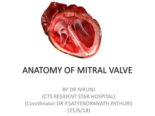
ANATOMY OF MITRAL VALVE
BY DR NIKUNJ
(CTS RESIDENT STAR HOSPITAL)
(Coordinator:DR P.SATYENDRANATH PATHURI)
(25/6/18)
THE ATRIO-VALVULAR JUNCTION
• The junction between the atrium and the valvular tissue is usually well delineated
by the different colors of these structures: the atrium is slightly pink, and the
leaflets are yellowish.
• This junction defines the hinge where the motion of the leaflets is initiated.
• The hinge allows demar- cation of the annulus fibrosus, which is not visible from
an atrial view.
ANNULUS FIBROSUS
• actually a discontinuous band of connective tissue that exists only in some parts of the
attachment of the posterior leaflet.
• The annulus in actuality does not exist at the attachment of the anterior leaflet because the
leaflet tissue is continuous with the aorto- mitral curtain that extends from the aortic valve
annulus to the base of the anterior leaflet
• At each extremity of the base of the anterior leaflet, the atrio-valvular junction is
reinforced by two dense triangular fibrous structures: the anterolateral and
posteromedial fibrous trigones.
• The shape of the annulus varies throughout the cardiac cycle
• During diastole, the shape is grossly circular . During systole, the annulus has a kidney
shape
• Instead of having a planar configuration, the annulus has a three-dimensional saddle-
shape configuration. The two lowest points are located at the fibrous trigones and the
• two highest points are located at the midpoints of the anterior and posterior annuli.
• The plane of the mitral valve annulus makes a 120° angle with the plane of the aortic valve
annulus.
The 26% ± 3% reduction of the mitral valve orifice area during systole results from the
contraction of the base of the heart and the displacement of the aorto-mitral curtain towards
the center of the orifice.
THE LEAFLETS
• The mitral valve comprises two
leaflets—1)anterior
• 2)posterior
• separated by two commissures.
These leaflets are the opening and
closing structures of the valve
• Optimal closure implies a precise
fitting between the surface area
of the leaflets and the orifice area
of the mitral valve.
• Although the anterior and
posterior leaflets have a dif-
ferent size and shape , with the
anterior leaflet more extended
vertically and the posterior leaflet
more extended transversally, they
have a similar surface area.
• The basal insertion of the
anterior leaflet occupies approx-
imately one third of the
circumference of the mitral
valve
• The remaining two thirds of the
circumference attaches to the
posterior leaflet and the
commissural tissue.
• The anterior leaflet is primarily
related to the left ventricular
outflow tract via the aorto-
mitral curtain whereas the
posterior leaflet is related to the
muscular parietal base of the
left ventricle. As a result of this
configuration, the maximum
stress during systole is concen-
trated at the midline of the
posterior leaflet
ANTERIOR LEAFLET
• ANTERIOR LEAFLET, also called the aortic leaflet, has a trapezoidal shape. Its base,
which measures 32 ± 1.3 mm, is inserted on the aorto-mitral curtain and the
adjacent fibrous trigones. The free edge presents with a slightly convex curvature.
At the midline, the height of the anterior leaflet averages 23 mm.
• From the base to the margin, two zones are clearly appa ent.
• 1)The proximal zone, called the atrial zone, is regular, thin, and translucent.
• 2)The distal zone, called the zone of coaptation or rough zone, is irregular and
thicker because of the numerous chordae attached to its ventricular side. The two
zones have a similar surface area.
• The surface of coaptation, which has a height of 7 to 9 mm,
• During diastole, the anterior leaflet divides the left ventricle into two functional
areas, the inflow chamber and the outflow tract.
• The surface of coaptation, which has a height of 7 to 9 mm
POSTERIOR LEAFLET
• The posterior leaflet is inserted approximately
on two thirds of the annulus, to the crest of
the ventricular wall. The free edge is deeply
scalloped by two indentations ,separating
three segments: the anterior, middle, and
posterior scallops are also called P1, P2, and
P3, respectively, to facilitate valve analysis. By
convention, the corresponding areas of the
anterior leaflet are called A1, A2, and A3, and
the commissures AC and PC.
• The size of the scallops of the posterior leaflet
differs. The largest is the middle scallop (P2)
and the smallest is the anterior scallop (P1)
• the posterior leaflet presents two zones from
its base to the free margin: the atrial zone is
smooth and translucent and the coaptation
zone is thicker
• THE COMMISSURES may be described
as a functional entity consisting of two
different structures: the commissural
leaflet, which provides continuity
between the anterior and posterior
leaflets, and the coaptation surfaces
with adjacent anterior and posterior
leaflets.
• The commissural leaflet is a small,
triangular segment of leaflet tissue. Its
base is attached to the annulus and its
free edge is supported by one or two
characteristic fanlike chordae.
• As a result of this configuration, the
junction between the anterior and
posterior leaflets does not reach the
annulus but forms a Y-shaped line of
coaptation.
THE SUSPENSION SYSTEM
• The leaflets are connected to the
ventricular cavity by a suspension system
called the subvalvular apparatus. The
suspension system has two functions:
• one is to facilitate he opening of the
leaflets during diastole (active opening);
• the other is to prevent the upward
displace- ment of the leaflets above the
plane of the annulus during systole.
• To accomplish these two functions, the
suspension system consists of two
structures with different charcteristics:
• THE PAPILLARY MUSCLES with contractile
properties and
• THE CHORDAE TENDINEAE with elastic
properties.
THE PAPILLARY MUSCLES
• insert on the ventricular wall, are usually
organized into two groups, designated
posteromedial and anterolateral, positioned
below the corresponding commissures
I—Large and bulky with a single head generating
numerous chordae.
II—Large and bulky with multiple heads
attaching numerous chordae.
III—Narrow and having few chordae.
IV—Arch shape from which arise several
chordae; the arch may form an arcade with
several trabeculations attached to the
myocardium.
V—Adherent to the ventricular wall and
generating multiple chordae
.
• The anterior papillary muscle most
often has a type I configuration
with occasionally an adjacent type
III papillary muscle attaching the
commissural chordae.
• The posterior papillary muscle
usually has a type II configuration
with one head attaching the
chordae of the anterior leaflet, one
head attaching the commissural
chordae, and one head attaching
chordae of the posterior leaflet.
• The papillary muscles are implanted on the muscular wall of
the left ventricle at a junction situated approximately 1/3
from the apex and 2/3 from the annulus
• The distance between the tip of the papillary muscle and the
plane of the mitral valve orifice also varies, with an average
of 22 ± 5 mm
THE CHORDAE TENDINEAE
THE CHORDAE TENDINEAE extend from the papillary
muscles to the leaflets . Three types can be described
depending upon their attachment on the leaflets
BASAL (OR TERTIARY) CHORDAE extend from the
papillary muscle or directly from the ventricular wall.
The basal chordae are attached to the base of the
posterior and commissural leaflets or to the annulus.
INTERMEDIARY (OR SECONDARY) CHORDAE extend
from the papillary muscles. The intermediary chordae
are attached to the ventricular side of the leaflets.
MARGINAL (OR PRIMARY) CHORDAE are attached to
the margin of the leaflets. The space between two
marginal chordae does not exceed 3 mm and their
attachment to the leaflet is often bifurcated or
trifurcated
FOUR ANATOMICAL STRUCTURES CLOSE TO THE ANNULUS ARE AT RISK
DURING SURGERY
• The circumflex artery runs
between the base of the left
atrial appendage and the
anterior commissure, 3 to 4 mm
from the leaflet attachment,
and then moves away from the
rest of the posterior annulus.
• The coronary sinus skirts the
attachment of the pos- terior
leaflet. It is initially in a lateral
position, and then crosses the
artery and becomes medial,
closerTo the posterior leaflet
attachment but 5 mm superior
to the annulus.
• The bundle of His is located
near the posteromedial trigone.
FOUR ANATOMICAL STRUCTURES CLOSE TO THE ANNULUS ARE AT RISK
DURING SURGERY
• The noncoronary and left
coronary aortic cusps are in
close relationship with the base
of the anterior leaflet
• .the nadir of these cusps is 6 to
10 mm away from the anterior
mitral valve annulus, a distance
that repre- sents a safety zone
for the placement of sutures in
this area provided that the
needle is properly oriented
towards the ventricle.
THANK YOU

More Related Content

What's hot

BMV balloons- FINAL.pptx
BMV balloons- FINAL.pptxBMV balloons- FINAL.pptx
BMV balloons- FINAL.pptxRohitWalse2
 
ECHOCARDIOGRAPHIC EVALUATION OF MITRAL VALVE DISEASE -MITRAL REGURGITATION
ECHOCARDIOGRAPHIC EVALUATION OF MITRAL VALVE DISEASE -MITRAL REGURGITATIONECHOCARDIOGRAPHIC EVALUATION OF MITRAL VALVE DISEASE -MITRAL REGURGITATION
ECHOCARDIOGRAPHIC EVALUATION OF MITRAL VALVE DISEASE -MITRAL REGURGITATIONPraveen Nagula
 
Coronary anatomy and angiographic views
Coronary anatomy and angiographic viewsCoronary anatomy and angiographic views
Coronary anatomy and angiographic viewsthanigai arasu
 
Anatomy tricuspid valve DR NIKUNJ R SHEKHADA (MBBS,MS GEN SURG ,DNB CTS SR)
Anatomy tricuspid valve DR NIKUNJ R SHEKHADA (MBBS,MS GEN SURG ,DNB CTS SR)Anatomy tricuspid valve DR NIKUNJ R SHEKHADA (MBBS,MS GEN SURG ,DNB CTS SR)
Anatomy tricuspid valve DR NIKUNJ R SHEKHADA (MBBS,MS GEN SURG ,DNB CTS SR)DR NIKUNJ SHEKHADA
 
Aortic root anatomy
Aortic root anatomyAortic root anatomy
Aortic root anatomyMohamed Gabr
 
Echo assessment of Aortic Regurgitation
Echo assessment of Aortic RegurgitationEcho assessment of Aortic Regurgitation
Echo assessment of Aortic RegurgitationMashiul Alam
 
Pressure, Damping and Ventricularization_Crimson Publishers
Pressure, Damping and Ventricularization_Crimson PublishersPressure, Damping and Ventricularization_Crimson Publishers
Pressure, Damping and Ventricularization_Crimson PublisherscrimsonpublishersOJCHD
 
Intracardiac shunts
Intracardiac shuntsIntracardiac shunts
Intracardiac shuntsFuad Farooq
 
Anatomy and embryology of valves
Anatomy and embryology of valvesAnatomy and embryology of valves
Anatomy and embryology of valvesDrvasanthi
 
Echo Mitral Stenosis
Echo Mitral StenosisEcho Mitral Stenosis
Echo Mitral StenosisMashiul Alam
 
Assessment of prosthetic valve function
Assessment of prosthetic valve functionAssessment of prosthetic valve function
Assessment of prosthetic valve functionSwapnil Garde
 
LEFT HEART CATHETERIZATION
LEFT HEART CATHETERIZATIONLEFT HEART CATHETERIZATION
LEFT HEART CATHETERIZATIONPraveen Nagula
 
Left ventricular pressure tracings
Left ventricular pressure tracingsLeft ventricular pressure tracings
Left ventricular pressure tracingsGOPAL GHOSH
 
Shunt quantification
Shunt quantificationShunt quantification
Shunt quantificationAnkur Gupta
 

What's hot (20)

BMV balloons- FINAL.pptx
BMV balloons- FINAL.pptxBMV balloons- FINAL.pptx
BMV balloons- FINAL.pptx
 
ECHOCARDIOGRAPHIC EVALUATION OF MITRAL VALVE DISEASE -MITRAL REGURGITATION
ECHOCARDIOGRAPHIC EVALUATION OF MITRAL VALVE DISEASE -MITRAL REGURGITATIONECHOCARDIOGRAPHIC EVALUATION OF MITRAL VALVE DISEASE -MITRAL REGURGITATION
ECHOCARDIOGRAPHIC EVALUATION OF MITRAL VALVE DISEASE -MITRAL REGURGITATION
 
Coronary anatomy and angiographic views
Coronary anatomy and angiographic viewsCoronary anatomy and angiographic views
Coronary anatomy and angiographic views
 
Anatomy tricuspid valve DR NIKUNJ R SHEKHADA (MBBS,MS GEN SURG ,DNB CTS SR)
Anatomy tricuspid valve DR NIKUNJ R SHEKHADA (MBBS,MS GEN SURG ,DNB CTS SR)Anatomy tricuspid valve DR NIKUNJ R SHEKHADA (MBBS,MS GEN SURG ,DNB CTS SR)
Anatomy tricuspid valve DR NIKUNJ R SHEKHADA (MBBS,MS GEN SURG ,DNB CTS SR)
 
Aortic root anatomy
Aortic root anatomyAortic root anatomy
Aortic root anatomy
 
Echo assessment of Aortic Regurgitation
Echo assessment of Aortic RegurgitationEcho assessment of Aortic Regurgitation
Echo assessment of Aortic Regurgitation
 
Pressure, Damping and Ventricularization_Crimson Publishers
Pressure, Damping and Ventricularization_Crimson PublishersPressure, Damping and Ventricularization_Crimson Publishers
Pressure, Damping and Ventricularization_Crimson Publishers
 
PVBD
PVBDPVBD
PVBD
 
Intracardiac shunts
Intracardiac shuntsIntracardiac shunts
Intracardiac shunts
 
PTMC/PBMC
PTMC/PBMCPTMC/PBMC
PTMC/PBMC
 
Echo assessment of mitral regurgitation
Echo assessment of mitral regurgitationEcho assessment of mitral regurgitation
Echo assessment of mitral regurgitation
 
Tricuspid valve
Tricuspid valveTricuspid valve
Tricuspid valve
 
Anatomy and embryology of valves
Anatomy and embryology of valvesAnatomy and embryology of valves
Anatomy and embryology of valves
 
Echo Mitral Stenosis
Echo Mitral StenosisEcho Mitral Stenosis
Echo Mitral Stenosis
 
PBMV:Tips and Tricks
PBMV:Tips and TricksPBMV:Tips and Tricks
PBMV:Tips and Tricks
 
Assessment of prosthetic valve function
Assessment of prosthetic valve functionAssessment of prosthetic valve function
Assessment of prosthetic valve function
 
LEFT HEART CATHETERIZATION
LEFT HEART CATHETERIZATIONLEFT HEART CATHETERIZATION
LEFT HEART CATHETERIZATION
 
TEE VIEWS
TEE VIEWSTEE VIEWS
TEE VIEWS
 
Left ventricular pressure tracings
Left ventricular pressure tracingsLeft ventricular pressure tracings
Left ventricular pressure tracings
 
Shunt quantification
Shunt quantificationShunt quantification
Shunt quantification
 

Similar to Mitral valve anatomy

Mitral valve surgical anatomy DR NIKUNJ R SHEKHADA (MBBS ,MS GRN SURG , DNB C...
Mitral valve surgical anatomy DR NIKUNJ R SHEKHADA (MBBS ,MS GRN SURG , DNB C...Mitral valve surgical anatomy DR NIKUNJ R SHEKHADA (MBBS ,MS GRN SURG , DNB C...
Mitral valve surgical anatomy DR NIKUNJ R SHEKHADA (MBBS ,MS GRN SURG , DNB C...DR NIKUNJ SHEKHADA
 
anatomy of mitral valve
anatomy of mitral valve  anatomy of mitral valve
anatomy of mitral valve India CTVS
 
Anatomy of heart
Anatomy of heartAnatomy of heart
Anatomy of heartnmonty02
 
Dr Garva Ameta anatomy Of Left Heart.pptx
Dr Garva Ameta anatomy Of Left Heart.pptxDr Garva Ameta anatomy Of Left Heart.pptx
Dr Garva Ameta anatomy Of Left Heart.pptxPooja Jakhar
 
Normal anatomy of tricuspid valve
Normal anatomy of tricuspid valve Normal anatomy of tricuspid valve
Normal anatomy of tricuspid valve India CTVS
 
Anatomy of mitral valve echo evaluation
Anatomy of mitral valve echo evaluationAnatomy of mitral valve echo evaluation
Anatomy of mitral valve echo evaluationmadhusiva03
 
Aortic root anatomy DR NIKUNJ R SHEKHADA (MBBS,MS GEN SURG,DNB CTS SR)
Aortic root anatomy DR NIKUNJ R SHEKHADA (MBBS,MS GEN SURG,DNB CTS SR)Aortic root anatomy DR NIKUNJ R SHEKHADA (MBBS,MS GEN SURG,DNB CTS SR)
Aortic root anatomy DR NIKUNJ R SHEKHADA (MBBS,MS GEN SURG,DNB CTS SR)DR NIKUNJ SHEKHADA
 
Arterial supply of head and neck
Arterial supply of head and neckArterial supply of head and neck
Arterial supply of head and neckAvinash Rathore
 
Blood supply of head and neck.pdf
Blood supply of head and neck.pdfBlood supply of head and neck.pdf
Blood supply of head and neck.pdfParulGarg894243
 
Cardiac anatomy and imaging techniques
Cardiac anatomy and imaging techniques Cardiac anatomy and imaging techniques
Cardiac anatomy and imaging techniques Hemanthvarma52
 
The valves of the heart
The valves of the heartThe valves of the heart
The valves of the heartIdris Siddiqui
 
Surface anatomy areas.pptx
Surface anatomy areas.pptxSurface anatomy areas.pptx
Surface anatomy areas.pptxMishiSoza
 
Anatomy of heart presenter dr.smita valani
Anatomy of heart  presenter dr.smita valaniAnatomy of heart  presenter dr.smita valani
Anatomy of heart presenter dr.smita valaniSmita Valani
 
Pmdc step 1 Review of CVS & Respiratory System
Pmdc step 1 Review of CVS & Respiratory SystemPmdc step 1 Review of CVS & Respiratory System
Pmdc step 1 Review of CVS & Respiratory SystemDrSaeed Shafi
 
PRESENTATION BOWEL.pptx
PRESENTATION BOWEL.pptxPRESENTATION BOWEL.pptx
PRESENTATION BOWEL.pptxShubham661884
 

Similar to Mitral valve anatomy (20)

Mitral valve surgical anatomy DR NIKUNJ R SHEKHADA (MBBS ,MS GRN SURG , DNB C...
Mitral valve surgical anatomy DR NIKUNJ R SHEKHADA (MBBS ,MS GRN SURG , DNB C...Mitral valve surgical anatomy DR NIKUNJ R SHEKHADA (MBBS ,MS GRN SURG , DNB C...
Mitral valve surgical anatomy DR NIKUNJ R SHEKHADA (MBBS ,MS GRN SURG , DNB C...
 
anatomy of mitral valve
anatomy of mitral valve  anatomy of mitral valve
anatomy of mitral valve
 
Anatomy of heart
Anatomy of heartAnatomy of heart
Anatomy of heart
 
Dr Garva Ameta anatomy Of Left Heart.pptx
Dr Garva Ameta anatomy Of Left Heart.pptxDr Garva Ameta anatomy Of Left Heart.pptx
Dr Garva Ameta anatomy Of Left Heart.pptx
 
Normal anatomy of tricuspid valve
Normal anatomy of tricuspid valve Normal anatomy of tricuspid valve
Normal anatomy of tricuspid valve
 
Anatomy of mitral valve echo evaluation
Anatomy of mitral valve echo evaluationAnatomy of mitral valve echo evaluation
Anatomy of mitral valve echo evaluation
 
Aortic valve disease
Aortic valve diseaseAortic valve disease
Aortic valve disease
 
Aortic valve disease
Aortic valve diseaseAortic valve disease
Aortic valve disease
 
Aortic root anatomy DR NIKUNJ R SHEKHADA (MBBS,MS GEN SURG,DNB CTS SR)
Aortic root anatomy DR NIKUNJ R SHEKHADA (MBBS,MS GEN SURG,DNB CTS SR)Aortic root anatomy DR NIKUNJ R SHEKHADA (MBBS,MS GEN SURG,DNB CTS SR)
Aortic root anatomy DR NIKUNJ R SHEKHADA (MBBS,MS GEN SURG,DNB CTS SR)
 
Arterial supply of head and neck
Arterial supply of head and neckArterial supply of head and neck
Arterial supply of head and neck
 
Mitral valve disease
Mitral valve diseaseMitral valve disease
Mitral valve disease
 
Blood supply of head and neck.pdf
Blood supply of head and neck.pdfBlood supply of head and neck.pdf
Blood supply of head and neck.pdf
 
Cardiac anatomy and imaging techniques
Cardiac anatomy and imaging techniques Cardiac anatomy and imaging techniques
Cardiac anatomy and imaging techniques
 
The valves of the heart
The valves of the heartThe valves of the heart
The valves of the heart
 
Anatomy of Heart..!!
Anatomy of Heart..!!Anatomy of Heart..!!
Anatomy of Heart..!!
 
Surface anatomy areas.pptx
Surface anatomy areas.pptxSurface anatomy areas.pptx
Surface anatomy areas.pptx
 
Anatomy of heart presenter dr.smita valani
Anatomy of heart  presenter dr.smita valaniAnatomy of heart  presenter dr.smita valani
Anatomy of heart presenter dr.smita valani
 
External carotid artery
External carotid arteryExternal carotid artery
External carotid artery
 
Pmdc step 1 Review of CVS & Respiratory System
Pmdc step 1 Review of CVS & Respiratory SystemPmdc step 1 Review of CVS & Respiratory System
Pmdc step 1 Review of CVS & Respiratory System
 
PRESENTATION BOWEL.pptx
PRESENTATION BOWEL.pptxPRESENTATION BOWEL.pptx
PRESENTATION BOWEL.pptx
 

More from DR NIKUNJ SHEKHADA

Mitral valve surgical treatment
Mitral valve surgical treatmentMitral valve surgical treatment
Mitral valve surgical treatmentDR NIKUNJ SHEKHADA
 
Cardiac cycle DR NIKUNJ R SHEKHADA (MBBS,MS GEN SURG ,DNB CTS SR)
Cardiac cycle DR  NIKUNJ R SHEKHADA (MBBS,MS GEN SURG ,DNB CTS SR)Cardiac cycle DR  NIKUNJ R SHEKHADA (MBBS,MS GEN SURG ,DNB CTS SR)
Cardiac cycle DR NIKUNJ R SHEKHADA (MBBS,MS GEN SURG ,DNB CTS SR)DR NIKUNJ SHEKHADA
 
History of cardiac surgery DR NIKUNJ R SHEKHADA (MBBS,MS GEN SURG ,DNB CTS SR)
History of cardiac surgery  DR  NIKUNJ R SHEKHADA (MBBS,MS GEN SURG ,DNB CTS SR)History of cardiac surgery  DR  NIKUNJ R SHEKHADA (MBBS,MS GEN SURG ,DNB CTS SR)
History of cardiac surgery DR NIKUNJ R SHEKHADA (MBBS,MS GEN SURG ,DNB CTS SR)DR NIKUNJ SHEKHADA
 
Cpb oxygenators DR NIKUNJ R SHEKHADA (MBBS,MS GEN SURG ,DNB CTS SR)
Cpb oxygenators  DR  NIKUNJ R SHEKHADA (MBBS,MS GEN SURG ,DNB CTS SR)Cpb oxygenators  DR  NIKUNJ R SHEKHADA (MBBS,MS GEN SURG ,DNB CTS SR)
Cpb oxygenators DR NIKUNJ R SHEKHADA (MBBS,MS GEN SURG ,DNB CTS SR)DR NIKUNJ SHEKHADA
 
Chordal preservation DR NIKUNJ R SHEKHADA (MBBS,MS GEN SURG ,DNB CTS SR)
Chordal preservation DR  NIKUNJ R SHEKHADA (MBBS,MS GEN SURG ,DNB CTS SR)Chordal preservation DR  NIKUNJ R SHEKHADA (MBBS,MS GEN SURG ,DNB CTS SR)
Chordal preservation DR NIKUNJ R SHEKHADA (MBBS,MS GEN SURG ,DNB CTS SR)DR NIKUNJ SHEKHADA
 
Post oprative care 1 DR NIKUNJ R SHEKHADA (MBBS,MS GEN SURG,DNB CTS SR)
Post oprative care 1 DR NIKUNJ R SHEKHADA (MBBS,MS GEN SURG,DNB CTS SR)Post oprative care 1 DR NIKUNJ R SHEKHADA (MBBS,MS GEN SURG,DNB CTS SR)
Post oprative care 1 DR NIKUNJ R SHEKHADA (MBBS,MS GEN SURG,DNB CTS SR)DR NIKUNJ SHEKHADA
 
Ecg1 DR NIKUNJ R SHEKHADA (MBBS,MS GEN SURG,DNB CTS SR)
Ecg1 DR NIKUNJ R SHEKHADA (MBBS,MS GEN SURG,DNB CTS SR)Ecg1 DR NIKUNJ R SHEKHADA (MBBS,MS GEN SURG,DNB CTS SR)
Ecg1 DR NIKUNJ R SHEKHADA (MBBS,MS GEN SURG,DNB CTS SR)DR NIKUNJ SHEKHADA
 
CARDIO PLEGIA DELIVERY DR NIKUNJ R SHEKHADA (MBBS,MS GEN SURG,DNB CTS SR
CARDIO PLEGIA DELIVERY  DR NIKUNJ R SHEKHADA (MBBS,MS GEN SURG,DNB CTS SRCARDIO PLEGIA DELIVERY  DR NIKUNJ R SHEKHADA (MBBS,MS GEN SURG,DNB CTS SR
CARDIO PLEGIA DELIVERY DR NIKUNJ R SHEKHADA (MBBS,MS GEN SURG,DNB CTS SRDR NIKUNJ SHEKHADA
 
Atrial septal defect DR NIKUNJ R SHEKHADA (MBBS,MS GEN SURG,DNB CTS SR
Atrial septal defect DR NIKUNJ R SHEKHADA (MBBS,MS GEN SURG,DNB CTS SRAtrial septal defect DR NIKUNJ R SHEKHADA (MBBS,MS GEN SURG,DNB CTS SR
Atrial septal defect DR NIKUNJ R SHEKHADA (MBBS,MS GEN SURG,DNB CTS SRDR NIKUNJ SHEKHADA
 
Asd managment DR NIKUNJ R SHEKHADA (MBBS,MS GEN SURG,DNB CTS SR)
Asd managment DR NIKUNJ R SHEKHADA (MBBS,MS GEN SURG,DNB CTS SR)Asd managment DR NIKUNJ R SHEKHADA (MBBS,MS GEN SURG,DNB CTS SR)
Asd managment DR NIKUNJ R SHEKHADA (MBBS,MS GEN SURG,DNB CTS SR)DR NIKUNJ SHEKHADA
 
Myocardial protection DR NIKUNJ R SHEKHADA (MBBS ,MS GRN SURG , DNB CTS SR
Myocardial protection DR NIKUNJ R SHEKHADA (MBBS ,MS GRN SURG , DNB CTS SRMyocardial protection DR NIKUNJ R SHEKHADA (MBBS ,MS GRN SURG , DNB CTS SR
Myocardial protection DR NIKUNJ R SHEKHADA (MBBS ,MS GRN SURG , DNB CTS SRDR NIKUNJ SHEKHADA
 
CARDIO PULMONARYBYPASS BLOOD PUMPS DR NIKUNJ R SHEKHADA (MBBS,MSGEN SURG ,DN...
CARDIO PULMONARYBYPASS BLOOD PUMPS  DR NIKUNJ R SHEKHADA (MBBS,MSGEN SURG ,DN...CARDIO PULMONARYBYPASS BLOOD PUMPS  DR NIKUNJ R SHEKHADA (MBBS,MSGEN SURG ,DN...
CARDIO PULMONARYBYPASS BLOOD PUMPS DR NIKUNJ R SHEKHADA (MBBS,MSGEN SURG ,DN...DR NIKUNJ SHEKHADA
 
Ventricular septal defect DR NIKUNJ .R .SHRKHADA (MBBS,MS GEN SURG DNB CTS SR)
Ventricular septal defect  DR NIKUNJ .R .SHRKHADA (MBBS,MS GEN SURG DNB CTS SR)Ventricular septal defect  DR NIKUNJ .R .SHRKHADA (MBBS,MS GEN SURG DNB CTS SR)
Ventricular septal defect DR NIKUNJ .R .SHRKHADA (MBBS,MS GEN SURG DNB CTS SR)DR NIKUNJ SHEKHADA
 
Coronary arteries and veins DR NIKUNJ .R .SHRKHADA (MBBS,MS GEN SURG DNB CTS SR)
Coronary arteries and veins DR NIKUNJ .R .SHRKHADA (MBBS,MS GEN SURG DNB CTS SR)Coronary arteries and veins DR NIKUNJ .R .SHRKHADA (MBBS,MS GEN SURG DNB CTS SR)
Coronary arteries and veins DR NIKUNJ .R .SHRKHADA (MBBS,MS GEN SURG DNB CTS SR)DR NIKUNJ SHEKHADA
 
Anatomy of lungs 3DR NIKUNJ R SHEKHADA (MBBS,MS GEN SURG DNB CTS SR)
Anatomy of lungs 3DR NIKUNJ R SHEKHADA (MBBS,MS GEN SURG DNB CTS SR)Anatomy of lungs 3DR NIKUNJ R SHEKHADA (MBBS,MS GEN SURG DNB CTS SR)
Anatomy of lungs 3DR NIKUNJ R SHEKHADA (MBBS,MS GEN SURG DNB CTS SR)DR NIKUNJ SHEKHADA
 

More from DR NIKUNJ SHEKHADA (20)

Aortic repair ppt
Aortic repair pptAortic repair ppt
Aortic repair ppt
 
Mitral valve surgical treatment
Mitral valve surgical treatmentMitral valve surgical treatment
Mitral valve surgical treatment
 
Mitral valve repair
Mitral valve repairMitral valve repair
Mitral valve repair
 
Aortic repair ppt
Aortic repair pptAortic repair ppt
Aortic repair ppt
 
Surgical emphsema
Surgical emphsemaSurgical emphsema
Surgical emphsema
 
Cardiac cycle DR NIKUNJ R SHEKHADA (MBBS,MS GEN SURG ,DNB CTS SR)
Cardiac cycle DR  NIKUNJ R SHEKHADA (MBBS,MS GEN SURG ,DNB CTS SR)Cardiac cycle DR  NIKUNJ R SHEKHADA (MBBS,MS GEN SURG ,DNB CTS SR)
Cardiac cycle DR NIKUNJ R SHEKHADA (MBBS,MS GEN SURG ,DNB CTS SR)
 
History of cardiac surgery DR NIKUNJ R SHEKHADA (MBBS,MS GEN SURG ,DNB CTS SR)
History of cardiac surgery  DR  NIKUNJ R SHEKHADA (MBBS,MS GEN SURG ,DNB CTS SR)History of cardiac surgery  DR  NIKUNJ R SHEKHADA (MBBS,MS GEN SURG ,DNB CTS SR)
History of cardiac surgery DR NIKUNJ R SHEKHADA (MBBS,MS GEN SURG ,DNB CTS SR)
 
Cpb oxygenators DR NIKUNJ R SHEKHADA (MBBS,MS GEN SURG ,DNB CTS SR)
Cpb oxygenators  DR  NIKUNJ R SHEKHADA (MBBS,MS GEN SURG ,DNB CTS SR)Cpb oxygenators  DR  NIKUNJ R SHEKHADA (MBBS,MS GEN SURG ,DNB CTS SR)
Cpb oxygenators DR NIKUNJ R SHEKHADA (MBBS,MS GEN SURG ,DNB CTS SR)
 
Chordal preservation DR NIKUNJ R SHEKHADA (MBBS,MS GEN SURG ,DNB CTS SR)
Chordal preservation DR  NIKUNJ R SHEKHADA (MBBS,MS GEN SURG ,DNB CTS SR)Chordal preservation DR  NIKUNJ R SHEKHADA (MBBS,MS GEN SURG ,DNB CTS SR)
Chordal preservation DR NIKUNJ R SHEKHADA (MBBS,MS GEN SURG ,DNB CTS SR)
 
Post oprative care 1 DR NIKUNJ R SHEKHADA (MBBS,MS GEN SURG,DNB CTS SR)
Post oprative care 1 DR NIKUNJ R SHEKHADA (MBBS,MS GEN SURG,DNB CTS SR)Post oprative care 1 DR NIKUNJ R SHEKHADA (MBBS,MS GEN SURG,DNB CTS SR)
Post oprative care 1 DR NIKUNJ R SHEKHADA (MBBS,MS GEN SURG,DNB CTS SR)
 
Ecg1 DR NIKUNJ R SHEKHADA (MBBS,MS GEN SURG,DNB CTS SR)
Ecg1 DR NIKUNJ R SHEKHADA (MBBS,MS GEN SURG,DNB CTS SR)Ecg1 DR NIKUNJ R SHEKHADA (MBBS,MS GEN SURG,DNB CTS SR)
Ecg1 DR NIKUNJ R SHEKHADA (MBBS,MS GEN SURG,DNB CTS SR)
 
CARDIO PLEGIA DELIVERY DR NIKUNJ R SHEKHADA (MBBS,MS GEN SURG,DNB CTS SR
CARDIO PLEGIA DELIVERY  DR NIKUNJ R SHEKHADA (MBBS,MS GEN SURG,DNB CTS SRCARDIO PLEGIA DELIVERY  DR NIKUNJ R SHEKHADA (MBBS,MS GEN SURG,DNB CTS SR
CARDIO PLEGIA DELIVERY DR NIKUNJ R SHEKHADA (MBBS,MS GEN SURG,DNB CTS SR
 
Atrial septal defect DR NIKUNJ R SHEKHADA (MBBS,MS GEN SURG,DNB CTS SR
Atrial septal defect DR NIKUNJ R SHEKHADA (MBBS,MS GEN SURG,DNB CTS SRAtrial septal defect DR NIKUNJ R SHEKHADA (MBBS,MS GEN SURG,DNB CTS SR
Atrial septal defect DR NIKUNJ R SHEKHADA (MBBS,MS GEN SURG,DNB CTS SR
 
Asd managment DR NIKUNJ R SHEKHADA (MBBS,MS GEN SURG,DNB CTS SR)
Asd managment DR NIKUNJ R SHEKHADA (MBBS,MS GEN SURG,DNB CTS SR)Asd managment DR NIKUNJ R SHEKHADA (MBBS,MS GEN SURG,DNB CTS SR)
Asd managment DR NIKUNJ R SHEKHADA (MBBS,MS GEN SURG,DNB CTS SR)
 
Myocardial protection DR NIKUNJ R SHEKHADA (MBBS ,MS GRN SURG , DNB CTS SR
Myocardial protection DR NIKUNJ R SHEKHADA (MBBS ,MS GRN SURG , DNB CTS SRMyocardial protection DR NIKUNJ R SHEKHADA (MBBS ,MS GRN SURG , DNB CTS SR
Myocardial protection DR NIKUNJ R SHEKHADA (MBBS ,MS GRN SURG , DNB CTS SR
 
CPB(CARDIO PULMONARY BYPASS)
CPB(CARDIO PULMONARY BYPASS)CPB(CARDIO PULMONARY BYPASS)
CPB(CARDIO PULMONARY BYPASS)
 
CARDIO PULMONARYBYPASS BLOOD PUMPS DR NIKUNJ R SHEKHADA (MBBS,MSGEN SURG ,DN...
CARDIO PULMONARYBYPASS BLOOD PUMPS  DR NIKUNJ R SHEKHADA (MBBS,MSGEN SURG ,DN...CARDIO PULMONARYBYPASS BLOOD PUMPS  DR NIKUNJ R SHEKHADA (MBBS,MSGEN SURG ,DN...
CARDIO PULMONARYBYPASS BLOOD PUMPS DR NIKUNJ R SHEKHADA (MBBS,MSGEN SURG ,DN...
 
Ventricular septal defect DR NIKUNJ .R .SHRKHADA (MBBS,MS GEN SURG DNB CTS SR)
Ventricular septal defect  DR NIKUNJ .R .SHRKHADA (MBBS,MS GEN SURG DNB CTS SR)Ventricular septal defect  DR NIKUNJ .R .SHRKHADA (MBBS,MS GEN SURG DNB CTS SR)
Ventricular septal defect DR NIKUNJ .R .SHRKHADA (MBBS,MS GEN SURG DNB CTS SR)
 
Coronary arteries and veins DR NIKUNJ .R .SHRKHADA (MBBS,MS GEN SURG DNB CTS SR)
Coronary arteries and veins DR NIKUNJ .R .SHRKHADA (MBBS,MS GEN SURG DNB CTS SR)Coronary arteries and veins DR NIKUNJ .R .SHRKHADA (MBBS,MS GEN SURG DNB CTS SR)
Coronary arteries and veins DR NIKUNJ .R .SHRKHADA (MBBS,MS GEN SURG DNB CTS SR)
 
Anatomy of lungs 3DR NIKUNJ R SHEKHADA (MBBS,MS GEN SURG DNB CTS SR)
Anatomy of lungs 3DR NIKUNJ R SHEKHADA (MBBS,MS GEN SURG DNB CTS SR)Anatomy of lungs 3DR NIKUNJ R SHEKHADA (MBBS,MS GEN SURG DNB CTS SR)
Anatomy of lungs 3DR NIKUNJ R SHEKHADA (MBBS,MS GEN SURG DNB CTS SR)
 

Recently uploaded

Introduction to ArtificiaI Intelligence in Higher Education
Introduction to ArtificiaI Intelligence in Higher EducationIntroduction to ArtificiaI Intelligence in Higher Education
Introduction to ArtificiaI Intelligence in Higher Educationpboyjonauth
 
Historical philosophical, theoretical, and legal foundations of special and i...
Historical philosophical, theoretical, and legal foundations of special and i...Historical philosophical, theoretical, and legal foundations of special and i...
Historical philosophical, theoretical, and legal foundations of special and i...jaredbarbolino94
 
Proudly South Africa powerpoint Thorisha.pptx
Proudly South Africa powerpoint Thorisha.pptxProudly South Africa powerpoint Thorisha.pptx
Proudly South Africa powerpoint Thorisha.pptxthorishapillay1
 
POINT- BIOCHEMISTRY SEM 2 ENZYMES UNIT 5.pptx
POINT- BIOCHEMISTRY SEM 2 ENZYMES UNIT 5.pptxPOINT- BIOCHEMISTRY SEM 2 ENZYMES UNIT 5.pptx
POINT- BIOCHEMISTRY SEM 2 ENZYMES UNIT 5.pptxSayali Powar
 
MARGINALIZATION (Different learners in Marginalized Group
MARGINALIZATION (Different learners in Marginalized GroupMARGINALIZATION (Different learners in Marginalized Group
MARGINALIZATION (Different learners in Marginalized GroupJonathanParaisoCruz
 
KSHARA STURA .pptx---KSHARA KARMA THERAPY (CAUSTIC THERAPY)————IMP.OF KSHARA ...
KSHARA STURA .pptx---KSHARA KARMA THERAPY (CAUSTIC THERAPY)————IMP.OF KSHARA ...KSHARA STURA .pptx---KSHARA KARMA THERAPY (CAUSTIC THERAPY)————IMP.OF KSHARA ...
KSHARA STURA .pptx---KSHARA KARMA THERAPY (CAUSTIC THERAPY)————IMP.OF KSHARA ...M56BOOKSTORE PRODUCT/SERVICE
 
Framing an Appropriate Research Question 6b9b26d93da94caf993c038d9efcdedb.pdf
Framing an Appropriate Research Question 6b9b26d93da94caf993c038d9efcdedb.pdfFraming an Appropriate Research Question 6b9b26d93da94caf993c038d9efcdedb.pdf
Framing an Appropriate Research Question 6b9b26d93da94caf993c038d9efcdedb.pdfUjwalaBharambe
 
DATA STRUCTURE AND ALGORITHM for beginners
DATA STRUCTURE AND ALGORITHM for beginnersDATA STRUCTURE AND ALGORITHM for beginners
DATA STRUCTURE AND ALGORITHM for beginnersSabitha Banu
 
Introduction to AI in Higher Education_draft.pptx
Introduction to AI in Higher Education_draft.pptxIntroduction to AI in Higher Education_draft.pptx
Introduction to AI in Higher Education_draft.pptxpboyjonauth
 
Biting mechanism of poisonous snakes.pdf
Biting mechanism of poisonous snakes.pdfBiting mechanism of poisonous snakes.pdf
Biting mechanism of poisonous snakes.pdfadityarao40181
 
Roles & Responsibilities in Pharmacovigilance
Roles & Responsibilities in PharmacovigilanceRoles & Responsibilities in Pharmacovigilance
Roles & Responsibilities in PharmacovigilanceSamikshaHamane
 
Full Stack Web Development Course for Beginners
Full Stack Web Development Course  for BeginnersFull Stack Web Development Course  for Beginners
Full Stack Web Development Course for BeginnersSabitha Banu
 
Computed Fields and api Depends in the Odoo 17
Computed Fields and api Depends in the Odoo 17Computed Fields and api Depends in the Odoo 17
Computed Fields and api Depends in the Odoo 17Celine George
 
How to Configure Email Server in Odoo 17
How to Configure Email Server in Odoo 17How to Configure Email Server in Odoo 17
How to Configure Email Server in Odoo 17Celine George
 
Final demo Grade 9 for demo Plan dessert.pptx
Final demo Grade 9 for demo Plan dessert.pptxFinal demo Grade 9 for demo Plan dessert.pptx
Final demo Grade 9 for demo Plan dessert.pptxAvyJaneVismanos
 
Crayon Activity Handout For the Crayon A
Crayon Activity Handout For the Crayon ACrayon Activity Handout For the Crayon A
Crayon Activity Handout For the Crayon AUnboundStockton
 
Alper Gobel In Media Res Media Component
Alper Gobel In Media Res Media ComponentAlper Gobel In Media Res Media Component
Alper Gobel In Media Res Media ComponentInMediaRes1
 
Capitol Tech U Doctoral Presentation - April 2024.pptx
Capitol Tech U Doctoral Presentation - April 2024.pptxCapitol Tech U Doctoral Presentation - April 2024.pptx
Capitol Tech U Doctoral Presentation - April 2024.pptxCapitolTechU
 

Recently uploaded (20)

Introduction to ArtificiaI Intelligence in Higher Education
Introduction to ArtificiaI Intelligence in Higher EducationIntroduction to ArtificiaI Intelligence in Higher Education
Introduction to ArtificiaI Intelligence in Higher Education
 
Historical philosophical, theoretical, and legal foundations of special and i...
Historical philosophical, theoretical, and legal foundations of special and i...Historical philosophical, theoretical, and legal foundations of special and i...
Historical philosophical, theoretical, and legal foundations of special and i...
 
Proudly South Africa powerpoint Thorisha.pptx
Proudly South Africa powerpoint Thorisha.pptxProudly South Africa powerpoint Thorisha.pptx
Proudly South Africa powerpoint Thorisha.pptx
 
POINT- BIOCHEMISTRY SEM 2 ENZYMES UNIT 5.pptx
POINT- BIOCHEMISTRY SEM 2 ENZYMES UNIT 5.pptxPOINT- BIOCHEMISTRY SEM 2 ENZYMES UNIT 5.pptx
POINT- BIOCHEMISTRY SEM 2 ENZYMES UNIT 5.pptx
 
MARGINALIZATION (Different learners in Marginalized Group
MARGINALIZATION (Different learners in Marginalized GroupMARGINALIZATION (Different learners in Marginalized Group
MARGINALIZATION (Different learners in Marginalized Group
 
KSHARA STURA .pptx---KSHARA KARMA THERAPY (CAUSTIC THERAPY)————IMP.OF KSHARA ...
KSHARA STURA .pptx---KSHARA KARMA THERAPY (CAUSTIC THERAPY)————IMP.OF KSHARA ...KSHARA STURA .pptx---KSHARA KARMA THERAPY (CAUSTIC THERAPY)————IMP.OF KSHARA ...
KSHARA STURA .pptx---KSHARA KARMA THERAPY (CAUSTIC THERAPY)————IMP.OF KSHARA ...
 
Framing an Appropriate Research Question 6b9b26d93da94caf993c038d9efcdedb.pdf
Framing an Appropriate Research Question 6b9b26d93da94caf993c038d9efcdedb.pdfFraming an Appropriate Research Question 6b9b26d93da94caf993c038d9efcdedb.pdf
Framing an Appropriate Research Question 6b9b26d93da94caf993c038d9efcdedb.pdf
 
ESSENTIAL of (CS/IT/IS) class 06 (database)
ESSENTIAL of (CS/IT/IS) class 06 (database)ESSENTIAL of (CS/IT/IS) class 06 (database)
ESSENTIAL of (CS/IT/IS) class 06 (database)
 
DATA STRUCTURE AND ALGORITHM for beginners
DATA STRUCTURE AND ALGORITHM for beginnersDATA STRUCTURE AND ALGORITHM for beginners
DATA STRUCTURE AND ALGORITHM for beginners
 
Introduction to AI in Higher Education_draft.pptx
Introduction to AI in Higher Education_draft.pptxIntroduction to AI in Higher Education_draft.pptx
Introduction to AI in Higher Education_draft.pptx
 
Biting mechanism of poisonous snakes.pdf
Biting mechanism of poisonous snakes.pdfBiting mechanism of poisonous snakes.pdf
Biting mechanism of poisonous snakes.pdf
 
Roles & Responsibilities in Pharmacovigilance
Roles & Responsibilities in PharmacovigilanceRoles & Responsibilities in Pharmacovigilance
Roles & Responsibilities in Pharmacovigilance
 
Full Stack Web Development Course for Beginners
Full Stack Web Development Course  for BeginnersFull Stack Web Development Course  for Beginners
Full Stack Web Development Course for Beginners
 
Computed Fields and api Depends in the Odoo 17
Computed Fields and api Depends in the Odoo 17Computed Fields and api Depends in the Odoo 17
Computed Fields and api Depends in the Odoo 17
 
How to Configure Email Server in Odoo 17
How to Configure Email Server in Odoo 17How to Configure Email Server in Odoo 17
How to Configure Email Server in Odoo 17
 
Final demo Grade 9 for demo Plan dessert.pptx
Final demo Grade 9 for demo Plan dessert.pptxFinal demo Grade 9 for demo Plan dessert.pptx
Final demo Grade 9 for demo Plan dessert.pptx
 
Crayon Activity Handout For the Crayon A
Crayon Activity Handout For the Crayon ACrayon Activity Handout For the Crayon A
Crayon Activity Handout For the Crayon A
 
Alper Gobel In Media Res Media Component
Alper Gobel In Media Res Media ComponentAlper Gobel In Media Res Media Component
Alper Gobel In Media Res Media Component
 
Capitol Tech U Doctoral Presentation - April 2024.pptx
Capitol Tech U Doctoral Presentation - April 2024.pptxCapitol Tech U Doctoral Presentation - April 2024.pptx
Capitol Tech U Doctoral Presentation - April 2024.pptx
 
9953330565 Low Rate Call Girls In Rohini Delhi NCR
9953330565 Low Rate Call Girls In Rohini  Delhi NCR9953330565 Low Rate Call Girls In Rohini  Delhi NCR
9953330565 Low Rate Call Girls In Rohini Delhi NCR
 

Mitral valve anatomy

  • 1. ANATOMY OF MITRAL VALVE BY DR NIKUNJ (CTS RESIDENT STAR HOSPITAL) (Coordinator:DR P.SATYENDRANATH PATHURI) (25/6/18)
  • 2.
  • 3.
  • 4. THE ATRIO-VALVULAR JUNCTION • The junction between the atrium and the valvular tissue is usually well delineated by the different colors of these structures: the atrium is slightly pink, and the leaflets are yellowish. • This junction defines the hinge where the motion of the leaflets is initiated. • The hinge allows demar- cation of the annulus fibrosus, which is not visible from an atrial view.
  • 5. ANNULUS FIBROSUS • actually a discontinuous band of connective tissue that exists only in some parts of the attachment of the posterior leaflet. • The annulus in actuality does not exist at the attachment of the anterior leaflet because the leaflet tissue is continuous with the aorto- mitral curtain that extends from the aortic valve annulus to the base of the anterior leaflet
  • 6. • At each extremity of the base of the anterior leaflet, the atrio-valvular junction is reinforced by two dense triangular fibrous structures: the anterolateral and posteromedial fibrous trigones.
  • 7. • The shape of the annulus varies throughout the cardiac cycle • During diastole, the shape is grossly circular . During systole, the annulus has a kidney shape
  • 8. • Instead of having a planar configuration, the annulus has a three-dimensional saddle- shape configuration. The two lowest points are located at the fibrous trigones and the • two highest points are located at the midpoints of the anterior and posterior annuli. • The plane of the mitral valve annulus makes a 120° angle with the plane of the aortic valve annulus.
  • 9. The 26% ± 3% reduction of the mitral valve orifice area during systole results from the contraction of the base of the heart and the displacement of the aorto-mitral curtain towards the center of the orifice.
  • 10. THE LEAFLETS • The mitral valve comprises two leaflets—1)anterior • 2)posterior • separated by two commissures. These leaflets are the opening and closing structures of the valve • Optimal closure implies a precise fitting between the surface area of the leaflets and the orifice area of the mitral valve. • Although the anterior and posterior leaflets have a dif- ferent size and shape , with the anterior leaflet more extended vertically and the posterior leaflet more extended transversally, they have a similar surface area.
  • 11. • The basal insertion of the anterior leaflet occupies approx- imately one third of the circumference of the mitral valve • The remaining two thirds of the circumference attaches to the posterior leaflet and the commissural tissue. • The anterior leaflet is primarily related to the left ventricular outflow tract via the aorto- mitral curtain whereas the posterior leaflet is related to the muscular parietal base of the left ventricle. As a result of this configuration, the maximum stress during systole is concen- trated at the midline of the posterior leaflet
  • 12. ANTERIOR LEAFLET • ANTERIOR LEAFLET, also called the aortic leaflet, has a trapezoidal shape. Its base, which measures 32 ± 1.3 mm, is inserted on the aorto-mitral curtain and the adjacent fibrous trigones. The free edge presents with a slightly convex curvature. At the midline, the height of the anterior leaflet averages 23 mm. • From the base to the margin, two zones are clearly appa ent. • 1)The proximal zone, called the atrial zone, is regular, thin, and translucent. • 2)The distal zone, called the zone of coaptation or rough zone, is irregular and thicker because of the numerous chordae attached to its ventricular side. The two zones have a similar surface area. • The surface of coaptation, which has a height of 7 to 9 mm, • During diastole, the anterior leaflet divides the left ventricle into two functional areas, the inflow chamber and the outflow tract.
  • 13. • The surface of coaptation, which has a height of 7 to 9 mm
  • 14. POSTERIOR LEAFLET • The posterior leaflet is inserted approximately on two thirds of the annulus, to the crest of the ventricular wall. The free edge is deeply scalloped by two indentations ,separating three segments: the anterior, middle, and posterior scallops are also called P1, P2, and P3, respectively, to facilitate valve analysis. By convention, the corresponding areas of the anterior leaflet are called A1, A2, and A3, and the commissures AC and PC. • The size of the scallops of the posterior leaflet differs. The largest is the middle scallop (P2) and the smallest is the anterior scallop (P1) • the posterior leaflet presents two zones from its base to the free margin: the atrial zone is smooth and translucent and the coaptation zone is thicker
  • 15. • THE COMMISSURES may be described as a functional entity consisting of two different structures: the commissural leaflet, which provides continuity between the anterior and posterior leaflets, and the coaptation surfaces with adjacent anterior and posterior leaflets. • The commissural leaflet is a small, triangular segment of leaflet tissue. Its base is attached to the annulus and its free edge is supported by one or two characteristic fanlike chordae. • As a result of this configuration, the junction between the anterior and posterior leaflets does not reach the annulus but forms a Y-shaped line of coaptation.
  • 16.
  • 17. THE SUSPENSION SYSTEM • The leaflets are connected to the ventricular cavity by a suspension system called the subvalvular apparatus. The suspension system has two functions: • one is to facilitate he opening of the leaflets during diastole (active opening); • the other is to prevent the upward displace- ment of the leaflets above the plane of the annulus during systole. • To accomplish these two functions, the suspension system consists of two structures with different charcteristics: • THE PAPILLARY MUSCLES with contractile properties and • THE CHORDAE TENDINEAE with elastic properties.
  • 18. THE PAPILLARY MUSCLES • insert on the ventricular wall, are usually organized into two groups, designated posteromedial and anterolateral, positioned below the corresponding commissures I—Large and bulky with a single head generating numerous chordae. II—Large and bulky with multiple heads attaching numerous chordae. III—Narrow and having few chordae. IV—Arch shape from which arise several chordae; the arch may form an arcade with several trabeculations attached to the myocardium. V—Adherent to the ventricular wall and generating multiple chordae .
  • 19. • The anterior papillary muscle most often has a type I configuration with occasionally an adjacent type III papillary muscle attaching the commissural chordae. • The posterior papillary muscle usually has a type II configuration with one head attaching the chordae of the anterior leaflet, one head attaching the commissural chordae, and one head attaching chordae of the posterior leaflet.
  • 20. • The papillary muscles are implanted on the muscular wall of the left ventricle at a junction situated approximately 1/3 from the apex and 2/3 from the annulus • The distance between the tip of the papillary muscle and the plane of the mitral valve orifice also varies, with an average of 22 ± 5 mm
  • 21. THE CHORDAE TENDINEAE THE CHORDAE TENDINEAE extend from the papillary muscles to the leaflets . Three types can be described depending upon their attachment on the leaflets BASAL (OR TERTIARY) CHORDAE extend from the papillary muscle or directly from the ventricular wall. The basal chordae are attached to the base of the posterior and commissural leaflets or to the annulus. INTERMEDIARY (OR SECONDARY) CHORDAE extend from the papillary muscles. The intermediary chordae are attached to the ventricular side of the leaflets. MARGINAL (OR PRIMARY) CHORDAE are attached to the margin of the leaflets. The space between two marginal chordae does not exceed 3 mm and their attachment to the leaflet is often bifurcated or trifurcated
  • 22.
  • 23.
  • 24. FOUR ANATOMICAL STRUCTURES CLOSE TO THE ANNULUS ARE AT RISK DURING SURGERY • The circumflex artery runs between the base of the left atrial appendage and the anterior commissure, 3 to 4 mm from the leaflet attachment, and then moves away from the rest of the posterior annulus. • The coronary sinus skirts the attachment of the pos- terior leaflet. It is initially in a lateral position, and then crosses the artery and becomes medial, closerTo the posterior leaflet attachment but 5 mm superior to the annulus. • The bundle of His is located near the posteromedial trigone.
  • 25. FOUR ANATOMICAL STRUCTURES CLOSE TO THE ANNULUS ARE AT RISK DURING SURGERY • The noncoronary and left coronary aortic cusps are in close relationship with the base of the anterior leaflet • .the nadir of these cusps is 6 to 10 mm away from the anterior mitral valve annulus, a distance that repre- sents a safety zone for the placement of sutures in this area provided that the needle is properly oriented towards the ventricle.