
Recommended
More Related Content
What's hot
What's hot (20)
Similar to Aortic root anatomy DR NIKUNJ R SHEKHADA (MBBS,MS GEN SURG,DNB CTS SR)
Similar to Aortic root anatomy DR NIKUNJ R SHEKHADA (MBBS,MS GEN SURG,DNB CTS SR) (20)
More from DR NIKUNJ SHEKHADA
More from DR NIKUNJ SHEKHADA (20)
Recently uploaded
Recently uploaded (20)
Aortic root anatomy DR NIKUNJ R SHEKHADA (MBBS,MS GEN SURG,DNB CTS SR)
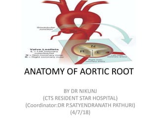
- 1. ANATOMY OF AORTIC ROOT BY DR NIKUNJ (CTS RESIDENT STAR HOSPITAL) (Coordinator:DR P.SATYENDRANATH PATHURI) (4/7/18)
- 3. AORTIC ROOT • The aortic root is the anatomic segment between the left ventricle and the ascending aorta. It contains the aortic valve and other anatomic elements, which function as a unit. The aortic root has several anatomic components: • subcommissural triangles, • aortic annulus, • aortic cusps, • aortic sinuses or sinuses of Valsalva, • sinotubular junction.
- 5. THE SUBCOMMISSURAL TRIANGLES • The subcommissural triangles are part of the left ventricular out fow tract, but they play an important role in the function of the aortic valve. • The subcommissural triangles of the noncoronary aortic cusp are fibrous extension of the intervalvular brous body and membranous septum, whereas the subcommissural triangle beneath the left and the right aortic cusps is an extension of the muscular interventricular septum.
- 6. THE AORTIC ANNULUS • The aortic annulus, a fibrous structure with a scalloped shape, attaches the aortic valve to the left ventricle. • It is attached directly to the myocardium in approximately 45% of its circumference, and to fibrous structures in the remaining 55% • The diameter of the aortic annulus is 10% to 20% larger than the diameter of the sinotubular junction of the aortic root in young patients . As the number of elastic fibers in the arterial wall decreases with age, the sinotubular junction dilates, and its diameter tends to become equal to that of the aortic annulus in older patients.
- 7. • With dilation of the aortic annulus, the subcommissural triangles of the noncoronary cusp tend to become more obtuse as the crescent shape of the aortic annulus along its fibrous insertion flattens.
- 10. CUSPS • The normal aortic valve has three cusps. Each cusp has a semilunar shape and has a base and a free margin. The base is attached to the aortic annulus in a crescent fashion. The point at which the free margin of a cusp joins its base is the commissure, and • the ridge in the aortic wall that lies immediately above the commissures is the sinotubular junction. • The free edge of each cusp is tougher consistency than the remainder of cusp. • At the mid point of each free edge is fibrou nodulus arantii on either side of nodulus is extremely thin. • The free margin of an aortic cusp extends from one of its commissures to the other. The length of the free margin of an aortic cusp is approximately 1.5 times the length of its base.
- 11. CUSPS • The three aortic cusps often have different sizes in a person, and the right and noncoronary cusps are usually larger than the left cusp. • The same cusp may have different sizes in individuals with the same body surface area • During diastole, the free margins and part of the body of the three cusps touch each other approximately in the center of the aortic root to seal the aortic orifice. • Thus, the average length of the free margins of three aortic cusps must exceed the diameter of the sinotubular junction to allow the cusps to coapt centrally and render the aortic valve competent .
- 12. • If a pathologic process causes shortening of the length of the free margin of a cusp, or if the sinotubular junction dilates, the cusps cannot coapt centrally, resulting in aortic insufciency . • If the length of a free margin is elongated, the cusp prolapses, and depending on the degree of prolapse, aortic insufficiency ensues
- 13. AORTIC SINUSES, OR SINUSES OF VALSALVA • The spaces contained between the aortic annulus and the sinotubular junction are the aortic sinuses. There are three cusps and three sinuses: • left cusp and sinus, • right cusp and sinus, • noncoronary cusp and sinus. • The left main coronary artery arises from the left aortic sinus, and the right coronary artery arises from the right aortic sinus. • There are three sinuses of the aortic valve, each related to the valve’s corresponding cusps. Each sinus is divided into three areas a central part and two adjacent parts, which are named according to the valve cusps they adjoin. • The noncoronary sinus is also refferred to as the posterior aortic sinus.
- 14. AORTIC SINUSES, OR SINUSES OF VALSALVA • The aortic sinuses facilitate closure of the aortic valve by creating eddies and currents between the cusps and arterial wall . • They also prevent the cusps from occluding the coronary artery orifices during systole, thus guaranteeing myocardial perfusion during the entire cardiac cycle.
- 16. RIGHT CORONARY SINUS • entire right coronary sinus lies adjacent to the RVOT. • central part lies adjacent to the crista supraventricularis, • left part is adjacent to the area of the RVOT in the angle between the crista supraventricularis and the pulmonary valve. • posterior (noncoronary) part of the right coronary sinus is related to the area of the right ventricle posteroinferior to the crista supraventricularis. • Inferiorly, the entire right coronary sinus is related to the interventricular septum; the muscular septum lies under the central and left parts, while either membranous or muscular septum may lie under the posterior part of the right coronary sinus.
- 17. NONCORONARY SINUS • The atrialchambers with the intervening atrial septum lie adjacent to the noncoronary sinus. • right and central parts of the noncoronary sinus are related to the right atrium and the interatrial septum, • left part is related to the left atrium. • Inferiorly, the right part, like the posterior part of the right coronary sinus, may be related either to the membranous or the muscular septum depending on the size of the membranous septum. However, beneath the central part of the noncoronary sinus, the membranous septum is a constant structure. The left part of the noncoronary sinus inserts into the anterior mitral leaflet
- 19. LEFT CORONARY SINUS • posterior part of the left coronary sinus shares the same relationship as the left part of the noncoronary sinus, that is, it is related to the left atrium posteriorly and to the anterior mitral leaflet inferiorly. • central part of the left aortic sinus is the only part of the aortic root that is not related to a cardiac chamber; it is adjacent to the epicardium only. • right part of the left coronary sinus lies adjacent to the pulmonary trunk at the level of the left pulmonary sinus. inferior to it lies the muscular interventricular septum.
- 22. • The aortic root of young individuals is elastic and very compliant. It expands and contracts during the cardiac cycle. • The normal aortic root has a fairly consistent shape, and the sizes of the cusps, the aortic annulus, the aortic sinuses, and the sinotubular junction are somewhat interdependent. • Thus, large cusps have a proportionally large annulus, sinus, and sinotubular junction.
- 23. THANK YOU
- 24. The ascending aorta • The ascending aorta begins at the distal extremity of the three aortic sinuses, the sinotubular junction, which lies at the line of opening of the free edge of the leaflets of the aortic valve. • It runs its short course passing superiorly obliquely to the right, and slightly forward toward the sternum. It is contained within the fibrous pericardial sac, so its surface is covered with serous pericardium. Its anterior surface abuts directly on the pulmonary trunk, which is also covered with serous pericardium. • the ascending aorta is related anteromedially to the right atrial appendage, and posterolaterally to the right ven-tricular out flow tract and the pulmonary trunk. • Extrapericardially, the thymus gland lies between it and the sternum.
- 25. The ascending aorta • The medial wall of the right atrium, the superior caval vein, and the right pleura relate to its right side. • On the left, its principal relationship is with the pulmonary trunk.
- 26. The arch of the aorta • The arch of the aorta begins at the superior attachment of the pericardial reflection just proximal to the origin of the brachiocephalic artery • It continues superiorly briefly before coursing posteriorly and to the left, crossing the lateral aspect of the distal trachea and finally terminating on the lateral aspect of the vertebral column. • Here it is tethered by the parietal pleura and the arterial ligament. • During its course, it gives off the brachiocephalic, the left common carotid, and the left subclavian arteries. •
- 27. • The descending, or thoracic, aorta continues from the arch, running an initial course lateral to the vertebral bodies and reaching an anterior position at its termination. It gives off many branches to the organs of the thorax throughout its course,