
Recommended
More Related Content
What's hot
What's hot (20)
Similar to Coronary arteries and veins DR NIKUNJ .R .SHRKHADA (MBBS,MS GEN SURG DNB CTS SR)
Similar to Coronary arteries and veins DR NIKUNJ .R .SHRKHADA (MBBS,MS GEN SURG DNB CTS SR) (20)
More from DR NIKUNJ SHEKHADA
More from DR NIKUNJ SHEKHADA (20)
Recently uploaded
Recently uploaded (20)
Coronary arteries and veins DR NIKUNJ .R .SHRKHADA (MBBS,MS GEN SURG DNB CTS SR)
- 1. ANATOMY OF CORONARY ARTERIES AND VEINS BY DR NIKUNJ (CTS RESIDENT STAR HOSPITAL) (Coordinator:DR P.SATYENDRANATH PATHURI) (18/6/18)
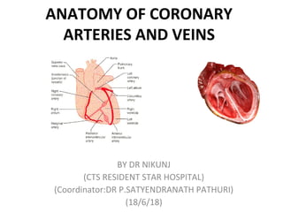
- 2. • The coronary circulation is composed of the coronary Arteries and veins together with the lymphatics of the heart. • The coronary arteries are the first branches of the ascending portion of the aorta, arising from the aortic root immediately above its attachment to the heart. • Normally, there are three sinuses at the aortic root The sinuses can be named, right coronary, left coronary, and noncoronary sinus
- 3. Right coronary artery • The right coronary artery emerges from the right- handfacing aortic sinus and immediately enters the right atrioventricular groove • Immediately after its origin, the artery gives rise to downgoing infundibular branches, one of which may also arise by a separate orifice • AV sulcus :Right Ant Ventricular A • Right Ant Atrial A - SA NODAL A • At acute marginal A upto the apex of heart. • RCA asit crosses the crux where it takes deep U turn and givingoff AV node A at the apex of turn. • RCA then terminates by biforcating in to the PDA & PLVB
- 5. • Angiographic segments of RCA • PROXIMAL RCA: Above sa nodal branch • MID RCA : sa nodal branch till acute marginal branch • DISTAL RCA: beyond acute marginal branch
- 7. Left coronary artery • LEFT MAIN CORONARY A • The main stem of the left coronary artery emerges from the left-hand-facing sinus between the pulmonary trunk and the left atrial appendage. It is a short structure, rarely extending beyond 1 cm before branching • In some hearts, the main stem trifurcates, with an intermediate branch present between the two main branches • LEFT ANTERIR DESCENDING A • LAD runs within the anterior interventricular groove, giving off diagonal branches to the obtuse margin, and the important perforating branches that pass inferiorly into the septum • The interventricular artery then continues toward the apex, and it frequently curves under the apex onto the diaphragmatic surface of the ventricles. • LOOP OF VIEUSSENS
- 9. • The circumflex branch of the left coronary artery • Originates from lmca at 90’ angle. • SA NODA A some time originates from first few cm • passes backward to run in relationship with the mitral orifice • Large branch originating from lcx continue around the LV in a AV groov termed as atrial circumflex A • Ventricular branch of lcx is Obtuse marginal A
- 10. •Type I : ends above the cardiac apex. •Type II :ends at cardiac apex. •Type III: ends beyond cardiac apex. Angiographic segments of LAD •PROXIMAL LAD: from LT coronary ostium to first major septal or diagonal •MID LAD :from first major septal or diagonal to second diagonal •DISTAL LAD :beyond second diagonal LAD
- 11. THE SINUS NODE A •Single 89% double11% •RCA :55-65% LCA:55-45%
- 13. AV NODE A •As RCA crosses the crux where it takes deep U turn and givingoff AV node A at the apex of turn. •AV node supplied by dominant coronary A. •KUGAL’S A.
- 14. VENTRICULAR SEPTUM •ANT : LAD. •POST: PDA. (small portion)
- 15. • 1. Great Cardiac vein • Begins near apex of heart; acc. LAD& more proximally cx artery • Terminates at lt end of coronary sinus • 2. Middle cardiac vein • •Accompanies PDA and opens at termination of coronary sinus • 3. Small Cardiac vein • Accompanies rt marginal artery Runs in AV groove to end into rt end of CS May open directly into rt atrium • 4. Oblique Vein of Lt Atrium Runs in the post surface of Lt Atrium and drains into Lt end of Coronary sinus • 5. Post Vein of Lt Ventricle • Runs on diaphragmatic surface of Lt ventricle and ends in middle of coronarysinus • 6. Rt Marginal vein • Accompanies Rt Marginal artery and drains into Small Cardiac vein or directly into the Rt Atrium • Valves are also found in the cardiac veins. That found in the great cardiac vein where it turns around the pulmonary surface is most constant and is called the valve of Vieussens
- 16. THANK YOU