SlideShare a Scribd company logo
1 of 44
SURGICAL ANATOMY
TRICUSPID VALVE &PULMONARY VALVE & RVOT
BY DR NIKUNJ
(CTS RESIDENT STAR HOSPITAL)
(Coordinator:DR P.SATYENDRANATH PATHURI)
(9/7/18)
SURGICAL ANATOMY
TRICUSPID VALVE
• The tricuspid valve is part of a complex functional system that also includes the
right atrium, the right ventricle, and the pulmonary circulation.
• With the tricuspid valve exposed, the surgeon observes sequentially
• right atrial cavity,
• atrio-valvular junction,
• tricuspid leaflets, and
• subvalvular apparatus.
RIGHT ATRIAL CAVITY
RIGHT ATRIAL CAVITY
• The right atrium consists of a curved
posterior groove continuous with the
superior and inferior vena cavae, a flat
interatrial septum, a trabeculated dome,
and the tricuspid valve
• The posterior groove is a smooth wall
separated from the trabeculated dome by a
ridge of muscle, the crista terminalis (a),
that extends from the superior vena cava to
the inferior vena cava.
• The interatrial septum has a central shallow
depression, the fossa ovalis (b),
• and the orifice of the coronary sinus (c)
guarded by a valvelike fold, the thebesian
valve (d).
• Another valvelike fold, the Eustachian valve
(e), guards the anterior extremity of the
inferior vena cava.
• The tendon of Todaro (f) is a fibrous structure formed by an extension of both the
Eustachian and thebesian valves.
• This tendon in conjunction with the orifice of the coronary sinus and the base of
the septal leaflet of the tricuspid valve delineate the triangle of Koch.This triangle
helps the surgeon locate two important components of the conduction system:
• the atrioventricular node (g), at the base of the triangle, and
• the bundle of His (h), which extends towards the apex of the triangle.
• The bundle of His crosses the septal segment of the tri- cuspid annulus
approximately 5 mm from the anteroseptal commissure. At this point the bundle
pierces the membranous septum or bypasses it posteriorly to reach the crest of
the muscular septum, where it bifurcates into multiple branches
THE ATRIO-VALVULAR JUNCTION
• The junction between the right atrium and the tricuspid valve is usually well-
delineated by the change in color and structure at the junction.
• In pathological valves,
• The annulus fibrosus is not visible from an atrial view because it is deeper and 2
mm external to the hinge
ANNULUS FIBROSUS
• This characteristic has important surgical implications
• sutures passed only through the hinge would compromise the motion of the
leaflets and would not reach the resistant fibrous body of the annulus; on the
contrary, sutures placed 2 mm external to the hinge and oriented towards the
ventricle reach the fibrous body of the annulus and preserve the free motion of
the leaflets.
• The annulus is a heterogeneous, almost virtual, structure composed of intermixed
fibrotic and elastic fibers in continuity with the leaflet tissue, the atrium, and the
ventricle. The posteromedial fibrous trigone reinforces the tricuspid annulus in the
area of the anteroseptal commissure.
• From the anteroseptal commissure, four annular segments can be described in a clockwise
fashion.
• The aortic segment is defined as the portion of the annulus localized at the proximity of the
neighboring aortic root. This identification is critically important because inadvertent suture
placement in this area during annuloplasty may lead to aortic valve or sinus injury.
• The anterior segment corresponds to the right outflow tract. It attaches the remaining part
of the anterior leaflet and the anteroposterior commissural leaflet.
• The posteriorsegment attaches the posterior leaflet and the postero- septal commissural
leaflet.
• The septal segment corresponds to the septum and attaches the septal leaflet and the
adjacent anteroseptal commissural leaflet .
• The septal attachment of the tricuspid valve is at a lower level than the septal
attachment of the mitral valve .
• As a result, a portion of the interventricular septum, the membranous septum,
separates the left ventricle from the right atrium , a configuration that explains the left
ventricular to right atrial shunts seen in congenital malformations.
• The annulus has a “wave form” instead of a planar configuration because of the bulging of
the right outflow tract, the nonlinear anterior and septal segments, and the lower position
(towards the apex) of the attachment of the septal leaflet.
• The two lowest positions (apically located) of the wave form are the anteroseptal
commissure,
• where the anterior leaflet attachment overrides the attachment of the septal leaflet (a)
and the posteroseptal commissure (b).
• The highest position is the portion of the aortic segment that corresponds to the right
outflow tract (c).
ANNULUS
• The shape of the “annulus” varies throughout the cardiac cycle. During diastole, it
is grossly circular (a) with a transverse diameter barely larger than the anteroseptal
diameter. During systole, the atrio- valvular junction has an ovoid shape with the
transverse diameter significantly larger than the anteroseptal diameter (b)
• The concentric reduction of the tricuspid valve orifice during systole results from the
contraction of the right and the left ventricles and the bulging of the aortic root. The
contraction of the orifice is not uniform because of the following underlying structures: the
septum, the right ventricle, and the aorta.
• In addition to concentric deformation, the tricuspid annulus displays three-dimensional (3D)
deformation at the junction of its different segments as well as rotational movement with
anterior displacement of its anterior segment in relation to its septal segment (c).
• This results in a helicoidal shape with a 3- to 7-mm gap at the anteroseptal commissure.
THE LEAFLETS
• he tricuspid valve consists of three leaflets, which are the opening and closing
structures of the tricuspid orifice.
• The three leaflets of the tricuspid valve have different sizes and shapes. The
anterior leaflet is larger than the posterior leaflet, which is larger than the septal
leaflet
THE ANTERIOR LEAFLET
• The anterior leaflet has a
semicircular shape and is attached
to the right ventricle and the
posteromedial trigone.
• The free edge of the valve may
present an indentation.
• The height of the anterior leaflet
averages 25 mm.
• Two zones,
• The proximal zone, or atrial zone, is
regular, thin, and translucent.
• The distal zone, or zone of
coaptation, is slightly irregular and
thicker than the proximal zone
THE POSTERIOR LEAFLET
• The posterior leaflet, which has a
roughly trapezoidal shape,
• is slightly smaller than the anterior
leaflet. It is inserted on one third of
the circumference of the annulus.
• This leaflet is often subdivided into
two or three segments by one or
two indentations.
• These indentations, sup- ported by
numerous chordae, facilitate the
opening of the leaflet during
diastole,
• the posterior leaflet has both
proximal and coaptation zones.
•
THE SEPTAL LEAFLET
• The septal leaflet is slightly
smaller than the posterior
leaflet and roughly
semicircular.
• A slight angle is visible at the
midline of its attachment to
the septum, which causes a
helicoidal shape of the
annulus
• the septal leaflet consists of
proximal and distal zones.
THE COMMISSURES
• The commissures, which separate the anterior, posterior, and septal leaflets, have
two zones the atrial zone and the coaptation zone.
• The commissural leaflets consist of semilunar leaflet tissue inserted on the annulus
with a free edge attaching characteristic fanlike chordae. The commissural leaflets
establish continuity between the three main leaflets of the tricuspid valve and
ensure the competency of the tricuspid valve.
• Since the commissural area is occupied by commissural tissue, the separation
between the leaflets does not reach the tricuspid annulus, an anatomical feature
that must be kept in mind when performing a commissurotomy.
•
THE SUBVALVULAR APPARATUS
• The tricuspid valve is connected to the
ventricular cavity by a suspension system—the
subvalvular apparatus—that has the following
two functions:
• one function is to limit the extent of the
upward displacement of the leafletsduring
systole;
• the other is to facilitate the opening of these
leaflets during diastole.
• To achieve these two goals, the suspension
system consists of the following two structures
that have different physical characteristics:
• papillary muscles, with contractile properties,
and
• chordae tendineae, with elastic properties.
THE PAPILLARY MUSCLES
• The papillary muscles inserted on the ventricular wall.
• The anterior papillary muscle is usually dominant and implanted on the anterior wall of the
ventricle near the apex. Its attachment may present two or three separate muscular
formations. The morphology of the anterior papillary muscle is usually long and bulky with
single or multiple heads.
• The posterior papillary muscle is either a single or a double entity implanted on the
posterior ventricular wall.
• The septal group comprises several small papillary muscles implanted on the septum. One
of the muscles near the anteroseptal commissure is implanted on the conus.
• The chordae tendineae, the extend from the papillary muscles to the leaflets, similar to the
suspension system of a “parachute”.
• On average, 25 chordae insert into the tricuspid valve.
• Three types of chordae can be described depending upon their attachment on the leaflets
• Basal chordae, attached to the base
• Intermediary or secondary chordae, attached to the
ventricular side of the belly of the leaflets.
• Marginal chordae, attached to the free edge.
• The attachment of the intermediary chordae to the leaflets may be bifurcated or
trifurcated but the marginal chordae are usually single, originating from various
levels of the papillary muscles.
• The interval between two adjacent chordae at the free edge rarely measures more
than 3 mm. This distribution prevents the prolapse of the free edge during systole.
•
TRICUSPID VALVE FUNCTION
• Two anatomical structures
close to the annulus are at risk
during tricuspid valve surgery
• (1) the non–coronary sinus of
Valsalva, especially the
commis- sure between the
non–coronary and right
coronary leaf- lets of the aortic
valve (asterisk)
• (2) the bundle of His, which
crosses the septal leaflet
attachment 3 to 5 mm from
the anteroseptal commissure
and then either perfo- rates or
circumscribes the membranous
septum before it bifurcates
into two branches.
RVOT
RIGHT VENTRICLE
• INLET :the right ventricle has a large sinus portion that surrounds and supports a
tricuspid atrioventricular (AV) valve.
• APEX
• OUTLET:smaller infundibulum (outlet portion) that supports a semilunar valve.
• The inlet and outlet valves of the right ventricle are thus widely separated.
• The entire sinus portion of the right ventricle and most of the infundibulum (both
free wall and septum) are coarsely trabeculated.
• The septal surface of the right ventricle is divided into
• an inlet portion:The inlet portion of the ventricular septum surrounds and
supports the tricuspid valve.
• a trabecular portion (sometimes called the apical trabecular portion), that portion
with the coarse trabecular pattern typical of the right ventricle
• The outlet portion of the right ventricular aspect of the ventricular septum is
smooth but complex and has three components
THE OUTLET PORTION OF THE RIGHT VENTRICULAR ASPECT OF THE
VENTRICULAR SEPTUM
• A) infundibular (conal) septum :which separates the pulmonary from the aortic
and tricuspid valves. Only part of the infundibular septum is interventricular , and
in some malformations (e.g., double outlet right ventricle)
• It must be emphasized that the most distal cepha lad portion of the infundibular
septum is not, strictly speaking, part of the ventricular septum, because in the
normal heart, the pulmonary valve arises from the apex of a cone of muscle and
does not have a septal attachment.
•
• B)outlet portion of the septum is the anterior (superior) extension, or division, of
the trabecula septomarginalis (septal band).
• C) A third small, very anterior portion is a narrow extension superior to the
trabecular septum.
• Laterally to the right, the infundibular septum imperceptibly merges with the free right
ventricular wall imme-diately beyond its attachment to the membranous septum; at that
point, it can be called the parietal extension of the infundibular septum .
• The parietal band lies anterior to the right aortic sinus , partially overlying that portion of
the free wall of the right ventricle termed the ventriculoinfundibular fold. (crista
superventricularis)
• Medially and to the left, the infundibular septum merges with the trabecular
portion of the septum between the limbs of the particularly prominent smooth, Y-
shaped muscle bundle called the trabecula septomarginalis.
• The trabecula septomarginalis extends apically to become continuous with the
moderator band, a prominent trabeculation running from septum to free wall
• The papillary muscle arrangement supporting the three leaflets of the tricuspid
valve is different from that of the mitral valve in the left ventricle.
• In the case of the tricuspid valve, in addition to a single large anterior papillary
muscle attached to the anterior free wall that fuses with the moderator band,
there are multiple smaller posterior papillary muscles attached partly to the
posterior (inferior) free wall and partly to the septum,
• group of small septal papillary muscles.
• The lowermost of these small septal muscles attaches posterior to the trabecula
septomarginalis.
• uppermost, called the medial (conal) papillary muscle (muscle of Lancisi or muscle
of Luschka), to the posterior limb of the septal band.
PULMONARY VALVE
• Pulmonary valve structure is similar to that of the
aortic valve.
• The pulmonary valve normally has three cusps, with a
nodule at the midpoint of each free edge, and lunulae
and thin, crescent-shaped coaptive surfaces on both
sides of the nodules.
• The pocket behind each cusp is the sinus.
• Pulmonary valve cusps have been described by several
terminologies,
• usually named by their relationships to the aortic
valve: right, left, and anterior
• as suggested by Anderson, right facing, left facing,
and nonfacing) in relationship to the adjacent
commissure of each valve.
• Major differences from the aortic valve are
• (1) lighter construction of pulmonary valve cusps.
• (2) normal absence of coronary artery origins, and
• (3) normal lack of fibrous continuity with the anterior tricuspid valve leaflet.
Pulmonary valve cusps are supported entirely by freestanding musculature, having
no direct relationship with the ventricular septum. The pulmonary valve is lifted
away from the ventricular septum by the subpulmonary infundibulum
• The first septal branch of the left anterior descending coronary artery pierces
theventricular septum below the shortest part of the subpulmonary infundibulum.
The artery is protected by the subpulmonary infundibulum
•
THANK YOU

More Related Content

What's hot

Aortic root anatomy DR NIKUNJ R SHEKHADA (MBBS,MS GEN SURG,DNB CTS SR)
Aortic root anatomy DR NIKUNJ R SHEKHADA (MBBS,MS GEN SURG,DNB CTS SR)Aortic root anatomy DR NIKUNJ R SHEKHADA (MBBS,MS GEN SURG,DNB CTS SR)
Aortic root anatomy DR NIKUNJ R SHEKHADA (MBBS,MS GEN SURG,DNB CTS SR)DR NIKUNJ SHEKHADA
 
Aortic stenosis Echo
Aortic stenosis Echo Aortic stenosis Echo
Aortic stenosis Echo madhusiva03
 
Echo assessment of Aortic Regurgitation
Echo assessment of Aortic RegurgitationEcho assessment of Aortic Regurgitation
Echo assessment of Aortic RegurgitationMashiul Alam
 
Mitral valve surgical anatomy DR NIKUNJ R SHEKHADA (MBBS ,MS GRN SURG , DNB C...
Mitral valve surgical anatomy DR NIKUNJ R SHEKHADA (MBBS ,MS GRN SURG , DNB C...Mitral valve surgical anatomy DR NIKUNJ R SHEKHADA (MBBS ,MS GRN SURG , DNB C...
Mitral valve surgical anatomy DR NIKUNJ R SHEKHADA (MBBS ,MS GRN SURG , DNB C...DR NIKUNJ SHEKHADA
 
Asd echo assessment
Asd echo assessmentAsd echo assessment
Asd echo assessmentMashiul Alam
 
Assessment of prosthetic valve function
Assessment of prosthetic valve functionAssessment of prosthetic valve function
Assessment of prosthetic valve functionSwapnil Garde
 
Echocardiography assessment of Aortic Regurgitation severity
Echocardiography assessment of Aortic Regurgitation severityEchocardiography assessment of Aortic Regurgitation severity
Echocardiography assessment of Aortic Regurgitation severityPRAVEEN GUPTA
 
ECHOCARDIOGRAPHIC EVALUATION OF MITRAL VALVE DISEASE -MITRAL REGURGITATION
ECHOCARDIOGRAPHIC EVALUATION OF MITRAL VALVE DISEASE -MITRAL REGURGITATIONECHOCARDIOGRAPHIC EVALUATION OF MITRAL VALVE DISEASE -MITRAL REGURGITATION
ECHOCARDIOGRAPHIC EVALUATION OF MITRAL VALVE DISEASE -MITRAL REGURGITATIONPraveen Nagula
 
Echo in restrictive cardiomyopathy
Echo in restrictive cardiomyopathyEcho in restrictive cardiomyopathy
Echo in restrictive cardiomyopathysruthiMeenaxshiSR
 

What's hot (20)

Mitral valve
Mitral valveMitral valve
Mitral valve
 
Aortic root anatomy DR NIKUNJ R SHEKHADA (MBBS,MS GEN SURG,DNB CTS SR)
Aortic root anatomy DR NIKUNJ R SHEKHADA (MBBS,MS GEN SURG,DNB CTS SR)Aortic root anatomy DR NIKUNJ R SHEKHADA (MBBS,MS GEN SURG,DNB CTS SR)
Aortic root anatomy DR NIKUNJ R SHEKHADA (MBBS,MS GEN SURG,DNB CTS SR)
 
Mitral valve anatomy
Mitral valve anatomyMitral valve anatomy
Mitral valve anatomy
 
Aortic stenosis Echo
Aortic stenosis Echo Aortic stenosis Echo
Aortic stenosis Echo
 
Echo assessment of Aortic Regurgitation
Echo assessment of Aortic RegurgitationEcho assessment of Aortic Regurgitation
Echo assessment of Aortic Regurgitation
 
Mitral valve surgical anatomy DR NIKUNJ R SHEKHADA (MBBS ,MS GRN SURG , DNB C...
Mitral valve surgical anatomy DR NIKUNJ R SHEKHADA (MBBS ,MS GRN SURG , DNB C...Mitral valve surgical anatomy DR NIKUNJ R SHEKHADA (MBBS ,MS GRN SURG , DNB C...
Mitral valve surgical anatomy DR NIKUNJ R SHEKHADA (MBBS ,MS GRN SURG , DNB C...
 
Asd echo assessment
Asd echo assessmentAsd echo assessment
Asd echo assessment
 
L-TGA or CCTGA
L-TGA or CCTGA L-TGA or CCTGA
L-TGA or CCTGA
 
Aortic repair ppt
Aortic repair pptAortic repair ppt
Aortic repair ppt
 
Assessment of prosthetic valve function
Assessment of prosthetic valve functionAssessment of prosthetic valve function
Assessment of prosthetic valve function
 
Echocardiography assessment of Aortic Regurgitation severity
Echocardiography assessment of Aortic Regurgitation severityEchocardiography assessment of Aortic Regurgitation severity
Echocardiography assessment of Aortic Regurgitation severity
 
Aortic stenosis
Aortic stenosis Aortic stenosis
Aortic stenosis
 
ECHO ASSESSMENT OF ASD FOR DEVICE CLOSURE
ECHO ASSESSMENT OF ASD FOR DEVICE CLOSURE ECHO ASSESSMENT OF ASD FOR DEVICE CLOSURE
ECHO ASSESSMENT OF ASD FOR DEVICE CLOSURE
 
Management of Small Aortic Root
Management of Small Aortic RootManagement of Small Aortic Root
Management of Small Aortic Root
 
ECHOCARDIOGRAPHIC EVALUATION OF MITRAL VALVE DISEASE -MITRAL REGURGITATION
ECHOCARDIOGRAPHIC EVALUATION OF MITRAL VALVE DISEASE -MITRAL REGURGITATIONECHOCARDIOGRAPHIC EVALUATION OF MITRAL VALVE DISEASE -MITRAL REGURGITATION
ECHOCARDIOGRAPHIC EVALUATION OF MITRAL VALVE DISEASE -MITRAL REGURGITATION
 
Echo in restrictive cardiomyopathy
Echo in restrictive cardiomyopathyEcho in restrictive cardiomyopathy
Echo in restrictive cardiomyopathy
 
Lv systolic function
Lv systolic functionLv systolic function
Lv systolic function
 
Echo assessment of mitral regurgitation
Echo assessment of mitral regurgitationEcho assessment of mitral regurgitation
Echo assessment of mitral regurgitation
 
Coronary physiology
Coronary physiologyCoronary physiology
Coronary physiology
 
Normal aortic valve echocardiogram
Normal aortic valve echocardiogramNormal aortic valve echocardiogram
Normal aortic valve echocardiogram
 

Similar to Anatomy tricuspid valve DR NIKUNJ R SHEKHADA (MBBS,MS GEN SURG ,DNB CTS SR)

Anatomy of heart
Anatomy of heartAnatomy of heart
Anatomy of heartnmonty02
 
anatomy of mitral valve
anatomy of mitral valve  anatomy of mitral valve
anatomy of mitral valve India CTVS
 
Mitral valve anatomy - ppt by kunwar sidharth
Mitral valve    anatomy - ppt by kunwar sidharthMitral valve    anatomy - ppt by kunwar sidharth
Mitral valve anatomy - ppt by kunwar sidharthkunwar sidharth
 
Dr Garva Ameta anatomy Of Left Heart.pptx
Dr Garva Ameta anatomy Of Left Heart.pptxDr Garva Ameta anatomy Of Left Heart.pptx
Dr Garva Ameta anatomy Of Left Heart.pptxPooja Jakhar
 
The valves of the heart
The valves of the heartThe valves of the heart
The valves of the heartIdris Siddiqui
 
2.Anatomy of oesophagus.ppt
2.Anatomy of oesophagus.ppt2.Anatomy of oesophagus.ppt
2.Anatomy of oesophagus.pptDr.arun Naragund
 
Morphological anatomy of heart
Morphological anatomy of heartMorphological anatomy of heart
Morphological anatomy of heartdrbashyal85
 
Anatomy of oesophagus.ppt
Anatomy of oesophagus.pptAnatomy of oesophagus.ppt
Anatomy of oesophagus.pptYtchechy
 
PRESENTATION BOWEL.pptx
PRESENTATION BOWEL.pptxPRESENTATION BOWEL.pptx
PRESENTATION BOWEL.pptxShubham661884
 
Cardiac anatomy and imaging techniques
Cardiac anatomy and imaging techniques Cardiac anatomy and imaging techniques
Cardiac anatomy and imaging techniques Hemanthvarma52
 
gastrointestinal system anatomy0406large pdf
gastrointestinal system anatomy0406large pdfgastrointestinal system anatomy0406large pdf
gastrointestinal system anatomy0406large pdfMBBS IMS MSU
 
Inner ear 2.pptx
Inner ear 2.pptxInner ear 2.pptx
Inner ear 2.pptxSruthiNaren
 
Anatomy and embryology of valves
Anatomy and embryology of valvesAnatomy and embryology of valves
Anatomy and embryology of valvesDrvasanthi
 
LARGE INTESTINE in the human body of a person
LARGE INTESTINE in the human  body of a personLARGE INTESTINE in the human  body of a person
LARGE INTESTINE in the human body of a personAtemJoshua
 
Large intestine .pdf
Large intestine .pdfLarge intestine .pdf
Large intestine .pdfssuserbf4af22
 
Surgery for atrial fibrillation abhijit presentation
Surgery for atrial fibrillation abhijit presentationSurgery for atrial fibrillation abhijit presentation
Surgery for atrial fibrillation abhijit presentationAbhijit Joshi
 

Similar to Anatomy tricuspid valve DR NIKUNJ R SHEKHADA (MBBS,MS GEN SURG ,DNB CTS SR) (20)

Aortic valve disease
Aortic valve diseaseAortic valve disease
Aortic valve disease
 
Aortic valve disease
Aortic valve diseaseAortic valve disease
Aortic valve disease
 
Anatomy of heart
Anatomy of heartAnatomy of heart
Anatomy of heart
 
anatomy of mitral valve
anatomy of mitral valve  anatomy of mitral valve
anatomy of mitral valve
 
Mitral valve anatomy - ppt by kunwar sidharth
Mitral valve    anatomy - ppt by kunwar sidharthMitral valve    anatomy - ppt by kunwar sidharth
Mitral valve anatomy - ppt by kunwar sidharth
 
Dr Garva Ameta anatomy Of Left Heart.pptx
Dr Garva Ameta anatomy Of Left Heart.pptxDr Garva Ameta anatomy Of Left Heart.pptx
Dr Garva Ameta anatomy Of Left Heart.pptx
 
The valves of the heart
The valves of the heartThe valves of the heart
The valves of the heart
 
2.Anatomy of oesophagus.ppt
2.Anatomy of oesophagus.ppt2.Anatomy of oesophagus.ppt
2.Anatomy of oesophagus.ppt
 
Morphological anatomy of heart
Morphological anatomy of heartMorphological anatomy of heart
Morphological anatomy of heart
 
Anatomy of oesophagus.ppt
Anatomy of oesophagus.pptAnatomy of oesophagus.ppt
Anatomy of oesophagus.ppt
 
PRESENTATION BOWEL.pptx
PRESENTATION BOWEL.pptxPRESENTATION BOWEL.pptx
PRESENTATION BOWEL.pptx
 
Cardiac anatomy and imaging techniques
Cardiac anatomy and imaging techniques Cardiac anatomy and imaging techniques
Cardiac anatomy and imaging techniques
 
gastrointestinal system anatomy0406large pdf
gastrointestinal system anatomy0406large pdfgastrointestinal system anatomy0406large pdf
gastrointestinal system anatomy0406large pdf
 
Inner ear 2.pptx
Inner ear 2.pptxInner ear 2.pptx
Inner ear 2.pptx
 
Anatomy and embryology of valves
Anatomy and embryology of valvesAnatomy and embryology of valves
Anatomy and embryology of valves
 
LARGE INTESTINE in the human body of a person
LARGE INTESTINE in the human  body of a personLARGE INTESTINE in the human  body of a person
LARGE INTESTINE in the human body of a person
 
Large intestine .pdf
Large intestine .pdfLarge intestine .pdf
Large intestine .pdf
 
Large intestine
Large intestineLarge intestine
Large intestine
 
Large intestine
Large intestineLarge intestine
Large intestine
 
Surgery for atrial fibrillation abhijit presentation
Surgery for atrial fibrillation abhijit presentationSurgery for atrial fibrillation abhijit presentation
Surgery for atrial fibrillation abhijit presentation
 

More from DR NIKUNJ SHEKHADA

Mitral valve surgical treatment
Mitral valve surgical treatmentMitral valve surgical treatment
Mitral valve surgical treatmentDR NIKUNJ SHEKHADA
 
Cardiac cycle DR NIKUNJ R SHEKHADA (MBBS,MS GEN SURG ,DNB CTS SR)
Cardiac cycle DR  NIKUNJ R SHEKHADA (MBBS,MS GEN SURG ,DNB CTS SR)Cardiac cycle DR  NIKUNJ R SHEKHADA (MBBS,MS GEN SURG ,DNB CTS SR)
Cardiac cycle DR NIKUNJ R SHEKHADA (MBBS,MS GEN SURG ,DNB CTS SR)DR NIKUNJ SHEKHADA
 
History of cardiac surgery DR NIKUNJ R SHEKHADA (MBBS,MS GEN SURG ,DNB CTS SR)
History of cardiac surgery  DR  NIKUNJ R SHEKHADA (MBBS,MS GEN SURG ,DNB CTS SR)History of cardiac surgery  DR  NIKUNJ R SHEKHADA (MBBS,MS GEN SURG ,DNB CTS SR)
History of cardiac surgery DR NIKUNJ R SHEKHADA (MBBS,MS GEN SURG ,DNB CTS SR)DR NIKUNJ SHEKHADA
 
Cpb oxygenators DR NIKUNJ R SHEKHADA (MBBS,MS GEN SURG ,DNB CTS SR)
Cpb oxygenators  DR  NIKUNJ R SHEKHADA (MBBS,MS GEN SURG ,DNB CTS SR)Cpb oxygenators  DR  NIKUNJ R SHEKHADA (MBBS,MS GEN SURG ,DNB CTS SR)
Cpb oxygenators DR NIKUNJ R SHEKHADA (MBBS,MS GEN SURG ,DNB CTS SR)DR NIKUNJ SHEKHADA
 
Chordal preservation DR NIKUNJ R SHEKHADA (MBBS,MS GEN SURG ,DNB CTS SR)
Chordal preservation DR  NIKUNJ R SHEKHADA (MBBS,MS GEN SURG ,DNB CTS SR)Chordal preservation DR  NIKUNJ R SHEKHADA (MBBS,MS GEN SURG ,DNB CTS SR)
Chordal preservation DR NIKUNJ R SHEKHADA (MBBS,MS GEN SURG ,DNB CTS SR)DR NIKUNJ SHEKHADA
 
Post oprative care 1 DR NIKUNJ R SHEKHADA (MBBS,MS GEN SURG,DNB CTS SR)
Post oprative care 1 DR NIKUNJ R SHEKHADA (MBBS,MS GEN SURG,DNB CTS SR)Post oprative care 1 DR NIKUNJ R SHEKHADA (MBBS,MS GEN SURG,DNB CTS SR)
Post oprative care 1 DR NIKUNJ R SHEKHADA (MBBS,MS GEN SURG,DNB CTS SR)DR NIKUNJ SHEKHADA
 
Ecg1 DR NIKUNJ R SHEKHADA (MBBS,MS GEN SURG,DNB CTS SR)
Ecg1 DR NIKUNJ R SHEKHADA (MBBS,MS GEN SURG,DNB CTS SR)Ecg1 DR NIKUNJ R SHEKHADA (MBBS,MS GEN SURG,DNB CTS SR)
Ecg1 DR NIKUNJ R SHEKHADA (MBBS,MS GEN SURG,DNB CTS SR)DR NIKUNJ SHEKHADA
 
CARDIO PLEGIA DELIVERY DR NIKUNJ R SHEKHADA (MBBS,MS GEN SURG,DNB CTS SR
CARDIO PLEGIA DELIVERY  DR NIKUNJ R SHEKHADA (MBBS,MS GEN SURG,DNB CTS SRCARDIO PLEGIA DELIVERY  DR NIKUNJ R SHEKHADA (MBBS,MS GEN SURG,DNB CTS SR
CARDIO PLEGIA DELIVERY DR NIKUNJ R SHEKHADA (MBBS,MS GEN SURG,DNB CTS SRDR NIKUNJ SHEKHADA
 
Atrial septal defect DR NIKUNJ R SHEKHADA (MBBS,MS GEN SURG,DNB CTS SR
Atrial septal defect DR NIKUNJ R SHEKHADA (MBBS,MS GEN SURG,DNB CTS SRAtrial septal defect DR NIKUNJ R SHEKHADA (MBBS,MS GEN SURG,DNB CTS SR
Atrial septal defect DR NIKUNJ R SHEKHADA (MBBS,MS GEN SURG,DNB CTS SRDR NIKUNJ SHEKHADA
 
Asd managment DR NIKUNJ R SHEKHADA (MBBS,MS GEN SURG,DNB CTS SR)
Asd managment DR NIKUNJ R SHEKHADA (MBBS,MS GEN SURG,DNB CTS SR)Asd managment DR NIKUNJ R SHEKHADA (MBBS,MS GEN SURG,DNB CTS SR)
Asd managment DR NIKUNJ R SHEKHADA (MBBS,MS GEN SURG,DNB CTS SR)DR NIKUNJ SHEKHADA
 
Myocardial protection DR NIKUNJ R SHEKHADA (MBBS ,MS GRN SURG , DNB CTS SR
Myocardial protection DR NIKUNJ R SHEKHADA (MBBS ,MS GRN SURG , DNB CTS SRMyocardial protection DR NIKUNJ R SHEKHADA (MBBS ,MS GRN SURG , DNB CTS SR
Myocardial protection DR NIKUNJ R SHEKHADA (MBBS ,MS GRN SURG , DNB CTS SRDR NIKUNJ SHEKHADA
 
CARDIO PULMONARYBYPASS BLOOD PUMPS DR NIKUNJ R SHEKHADA (MBBS,MSGEN SURG ,DN...
CARDIO PULMONARYBYPASS BLOOD PUMPS  DR NIKUNJ R SHEKHADA (MBBS,MSGEN SURG ,DN...CARDIO PULMONARYBYPASS BLOOD PUMPS  DR NIKUNJ R SHEKHADA (MBBS,MSGEN SURG ,DN...
CARDIO PULMONARYBYPASS BLOOD PUMPS DR NIKUNJ R SHEKHADA (MBBS,MSGEN SURG ,DN...DR NIKUNJ SHEKHADA
 
Ventricular septal defect DR NIKUNJ .R .SHRKHADA (MBBS,MS GEN SURG DNB CTS SR)
Ventricular septal defect  DR NIKUNJ .R .SHRKHADA (MBBS,MS GEN SURG DNB CTS SR)Ventricular septal defect  DR NIKUNJ .R .SHRKHADA (MBBS,MS GEN SURG DNB CTS SR)
Ventricular septal defect DR NIKUNJ .R .SHRKHADA (MBBS,MS GEN SURG DNB CTS SR)DR NIKUNJ SHEKHADA
 
Coronary arteries and veins DR NIKUNJ .R .SHRKHADA (MBBS,MS GEN SURG DNB CTS SR)
Coronary arteries and veins DR NIKUNJ .R .SHRKHADA (MBBS,MS GEN SURG DNB CTS SR)Coronary arteries and veins DR NIKUNJ .R .SHRKHADA (MBBS,MS GEN SURG DNB CTS SR)
Coronary arteries and veins DR NIKUNJ .R .SHRKHADA (MBBS,MS GEN SURG DNB CTS SR)DR NIKUNJ SHEKHADA
 
Anatomy of lungs 3DR NIKUNJ R SHEKHADA (MBBS,MS GEN SURG DNB CTS SR)
Anatomy of lungs 3DR NIKUNJ R SHEKHADA (MBBS,MS GEN SURG DNB CTS SR)Anatomy of lungs 3DR NIKUNJ R SHEKHADA (MBBS,MS GEN SURG DNB CTS SR)
Anatomy of lungs 3DR NIKUNJ R SHEKHADA (MBBS,MS GEN SURG DNB CTS SR)DR NIKUNJ SHEKHADA
 

More from DR NIKUNJ SHEKHADA (20)

Mitral valve surgical treatment
Mitral valve surgical treatmentMitral valve surgical treatment
Mitral valve surgical treatment
 
Mitral valve repair
Mitral valve repairMitral valve repair
Mitral valve repair
 
Mitral valve disease
Mitral valve diseaseMitral valve disease
Mitral valve disease
 
Aortic repair ppt
Aortic repair pptAortic repair ppt
Aortic repair ppt
 
Surgical emphsema
Surgical emphsemaSurgical emphsema
Surgical emphsema
 
Cardiac cycle DR NIKUNJ R SHEKHADA (MBBS,MS GEN SURG ,DNB CTS SR)
Cardiac cycle DR  NIKUNJ R SHEKHADA (MBBS,MS GEN SURG ,DNB CTS SR)Cardiac cycle DR  NIKUNJ R SHEKHADA (MBBS,MS GEN SURG ,DNB CTS SR)
Cardiac cycle DR NIKUNJ R SHEKHADA (MBBS,MS GEN SURG ,DNB CTS SR)
 
History of cardiac surgery DR NIKUNJ R SHEKHADA (MBBS,MS GEN SURG ,DNB CTS SR)
History of cardiac surgery  DR  NIKUNJ R SHEKHADA (MBBS,MS GEN SURG ,DNB CTS SR)History of cardiac surgery  DR  NIKUNJ R SHEKHADA (MBBS,MS GEN SURG ,DNB CTS SR)
History of cardiac surgery DR NIKUNJ R SHEKHADA (MBBS,MS GEN SURG ,DNB CTS SR)
 
Cpb oxygenators DR NIKUNJ R SHEKHADA (MBBS,MS GEN SURG ,DNB CTS SR)
Cpb oxygenators  DR  NIKUNJ R SHEKHADA (MBBS,MS GEN SURG ,DNB CTS SR)Cpb oxygenators  DR  NIKUNJ R SHEKHADA (MBBS,MS GEN SURG ,DNB CTS SR)
Cpb oxygenators DR NIKUNJ R SHEKHADA (MBBS,MS GEN SURG ,DNB CTS SR)
 
Chordal preservation DR NIKUNJ R SHEKHADA (MBBS,MS GEN SURG ,DNB CTS SR)
Chordal preservation DR  NIKUNJ R SHEKHADA (MBBS,MS GEN SURG ,DNB CTS SR)Chordal preservation DR  NIKUNJ R SHEKHADA (MBBS,MS GEN SURG ,DNB CTS SR)
Chordal preservation DR NIKUNJ R SHEKHADA (MBBS,MS GEN SURG ,DNB CTS SR)
 
Post oprative care 1 DR NIKUNJ R SHEKHADA (MBBS,MS GEN SURG,DNB CTS SR)
Post oprative care 1 DR NIKUNJ R SHEKHADA (MBBS,MS GEN SURG,DNB CTS SR)Post oprative care 1 DR NIKUNJ R SHEKHADA (MBBS,MS GEN SURG,DNB CTS SR)
Post oprative care 1 DR NIKUNJ R SHEKHADA (MBBS,MS GEN SURG,DNB CTS SR)
 
Ecg1 DR NIKUNJ R SHEKHADA (MBBS,MS GEN SURG,DNB CTS SR)
Ecg1 DR NIKUNJ R SHEKHADA (MBBS,MS GEN SURG,DNB CTS SR)Ecg1 DR NIKUNJ R SHEKHADA (MBBS,MS GEN SURG,DNB CTS SR)
Ecg1 DR NIKUNJ R SHEKHADA (MBBS,MS GEN SURG,DNB CTS SR)
 
CARDIO PLEGIA DELIVERY DR NIKUNJ R SHEKHADA (MBBS,MS GEN SURG,DNB CTS SR
CARDIO PLEGIA DELIVERY  DR NIKUNJ R SHEKHADA (MBBS,MS GEN SURG,DNB CTS SRCARDIO PLEGIA DELIVERY  DR NIKUNJ R SHEKHADA (MBBS,MS GEN SURG,DNB CTS SR
CARDIO PLEGIA DELIVERY DR NIKUNJ R SHEKHADA (MBBS,MS GEN SURG,DNB CTS SR
 
Atrial septal defect DR NIKUNJ R SHEKHADA (MBBS,MS GEN SURG,DNB CTS SR
Atrial septal defect DR NIKUNJ R SHEKHADA (MBBS,MS GEN SURG,DNB CTS SRAtrial septal defect DR NIKUNJ R SHEKHADA (MBBS,MS GEN SURG,DNB CTS SR
Atrial septal defect DR NIKUNJ R SHEKHADA (MBBS,MS GEN SURG,DNB CTS SR
 
Asd managment DR NIKUNJ R SHEKHADA (MBBS,MS GEN SURG,DNB CTS SR)
Asd managment DR NIKUNJ R SHEKHADA (MBBS,MS GEN SURG,DNB CTS SR)Asd managment DR NIKUNJ R SHEKHADA (MBBS,MS GEN SURG,DNB CTS SR)
Asd managment DR NIKUNJ R SHEKHADA (MBBS,MS GEN SURG,DNB CTS SR)
 
Myocardial protection DR NIKUNJ R SHEKHADA (MBBS ,MS GRN SURG , DNB CTS SR
Myocardial protection DR NIKUNJ R SHEKHADA (MBBS ,MS GRN SURG , DNB CTS SRMyocardial protection DR NIKUNJ R SHEKHADA (MBBS ,MS GRN SURG , DNB CTS SR
Myocardial protection DR NIKUNJ R SHEKHADA (MBBS ,MS GRN SURG , DNB CTS SR
 
CPB(CARDIO PULMONARY BYPASS)
CPB(CARDIO PULMONARY BYPASS)CPB(CARDIO PULMONARY BYPASS)
CPB(CARDIO PULMONARY BYPASS)
 
CARDIO PULMONARYBYPASS BLOOD PUMPS DR NIKUNJ R SHEKHADA (MBBS,MSGEN SURG ,DN...
CARDIO PULMONARYBYPASS BLOOD PUMPS  DR NIKUNJ R SHEKHADA (MBBS,MSGEN SURG ,DN...CARDIO PULMONARYBYPASS BLOOD PUMPS  DR NIKUNJ R SHEKHADA (MBBS,MSGEN SURG ,DN...
CARDIO PULMONARYBYPASS BLOOD PUMPS DR NIKUNJ R SHEKHADA (MBBS,MSGEN SURG ,DN...
 
Ventricular septal defect DR NIKUNJ .R .SHRKHADA (MBBS,MS GEN SURG DNB CTS SR)
Ventricular septal defect  DR NIKUNJ .R .SHRKHADA (MBBS,MS GEN SURG DNB CTS SR)Ventricular septal defect  DR NIKUNJ .R .SHRKHADA (MBBS,MS GEN SURG DNB CTS SR)
Ventricular septal defect DR NIKUNJ .R .SHRKHADA (MBBS,MS GEN SURG DNB CTS SR)
 
Coronary arteries and veins DR NIKUNJ .R .SHRKHADA (MBBS,MS GEN SURG DNB CTS SR)
Coronary arteries and veins DR NIKUNJ .R .SHRKHADA (MBBS,MS GEN SURG DNB CTS SR)Coronary arteries and veins DR NIKUNJ .R .SHRKHADA (MBBS,MS GEN SURG DNB CTS SR)
Coronary arteries and veins DR NIKUNJ .R .SHRKHADA (MBBS,MS GEN SURG DNB CTS SR)
 
Anatomy of lungs 3DR NIKUNJ R SHEKHADA (MBBS,MS GEN SURG DNB CTS SR)
Anatomy of lungs 3DR NIKUNJ R SHEKHADA (MBBS,MS GEN SURG DNB CTS SR)Anatomy of lungs 3DR NIKUNJ R SHEKHADA (MBBS,MS GEN SURG DNB CTS SR)
Anatomy of lungs 3DR NIKUNJ R SHEKHADA (MBBS,MS GEN SURG DNB CTS SR)
 

Recently uploaded

ANATOMY AND PHYSIOLOGY OF RESPIRATORY SYSTEM.pptx
ANATOMY AND PHYSIOLOGY OF RESPIRATORY SYSTEM.pptxANATOMY AND PHYSIOLOGY OF RESPIRATORY SYSTEM.pptx
ANATOMY AND PHYSIOLOGY OF RESPIRATORY SYSTEM.pptxSwetaba Besh
 
ANATOMY AND PHYSIOLOGY OF REPRODUCTIVE SYSTEM.pptx
ANATOMY AND PHYSIOLOGY OF REPRODUCTIVE SYSTEM.pptxANATOMY AND PHYSIOLOGY OF REPRODUCTIVE SYSTEM.pptx
ANATOMY AND PHYSIOLOGY OF REPRODUCTIVE SYSTEM.pptxSwetaba Besh
 
Call Girls Rishikesh Just Call 9667172968 Top Class Call Girl Service Available
Call Girls Rishikesh Just Call 9667172968 Top Class Call Girl Service AvailableCall Girls Rishikesh Just Call 9667172968 Top Class Call Girl Service Available
Call Girls Rishikesh Just Call 9667172968 Top Class Call Girl Service AvailableSteve Davis
 
Call Girls in Lucknow Just Call 👉👉 8875999948 Top Class Call Girl Service Ava...
Call Girls in Lucknow Just Call 👉👉 8875999948 Top Class Call Girl Service Ava...Call Girls in Lucknow Just Call 👉👉 8875999948 Top Class Call Girl Service Ava...
Call Girls in Lucknow Just Call 👉👉 8875999948 Top Class Call Girl Service Ava...Janvi Singh
 
Cardiac Output, Venous Return, and Their Regulation
Cardiac Output, Venous Return, and Their RegulationCardiac Output, Venous Return, and Their Regulation
Cardiac Output, Venous Return, and Their RegulationMedicoseAcademics
 
Call Girls in Lucknow Just Call 👉👉8630512678 Top Class Call Girl Service Avai...
Call Girls in Lucknow Just Call 👉👉8630512678 Top Class Call Girl Service Avai...Call Girls in Lucknow Just Call 👉👉8630512678 Top Class Call Girl Service Avai...
Call Girls in Lucknow Just Call 👉👉8630512678 Top Class Call Girl Service Avai...soniyagrag336
 
Call Girl in Chennai | Whatsapp No 📞 7427069034 📞 VIP Escorts Service Availab...
Call Girl in Chennai | Whatsapp No 📞 7427069034 📞 VIP Escorts Service Availab...Call Girl in Chennai | Whatsapp No 📞 7427069034 📞 VIP Escorts Service Availab...
Call Girl in Chennai | Whatsapp No 📞 7427069034 📞 VIP Escorts Service Availab...amritaverma53
 
Indore Call Girls ❤️🍑7718850664❤️🍑 Call Girl service in Indore ☎️ Indore Call...
Indore Call Girls ❤️🍑7718850664❤️🍑 Call Girl service in Indore ☎️ Indore Call...Indore Call Girls ❤️🍑7718850664❤️🍑 Call Girl service in Indore ☎️ Indore Call...
Indore Call Girls ❤️🍑7718850664❤️🍑 Call Girl service in Indore ☎️ Indore Call...minkseocompany
 
Bhopal❤CALL GIRL 9352988975 ❤CALL GIRLS IN Bhopal ESCORT SERVICE
Bhopal❤CALL GIRL 9352988975 ❤CALL GIRLS IN Bhopal ESCORT SERVICEBhopal❤CALL GIRL 9352988975 ❤CALL GIRLS IN Bhopal ESCORT SERVICE
Bhopal❤CALL GIRL 9352988975 ❤CALL GIRLS IN Bhopal ESCORT SERVICErahuljha3240
 
Call Girls Kathua Just Call 8250077686 Top Class Call Girl Service Available
Call Girls Kathua Just Call 8250077686 Top Class Call Girl Service AvailableCall Girls Kathua Just Call 8250077686 Top Class Call Girl Service Available
Call Girls Kathua Just Call 8250077686 Top Class Call Girl Service AvailableDipal Arora
 
Call Girls Mussoorie Just Call 8854095900 Top Class Call Girl Service Available
Call Girls Mussoorie Just Call 8854095900 Top Class Call Girl Service AvailableCall Girls Mussoorie Just Call 8854095900 Top Class Call Girl Service Available
Call Girls Mussoorie Just Call 8854095900 Top Class Call Girl Service AvailableJanvi Singh
 
Lucknow Call Girls Service { 9984666624 } ❤️VVIP ROCKY Call Girl in Lucknow U...
Lucknow Call Girls Service { 9984666624 } ❤️VVIP ROCKY Call Girl in Lucknow U...Lucknow Call Girls Service { 9984666624 } ❤️VVIP ROCKY Call Girl in Lucknow U...
Lucknow Call Girls Service { 9984666624 } ❤️VVIP ROCKY Call Girl in Lucknow U...Janvi Singh
 
Cara Menggugurkan Kandungan Dengan Cepat Selesai Dalam 24 Jam Secara Alami Bu...
Cara Menggugurkan Kandungan Dengan Cepat Selesai Dalam 24 Jam Secara Alami Bu...Cara Menggugurkan Kandungan Dengan Cepat Selesai Dalam 24 Jam Secara Alami Bu...
Cara Menggugurkan Kandungan Dengan Cepat Selesai Dalam 24 Jam Secara Alami Bu...Cara Menggugurkan Kandungan 087776558899
 
Russian Call Girls In Pune 👉 Just CALL ME: 9352988975 ✅❤️💯low cost unlimited ...
Russian Call Girls In Pune 👉 Just CALL ME: 9352988975 ✅❤️💯low cost unlimited ...Russian Call Girls In Pune 👉 Just CALL ME: 9352988975 ✅❤️💯low cost unlimited ...
Russian Call Girls In Pune 👉 Just CALL ME: 9352988975 ✅❤️💯low cost unlimited ...chanderprakash5506
 
Call Girls Bangalore - 450+ Call Girl Cash Payment 💯Call Us 🔝 6378878445 🔝 💃 ...
Call Girls Bangalore - 450+ Call Girl Cash Payment 💯Call Us 🔝 6378878445 🔝 💃 ...Call Girls Bangalore - 450+ Call Girl Cash Payment 💯Call Us 🔝 6378878445 🔝 💃 ...
Call Girls Bangalore - 450+ Call Girl Cash Payment 💯Call Us 🔝 6378878445 🔝 💃 ...gragneelam30
 
Call girls Service Phullen / 9332606886 Genuine Call girls with real Photos a...
Call girls Service Phullen / 9332606886 Genuine Call girls with real Photos a...Call girls Service Phullen / 9332606886 Genuine Call girls with real Photos a...
Call girls Service Phullen / 9332606886 Genuine Call girls with real Photos a...call girls hydrabad
 
7 steps How to prevent Thalassemia : Dr Sharda Jain & Vandana Gupta
7 steps How to prevent Thalassemia : Dr Sharda Jain & Vandana Gupta7 steps How to prevent Thalassemia : Dr Sharda Jain & Vandana Gupta
7 steps How to prevent Thalassemia : Dr Sharda Jain & Vandana GuptaLifecare Centre
 
Bhawanipatna Call Girls 📞9332606886 Call Girls in Bhawanipatna Escorts servic...
Bhawanipatna Call Girls 📞9332606886 Call Girls in Bhawanipatna Escorts servic...Bhawanipatna Call Girls 📞9332606886 Call Girls in Bhawanipatna Escorts servic...
Bhawanipatna Call Girls 📞9332606886 Call Girls in Bhawanipatna Escorts servic...Dipal Arora
 
💰Call Girl In Bangalore☎️63788-78445💰 Call Girl service in Bangalore☎️Bangalo...
💰Call Girl In Bangalore☎️63788-78445💰 Call Girl service in Bangalore☎️Bangalo...💰Call Girl In Bangalore☎️63788-78445💰 Call Girl service in Bangalore☎️Bangalo...
💰Call Girl In Bangalore☎️63788-78445💰 Call Girl service in Bangalore☎️Bangalo...gragneelam30
 
Call Girls Service Jaipur {9521753030 } ❤️VVIP BHAWNA Call Girl in Jaipur Raj...
Call Girls Service Jaipur {9521753030 } ❤️VVIP BHAWNA Call Girl in Jaipur Raj...Call Girls Service Jaipur {9521753030 } ❤️VVIP BHAWNA Call Girl in Jaipur Raj...
Call Girls Service Jaipur {9521753030 } ❤️VVIP BHAWNA Call Girl in Jaipur Raj...Janvi Singh
 

Recently uploaded (20)

ANATOMY AND PHYSIOLOGY OF RESPIRATORY SYSTEM.pptx
ANATOMY AND PHYSIOLOGY OF RESPIRATORY SYSTEM.pptxANATOMY AND PHYSIOLOGY OF RESPIRATORY SYSTEM.pptx
ANATOMY AND PHYSIOLOGY OF RESPIRATORY SYSTEM.pptx
 
ANATOMY AND PHYSIOLOGY OF REPRODUCTIVE SYSTEM.pptx
ANATOMY AND PHYSIOLOGY OF REPRODUCTIVE SYSTEM.pptxANATOMY AND PHYSIOLOGY OF REPRODUCTIVE SYSTEM.pptx
ANATOMY AND PHYSIOLOGY OF REPRODUCTIVE SYSTEM.pptx
 
Call Girls Rishikesh Just Call 9667172968 Top Class Call Girl Service Available
Call Girls Rishikesh Just Call 9667172968 Top Class Call Girl Service AvailableCall Girls Rishikesh Just Call 9667172968 Top Class Call Girl Service Available
Call Girls Rishikesh Just Call 9667172968 Top Class Call Girl Service Available
 
Call Girls in Lucknow Just Call 👉👉 8875999948 Top Class Call Girl Service Ava...
Call Girls in Lucknow Just Call 👉👉 8875999948 Top Class Call Girl Service Ava...Call Girls in Lucknow Just Call 👉👉 8875999948 Top Class Call Girl Service Ava...
Call Girls in Lucknow Just Call 👉👉 8875999948 Top Class Call Girl Service Ava...
 
Cardiac Output, Venous Return, and Their Regulation
Cardiac Output, Venous Return, and Their RegulationCardiac Output, Venous Return, and Their Regulation
Cardiac Output, Venous Return, and Their Regulation
 
Call Girls in Lucknow Just Call 👉👉8630512678 Top Class Call Girl Service Avai...
Call Girls in Lucknow Just Call 👉👉8630512678 Top Class Call Girl Service Avai...Call Girls in Lucknow Just Call 👉👉8630512678 Top Class Call Girl Service Avai...
Call Girls in Lucknow Just Call 👉👉8630512678 Top Class Call Girl Service Avai...
 
Call Girl in Chennai | Whatsapp No 📞 7427069034 📞 VIP Escorts Service Availab...
Call Girl in Chennai | Whatsapp No 📞 7427069034 📞 VIP Escorts Service Availab...Call Girl in Chennai | Whatsapp No 📞 7427069034 📞 VIP Escorts Service Availab...
Call Girl in Chennai | Whatsapp No 📞 7427069034 📞 VIP Escorts Service Availab...
 
Indore Call Girls ❤️🍑7718850664❤️🍑 Call Girl service in Indore ☎️ Indore Call...
Indore Call Girls ❤️🍑7718850664❤️🍑 Call Girl service in Indore ☎️ Indore Call...Indore Call Girls ❤️🍑7718850664❤️🍑 Call Girl service in Indore ☎️ Indore Call...
Indore Call Girls ❤️🍑7718850664❤️🍑 Call Girl service in Indore ☎️ Indore Call...
 
Bhopal❤CALL GIRL 9352988975 ❤CALL GIRLS IN Bhopal ESCORT SERVICE
Bhopal❤CALL GIRL 9352988975 ❤CALL GIRLS IN Bhopal ESCORT SERVICEBhopal❤CALL GIRL 9352988975 ❤CALL GIRLS IN Bhopal ESCORT SERVICE
Bhopal❤CALL GIRL 9352988975 ❤CALL GIRLS IN Bhopal ESCORT SERVICE
 
Call Girls Kathua Just Call 8250077686 Top Class Call Girl Service Available
Call Girls Kathua Just Call 8250077686 Top Class Call Girl Service AvailableCall Girls Kathua Just Call 8250077686 Top Class Call Girl Service Available
Call Girls Kathua Just Call 8250077686 Top Class Call Girl Service Available
 
Call Girls Mussoorie Just Call 8854095900 Top Class Call Girl Service Available
Call Girls Mussoorie Just Call 8854095900 Top Class Call Girl Service AvailableCall Girls Mussoorie Just Call 8854095900 Top Class Call Girl Service Available
Call Girls Mussoorie Just Call 8854095900 Top Class Call Girl Service Available
 
Lucknow Call Girls Service { 9984666624 } ❤️VVIP ROCKY Call Girl in Lucknow U...
Lucknow Call Girls Service { 9984666624 } ❤️VVIP ROCKY Call Girl in Lucknow U...Lucknow Call Girls Service { 9984666624 } ❤️VVIP ROCKY Call Girl in Lucknow U...
Lucknow Call Girls Service { 9984666624 } ❤️VVIP ROCKY Call Girl in Lucknow U...
 
Cara Menggugurkan Kandungan Dengan Cepat Selesai Dalam 24 Jam Secara Alami Bu...
Cara Menggugurkan Kandungan Dengan Cepat Selesai Dalam 24 Jam Secara Alami Bu...Cara Menggugurkan Kandungan Dengan Cepat Selesai Dalam 24 Jam Secara Alami Bu...
Cara Menggugurkan Kandungan Dengan Cepat Selesai Dalam 24 Jam Secara Alami Bu...
 
Russian Call Girls In Pune 👉 Just CALL ME: 9352988975 ✅❤️💯low cost unlimited ...
Russian Call Girls In Pune 👉 Just CALL ME: 9352988975 ✅❤️💯low cost unlimited ...Russian Call Girls In Pune 👉 Just CALL ME: 9352988975 ✅❤️💯low cost unlimited ...
Russian Call Girls In Pune 👉 Just CALL ME: 9352988975 ✅❤️💯low cost unlimited ...
 
Call Girls Bangalore - 450+ Call Girl Cash Payment 💯Call Us 🔝 6378878445 🔝 💃 ...
Call Girls Bangalore - 450+ Call Girl Cash Payment 💯Call Us 🔝 6378878445 🔝 💃 ...Call Girls Bangalore - 450+ Call Girl Cash Payment 💯Call Us 🔝 6378878445 🔝 💃 ...
Call Girls Bangalore - 450+ Call Girl Cash Payment 💯Call Us 🔝 6378878445 🔝 💃 ...
 
Call girls Service Phullen / 9332606886 Genuine Call girls with real Photos a...
Call girls Service Phullen / 9332606886 Genuine Call girls with real Photos a...Call girls Service Phullen / 9332606886 Genuine Call girls with real Photos a...
Call girls Service Phullen / 9332606886 Genuine Call girls with real Photos a...
 
7 steps How to prevent Thalassemia : Dr Sharda Jain & Vandana Gupta
7 steps How to prevent Thalassemia : Dr Sharda Jain & Vandana Gupta7 steps How to prevent Thalassemia : Dr Sharda Jain & Vandana Gupta
7 steps How to prevent Thalassemia : Dr Sharda Jain & Vandana Gupta
 
Bhawanipatna Call Girls 📞9332606886 Call Girls in Bhawanipatna Escorts servic...
Bhawanipatna Call Girls 📞9332606886 Call Girls in Bhawanipatna Escorts servic...Bhawanipatna Call Girls 📞9332606886 Call Girls in Bhawanipatna Escorts servic...
Bhawanipatna Call Girls 📞9332606886 Call Girls in Bhawanipatna Escorts servic...
 
💰Call Girl In Bangalore☎️63788-78445💰 Call Girl service in Bangalore☎️Bangalo...
💰Call Girl In Bangalore☎️63788-78445💰 Call Girl service in Bangalore☎️Bangalo...💰Call Girl In Bangalore☎️63788-78445💰 Call Girl service in Bangalore☎️Bangalo...
💰Call Girl In Bangalore☎️63788-78445💰 Call Girl service in Bangalore☎️Bangalo...
 
Call Girls Service Jaipur {9521753030 } ❤️VVIP BHAWNA Call Girl in Jaipur Raj...
Call Girls Service Jaipur {9521753030 } ❤️VVIP BHAWNA Call Girl in Jaipur Raj...Call Girls Service Jaipur {9521753030 } ❤️VVIP BHAWNA Call Girl in Jaipur Raj...
Call Girls Service Jaipur {9521753030 } ❤️VVIP BHAWNA Call Girl in Jaipur Raj...
 

Anatomy tricuspid valve DR NIKUNJ R SHEKHADA (MBBS,MS GEN SURG ,DNB CTS SR)

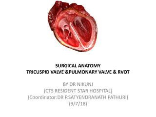
  • 1. SURGICAL ANATOMY TRICUSPID VALVE &PULMONARY VALVE & RVOT BY DR NIKUNJ (CTS RESIDENT STAR HOSPITAL) (Coordinator:DR P.SATYENDRANATH PATHURI) (9/7/18)
  • 2. SURGICAL ANATOMY TRICUSPID VALVE • The tricuspid valve is part of a complex functional system that also includes the right atrium, the right ventricle, and the pulmonary circulation. • With the tricuspid valve exposed, the surgeon observes sequentially • right atrial cavity, • atrio-valvular junction, • tricuspid leaflets, and • subvalvular apparatus.
  • 3.
  • 5. RIGHT ATRIAL CAVITY • The right atrium consists of a curved posterior groove continuous with the superior and inferior vena cavae, a flat interatrial septum, a trabeculated dome, and the tricuspid valve • The posterior groove is a smooth wall separated from the trabeculated dome by a ridge of muscle, the crista terminalis (a), that extends from the superior vena cava to the inferior vena cava. • The interatrial septum has a central shallow depression, the fossa ovalis (b), • and the orifice of the coronary sinus (c) guarded by a valvelike fold, the thebesian valve (d). • Another valvelike fold, the Eustachian valve (e), guards the anterior extremity of the inferior vena cava.
  • 6. • The tendon of Todaro (f) is a fibrous structure formed by an extension of both the Eustachian and thebesian valves. • This tendon in conjunction with the orifice of the coronary sinus and the base of the septal leaflet of the tricuspid valve delineate the triangle of Koch.This triangle helps the surgeon locate two important components of the conduction system: • the atrioventricular node (g), at the base of the triangle, and • the bundle of His (h), which extends towards the apex of the triangle. • The bundle of His crosses the septal segment of the tri- cuspid annulus approximately 5 mm from the anteroseptal commissure. At this point the bundle pierces the membranous septum or bypasses it posteriorly to reach the crest of the muscular septum, where it bifurcates into multiple branches
  • 7. THE ATRIO-VALVULAR JUNCTION • The junction between the right atrium and the tricuspid valve is usually well- delineated by the change in color and structure at the junction. • In pathological valves, • The annulus fibrosus is not visible from an atrial view because it is deeper and 2 mm external to the hinge
  • 8. ANNULUS FIBROSUS • This characteristic has important surgical implications • sutures passed only through the hinge would compromise the motion of the leaflets and would not reach the resistant fibrous body of the annulus; on the contrary, sutures placed 2 mm external to the hinge and oriented towards the ventricle reach the fibrous body of the annulus and preserve the free motion of the leaflets.
  • 9. • The annulus is a heterogeneous, almost virtual, structure composed of intermixed fibrotic and elastic fibers in continuity with the leaflet tissue, the atrium, and the ventricle. The posteromedial fibrous trigone reinforces the tricuspid annulus in the area of the anteroseptal commissure.
  • 10.
  • 11. • From the anteroseptal commissure, four annular segments can be described in a clockwise fashion. • The aortic segment is defined as the portion of the annulus localized at the proximity of the neighboring aortic root. This identification is critically important because inadvertent suture placement in this area during annuloplasty may lead to aortic valve or sinus injury. • The anterior segment corresponds to the right outflow tract. It attaches the remaining part of the anterior leaflet and the anteroposterior commissural leaflet. • The posteriorsegment attaches the posterior leaflet and the postero- septal commissural leaflet. • The septal segment corresponds to the septum and attaches the septal leaflet and the adjacent anteroseptal commissural leaflet .
  • 12. • The septal attachment of the tricuspid valve is at a lower level than the septal attachment of the mitral valve . • As a result, a portion of the interventricular septum, the membranous septum, separates the left ventricle from the right atrium , a configuration that explains the left ventricular to right atrial shunts seen in congenital malformations.
  • 13. • The annulus has a “wave form” instead of a planar configuration because of the bulging of the right outflow tract, the nonlinear anterior and septal segments, and the lower position (towards the apex) of the attachment of the septal leaflet. • The two lowest positions (apically located) of the wave form are the anteroseptal commissure, • where the anterior leaflet attachment overrides the attachment of the septal leaflet (a) and the posteroseptal commissure (b). • The highest position is the portion of the aortic segment that corresponds to the right outflow tract (c).
  • 14. ANNULUS • The shape of the “annulus” varies throughout the cardiac cycle. During diastole, it is grossly circular (a) with a transverse diameter barely larger than the anteroseptal diameter. During systole, the atrio- valvular junction has an ovoid shape with the transverse diameter significantly larger than the anteroseptal diameter (b)
  • 15. • The concentric reduction of the tricuspid valve orifice during systole results from the contraction of the right and the left ventricles and the bulging of the aortic root. The contraction of the orifice is not uniform because of the following underlying structures: the septum, the right ventricle, and the aorta. • In addition to concentric deformation, the tricuspid annulus displays three-dimensional (3D) deformation at the junction of its different segments as well as rotational movement with anterior displacement of its anterior segment in relation to its septal segment (c). • This results in a helicoidal shape with a 3- to 7-mm gap at the anteroseptal commissure.
  • 16.
  • 17. THE LEAFLETS • he tricuspid valve consists of three leaflets, which are the opening and closing structures of the tricuspid orifice. • The three leaflets of the tricuspid valve have different sizes and shapes. The anterior leaflet is larger than the posterior leaflet, which is larger than the septal leaflet
  • 18.
  • 19. THE ANTERIOR LEAFLET • The anterior leaflet has a semicircular shape and is attached to the right ventricle and the posteromedial trigone. • The free edge of the valve may present an indentation. • The height of the anterior leaflet averages 25 mm. • Two zones, • The proximal zone, or atrial zone, is regular, thin, and translucent. • The distal zone, or zone of coaptation, is slightly irregular and thicker than the proximal zone
  • 20. THE POSTERIOR LEAFLET • The posterior leaflet, which has a roughly trapezoidal shape, • is slightly smaller than the anterior leaflet. It is inserted on one third of the circumference of the annulus. • This leaflet is often subdivided into two or three segments by one or two indentations. • These indentations, sup- ported by numerous chordae, facilitate the opening of the leaflet during diastole, • the posterior leaflet has both proximal and coaptation zones. •
  • 21. THE SEPTAL LEAFLET • The septal leaflet is slightly smaller than the posterior leaflet and roughly semicircular. • A slight angle is visible at the midline of its attachment to the septum, which causes a helicoidal shape of the annulus • the septal leaflet consists of proximal and distal zones.
  • 22. THE COMMISSURES • The commissures, which separate the anterior, posterior, and septal leaflets, have two zones the atrial zone and the coaptation zone. • The commissural leaflets consist of semilunar leaflet tissue inserted on the annulus with a free edge attaching characteristic fanlike chordae. The commissural leaflets establish continuity between the three main leaflets of the tricuspid valve and ensure the competency of the tricuspid valve. • Since the commissural area is occupied by commissural tissue, the separation between the leaflets does not reach the tricuspid annulus, an anatomical feature that must be kept in mind when performing a commissurotomy. •
  • 23.
  • 24. THE SUBVALVULAR APPARATUS • The tricuspid valve is connected to the ventricular cavity by a suspension system—the subvalvular apparatus—that has the following two functions: • one function is to limit the extent of the upward displacement of the leafletsduring systole; • the other is to facilitate the opening of these leaflets during diastole. • To achieve these two goals, the suspension system consists of the following two structures that have different physical characteristics: • papillary muscles, with contractile properties, and • chordae tendineae, with elastic properties.
  • 25. THE PAPILLARY MUSCLES • The papillary muscles inserted on the ventricular wall. • The anterior papillary muscle is usually dominant and implanted on the anterior wall of the ventricle near the apex. Its attachment may present two or three separate muscular formations. The morphology of the anterior papillary muscle is usually long and bulky with single or multiple heads. • The posterior papillary muscle is either a single or a double entity implanted on the posterior ventricular wall. • The septal group comprises several small papillary muscles implanted on the septum. One of the muscles near the anteroseptal commissure is implanted on the conus.
  • 26. • The chordae tendineae, the extend from the papillary muscles to the leaflets, similar to the suspension system of a “parachute”. • On average, 25 chordae insert into the tricuspid valve. • Three types of chordae can be described depending upon their attachment on the leaflets • Basal chordae, attached to the base • Intermediary or secondary chordae, attached to the ventricular side of the belly of the leaflets. • Marginal chordae, attached to the free edge.
  • 27. • The attachment of the intermediary chordae to the leaflets may be bifurcated or trifurcated but the marginal chordae are usually single, originating from various levels of the papillary muscles. • The interval between two adjacent chordae at the free edge rarely measures more than 3 mm. This distribution prevents the prolapse of the free edge during systole. •
  • 28.
  • 29.
  • 31. • Two anatomical structures close to the annulus are at risk during tricuspid valve surgery • (1) the non–coronary sinus of Valsalva, especially the commis- sure between the non–coronary and right coronary leaf- lets of the aortic valve (asterisk) • (2) the bundle of His, which crosses the septal leaflet attachment 3 to 5 mm from the anteroseptal commissure and then either perfo- rates or circumscribes the membranous septum before it bifurcates into two branches.
  • 32. RVOT
  • 33.
  • 34. RIGHT VENTRICLE • INLET :the right ventricle has a large sinus portion that surrounds and supports a tricuspid atrioventricular (AV) valve. • APEX • OUTLET:smaller infundibulum (outlet portion) that supports a semilunar valve. • The inlet and outlet valves of the right ventricle are thus widely separated. • The entire sinus portion of the right ventricle and most of the infundibulum (both free wall and septum) are coarsely trabeculated.
  • 35. • The septal surface of the right ventricle is divided into • an inlet portion:The inlet portion of the ventricular septum surrounds and supports the tricuspid valve. • a trabecular portion (sometimes called the apical trabecular portion), that portion with the coarse trabecular pattern typical of the right ventricle • The outlet portion of the right ventricular aspect of the ventricular septum is smooth but complex and has three components
  • 36. THE OUTLET PORTION OF THE RIGHT VENTRICULAR ASPECT OF THE VENTRICULAR SEPTUM • A) infundibular (conal) septum :which separates the pulmonary from the aortic and tricuspid valves. Only part of the infundibular septum is interventricular , and in some malformations (e.g., double outlet right ventricle) • It must be emphasized that the most distal cepha lad portion of the infundibular septum is not, strictly speaking, part of the ventricular septum, because in the normal heart, the pulmonary valve arises from the apex of a cone of muscle and does not have a septal attachment. •
  • 37. • B)outlet portion of the septum is the anterior (superior) extension, or division, of the trabecula septomarginalis (septal band). • C) A third small, very anterior portion is a narrow extension superior to the trabecular septum.
  • 38. • Laterally to the right, the infundibular septum imperceptibly merges with the free right ventricular wall imme-diately beyond its attachment to the membranous septum; at that point, it can be called the parietal extension of the infundibular septum . • The parietal band lies anterior to the right aortic sinus , partially overlying that portion of the free wall of the right ventricle termed the ventriculoinfundibular fold. (crista superventricularis)
  • 39. • Medially and to the left, the infundibular septum merges with the trabecular portion of the septum between the limbs of the particularly prominent smooth, Y- shaped muscle bundle called the trabecula septomarginalis. • The trabecula septomarginalis extends apically to become continuous with the moderator band, a prominent trabeculation running from septum to free wall
  • 40. • The papillary muscle arrangement supporting the three leaflets of the tricuspid valve is different from that of the mitral valve in the left ventricle. • In the case of the tricuspid valve, in addition to a single large anterior papillary muscle attached to the anterior free wall that fuses with the moderator band, there are multiple smaller posterior papillary muscles attached partly to the posterior (inferior) free wall and partly to the septum, • group of small septal papillary muscles. • The lowermost of these small septal muscles attaches posterior to the trabecula septomarginalis. • uppermost, called the medial (conal) papillary muscle (muscle of Lancisi or muscle of Luschka), to the posterior limb of the septal band.
  • 41. PULMONARY VALVE • Pulmonary valve structure is similar to that of the aortic valve. • The pulmonary valve normally has three cusps, with a nodule at the midpoint of each free edge, and lunulae and thin, crescent-shaped coaptive surfaces on both sides of the nodules. • The pocket behind each cusp is the sinus. • Pulmonary valve cusps have been described by several terminologies, • usually named by their relationships to the aortic valve: right, left, and anterior • as suggested by Anderson, right facing, left facing, and nonfacing) in relationship to the adjacent commissure of each valve.
  • 42. • Major differences from the aortic valve are • (1) lighter construction of pulmonary valve cusps. • (2) normal absence of coronary artery origins, and • (3) normal lack of fibrous continuity with the anterior tricuspid valve leaflet. Pulmonary valve cusps are supported entirely by freestanding musculature, having no direct relationship with the ventricular septum. The pulmonary valve is lifted away from the ventricular septum by the subpulmonary infundibulum
  • 43. • The first septal branch of the left anterior descending coronary artery pierces theventricular septum below the shortest part of the subpulmonary infundibulum. The artery is protected by the subpulmonary infundibulum •