
Recommended
Recommended
More Related Content
What's hot
What's hot (20)
Similar to Adaptations of Melanosomes & Neuroendocrine Cells
Similar to Adaptations of Melanosomes & Neuroendocrine Cells (20)
More from Muunda Mudenda
More from Muunda Mudenda (13)
Recently uploaded
Recently uploaded (20)
Adaptations of Melanosomes & Neuroendocrine Cells
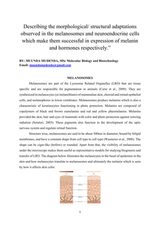
- 1. 1 Describing the morphological/ structural adaptations observed in the melanosomes and neuroendocrine cells which make them successful in expression of melanin and hormones respectively.” BY: MUUNDA MUDENDA, MSc Molecular Biology and Biotechnology Email: muundamudenda@gmail.com MELANOSOMES Melanosomes are part of the Lysosome Related Organelles (LRO) that are tissue specific and are responsible for pigmentation in animals (Curie et al., 2009). They are synthesized in melanocytes (or melanoblasts) of mammalian skin, choroid and retinal epithelial cells, and melanophores in lower vertebrates. Melanosomes produce melanins which is also a characteristic of keratinocytes functioning in photo protection. Melanins are composed of copolymers of black and brown eumelanins and red and yellow pheomelanins. Melanins provided the skin, hair and eyes of mammals with color and photo protection against ionizing radiation (Setaluri, 2003). These pigments also function in the development of the optic nervous system and regulate retinal function. Structure wise, melanosomes are said to be about 500nm in diameter, bound by bilipid membranes, and have a constant shape from cell type to cell type (Wasmeier et al., 2008). The shape can be cigar-like (hollow) or rounded. Apart from that, the visibility of melanosomes under the microscope makes them useful as representative models for studying biogenesis and transfer of LRO. The diagram below illustrates the melanocytes in the basal of epidermis in the skin and how melanocytes translate to melanosomes and ultimately the melanin which is seen by how it affects skin color.
- 2. 2 Source of Image: courses.lumenlearning.com Distinct Morphological Stages of Melanosome Maturation. The following stages are excerpts from (Setaluri, 2003) Stage 1: Membrane vesicles containing no visible pigment but irregular internal membrane structures are called premelanosomes. Stage 2: Premelanosomes of stage 1 results in elongation of the vesicles, and ordering of the internal membranes into parallel structures. Stage 3: Melanosomes can be distinguished by the presence of ordered deposition of melanin on the internal fibers. Stage 4: Melanosomes filled with melanin pigment no luminal structures are distinguishable. The diagram below illustrates the morphological stages of melanosomes. The diagram also illustrates the morphological differences between eumelanosomes and pheomelanosomes. Source of Image: (D’Alba & Shawkey, 2019) Structural adaption of Melanosomes In essence, melanin is synthesized in the melanocytes and the melanosomes are the vehicles by which melanin is transported from the melanocytes of basal epidermis to the lower and upper keratinocytes. As such, the size and shape of the melanocytes play a major role when it comes to the transportation of melanocytes. To begin with, the fairly large size of melanosomes helps to make readily available enough melanin pigments where they are needed. According to D’Alba & Shawkey, (2019), melanin is composed of 30–50 nm Nano- particles, in which melanin monomers are cross-linked together and form stacking structures by pi-pi interactions. This implies that the shape of melanosomes is well adapted to
- 3. 3 accommodate the stacking structure organization made by melanin during its transportation to the action regions of the body. Moreover, melanosomes with ciger-like structures easily navigate the cell matrix and its components to the regions where they are needed to deliver melanin pigment. The different biological molecules such as proteins and lipids, found in melanosomes and the physical and chemical properties of their surfaces determine the properties and biological functions of melanins (Simon et al., 2008). NEUROENDOCRINE CELLS Neuroendocrine cells are the connecting link between the nervous system and the endocrine system. The two systems, together, form what is called the neuroendocrine system. The neuroendocrine cells are sometimes referred to as Neurosecretory Cells (NSC) because of attributes like membrane excitability which is similar to neurons. However, unlink neurons which release neurotransmitters into the synaptic clefts, neuroendocrine cells secret hormones (Volume et al., 2009). How the neuroendocrine system works is that, the nervous system release signals (neurotransmitters) which are sent to the neuroendocrine cells and these release (secret) neurohormones (peptides, proteins) into the blood leading to action to the target site. Neuroendocrine cells are located in multiple organs (Figure 1) of the body which enables them to control different cellular and physiological processes in the body. Because of their scattered or dispersed kind of organization, these are often called dispersed or diffused endocrine cells. A classic example of neuroendocrine cells is the Adrenal Medulla cells in the inner part of the Adrenal glands. These release the hormone adrenaline to the blood. Other hormones related to the neuroendocrine system include; gastrin, serotonin, epinephrine, insulin, growth hormone, vasopressin, and oxytocin among several others. Figure 1.1: Distribution of neuroendocrine cells in the neuroendocrine system. Source of Image: neuroendocrinecancer.org.uk
- 4. 4 Cancer.ca highlights some functions of neuroendocrine cells as follows; Regulation of air and blood flow through the lungs. Regulating blood pressure and heart rates. Release of digestive enzymes to break down food. Controlling movement of food in the Gastro Intestinal Track. Controlling bone and muscle growth and development. Structure and Function Following the diagram by (Hartenstein, 2006) below, the endocrine cells are seen to have long axons and flask-like shaped with two ends. The first end, called the neuronal input site, is where the neurotransmitter signals are received by the neuroendocrine cells. The second end is termed the neurohemal release site. It is the part linked to blood vessels. According to this structural description it can be noted that neuroendocrine cells are well adapted for their function as they connect the source of signals (nervous system), produce the hormones, and they connect to the endocrine system through the blood vessels and endocrine cells. At the neurohemal terminal, the neuroendocrine cells are able to release the neurohormones going to endocrine cells in endocrine glands. It should be noted that the diagram below illustrates the neuroendocrine cells in the normal prostate. Source of Image: (Hartenstein, 2006)
- 5. 5 REFERENCES Curie, I., Recherche, C. De, & France, F.-. (2009). Melanosomes – dark organelles enlighten endosomal mem. Nature Molecular Cell Biology, 8(10), 786–797. https://doi.org/10.1038/nrm2258.Melanosomes D’Alba, L., & Shawkey, M. D. (2019). Melanosomes: Biogenesis, properties, and evolution of an ancient organelle. Physiological Reviews, 99(1), 1–19. https://doi.org/10.1152/physrev.00059.2017 Hartenstein, V. (2006). The neuroendocrine system of invertebrates: A developmental and evolutionary perspective. Journal of Endocrinology, 190(3), 555–570. https://doi.org/10.1677/joe.1.06964 Setaluri, V. (2003). The Melanosome: Dark Pigment Granule Shines Bright Light on Vesicle Biogenesis and More. Journal of Investigative Dermatology, 121(4), 650–660. https://doi.org/10.1046/j.1523-1747.2003.12500.x Simon, J. D., Hong, L., & Peles, D. N. (2008). Insights into melanosomes and melanin from some interesting spatial and temporal properties. Journal of Physical Chemistry B, 112(42), 13201–13217. https://doi.org/10.1021/jp804248h Volume, I., Sirovich, L., & Marsden, J. E. (2009). Mathematical physiology: v.1: Cellular physiology. In Choice Reviews Online (Vol. 46, Issue 10). https://doi.org/10.5860/choice.46-5592 Wasmeier, C., Hume, A. N., Bolasco, G., & Seabra, M. C. (2008). Melanosomes at a glance. Journal of Cell Science, 121(24), 3995–3999. https://doi.org/10.1242/jcs.040667