
Recommended
Recommended
More Related Content
What's hot
What's hot (20)
Similar to SOUNDNESS .pptx
Similar to SOUNDNESS .pptx (20)
More from Hajee Mohammad Danesh Science and Technology University, Dinajpur-5200,Bangladesh
More from Hajee Mohammad Danesh Science and Technology University, Dinajpur-5200,Bangladesh (17)
Recently uploaded
Recently uploaded (20)
SOUNDNESS .pptx
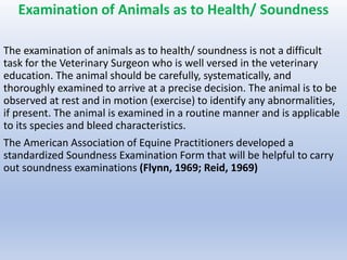
- 1. Examination of Animals as to Health/ Soundness The examination of animals as to health/ soundness is not a difficult task for the Veterinary Surgeon who is well versed in the veterinary education. The animal should be carefully, systematically, and thoroughly examined to arrive at a precise decision. The animal is to be observed at rest and in motion (exercise) to identify any abnormalities, if present. The animal is examined in a routine manner and is applicable to its species and bleed characteristics. The American Association of Equine Practitioners developed a standardized Soundness Examination Form that will be helpful to carry out soundness examinations (Flynn, 1969; Reid, 1969)
- 2. Conformation is the physical appearance of an animal which is outlined by frameworks (bones) and that reflect their activities. Ideal or normal conformation depends on type, breed and intended use or the animal. Since, well conformation and movement of animals are most important features the veterinarians should have an understanding and differentiate between the normal and minor conformations of a particular animal. The following are the components of conformations generally applied in equine. 1. Balance is the relationship of movement among the forehand, hindquarters, limbs, body, right and left sides of the body. A well- balanced horse moves efficiently, the proportions and curvature of the topline. 2. Head should be symmetrical, functional with sufficient cranial space 3. Quality is the well-defined tendons or the cannon bones 4. Substance indicates the thickness, breadth and depth of bone, muscle and other tissues 5. Correctness of angles and structures of cranial and lateral views of forelimb 6. Platting means the flatness of the foot
- 5. Conformational Abnormalities Faults in conformation of the forelimbs — Base narrow, base wide, toe-in or pigeon-toed, toe-out or splay- footed, base wide Backward (palmar) deviation or the corpus (sheep knees/ goat knees/ calf knees) Forward (dorsal) deviation or the carpus (bucked knees/ knee sprung) Medial deviations or the carpus (knock knees/ carpus vulgus / knee narrow conformation) Lateral deviation of the carpus (Bandy-legged conformation / Bow-legged/ Carpus varus) Open knees Off-set knees / Bench- knees Tied-in knees Cut-out under the knees Standing under in front Camped in front Short upright pastern Long sloping pastern Long upright pastern
- 6. Faults in conformation of the hind limbs Standing under behind Sickle hocks Excessive angulation of the hock / small hock angles Straight behind / Excessive straight limbs Camped behind Base narrow Base narrow from fetlocks down Base wide Cow hocks / Tarsus vulgas / medial deviation of the hock Faults in conformation of the feet Flat foot Contracted heels or contracted foot Unilateral contracted foot Bull-nosed foot Buttress foot Thin wall and the sole Club foot Coon-footed
- 7. Movement Movement comprises Travel" and Action" of an animal. Travel means the way of flight or a single hoof in relation to the other limbs and is viewed from the front or rear Action refers to the joint flexion; stride length, suspension, and other qualities. It is evaluated from a side view.
- 8. Natural gaits of pony/horse The following are the natural gaits of pony/ horse: Walk is a fore-beat gait that must have an even rhythm as the feet land and take off in the subsequent order left hind, left fore, right hind and right fore. Pace is a two-beat lateral gait in which the two right limbs rise and land alternately with the two left limbs. It is a viable gait for a racehorse only. Trot is a two-beat diagonal gait, specially refers to an English gait with a moderate to great degree of impulsion. The right fore and left hind rise and fall together alternately with the opposite diagonal pair. Canter / Lope is a three-beat gait in the following way - one hind limb, then the other hind limb simultaneously with its diagonal fore limb, and finally the remaining fore limb Gallop/ Run is a Cote-beat gait with an increased impulsion and length of stride, when the diagonal pair breaks, resulting in fore- beats. The footfall sequence of a right Iead gallop is left hind, right hind, left fore and right fore.
- 10. Stride phases The stride of horse or pony comprises five phases: 1. Landing 2. Loading 3. Stance 4. Breakover 5. Swing Landing: In landing the hoofs touch the ground and the limbs start to receive the weight of the body. Loading: As the body advances forward and the horse's centre of gravity pass over the hoofs, the fetlocks descend to their lowest point, resulting in an almost horizontal pastern. Stance: It means the relative position of the legs in which an animal stands. The fetlock lifts to a relative position that it is comparable to the horse's stand at rest. The change between the loading phasic and the stance phase is very stressful to the internal structures of the hoof and the lower limb. The horse's centric of cavity advances ahead of the hoof.
- 11. Breakover: It refers to the phase when the hoofs Ieave the ground. It begins with the heels lift and the hoofs start to pivot at the toe. The knees or hocks relax and start to flex. Breakover is measured from the time the heels Ieave the ground to the time the toes leave the ground. The deep digital flexor tendon is still stretched just prior to the starting of breakover to counter at the downward pressure of the weight of the body. Swing: The swing is the advancement of the limbs through the air and straightens out in preparation for landing. Normal movement: The normal movement refers to the straight root flight pattern of a horse. The "ideal" or "Standard" movement is the basis (or comparison with deviation that depends upon the ideal body and limb conformation, the standard for forelimb movement begins with straight frameworks and a series or hinge joints symmetrically alike and advancing in a true forward and backward level. The standard foot flight for the hind limb is slightly dissimilar from the forelimb due to turning out of the hind limb to some degree.
- 12. Abnormalities of movement: Movement abnormalities are the defects of the gaits, which occur at the time of regular work. The following are the movement / gait abnormalities: forging, lateral gait defects and interfering Factors that alter movements are given below: Conformation Imbalance Traction Footing Defective training Faulty shoeing Track Pain due to improper fitting or harness or various affections Improper level of fitness Age Dental problems In female during oestrous cycle
- 13. Sign of Stride: The following strides are to be observed carefully- the phases of the stride the path of the foot in flight the arc of the foot flight the foot landing the joint flexion angles the joint extension the symmetry the duration
- 14. The veterinary surgeon is to confirm that the animal is not lame by close observation. "Mechanical lameness" resulting from painful and non-painful alteration in gait should be differentiated from "Neurologic dysfunction". If it is found lame, but not sufficiently to interfere with its usefulness, the following questions will be asked to the owner Questions to be asked How long has the animal (horse / pony) been lame? Did the owner know what caused the lameness? Did the animal warn out of the lameness? Did it stumble? What treatment has been given and it has been found helpful? When was the animal shod?
- 15. Diagnostic aids and application examination of an animal The following instruments and appliances are required for examination of an animal: Stethoscope / Phonendoscope, clinical thermometer, measuring tape, measuring staff, weight box, weybridge, rubber or wooden hammer with pleximeter, torchlight, halter, ear- twitch, lip-twitch, mouth gag, hoof-tester, protector, magnifying glass, metal detector, ear speculum, vaginal speculum, ophthalmoscope, retinoscope, thermograph, compound microscope with necessary materials, X-ray machine and X- ray imaging unit, Fluoroscopy, Fluoroscopy with daylight image intensifier, Ultrasonography, X-ray cinematography, Videography, Computed tomography.
- 16. Thermography as a diagnostic aid: A thermograph has been described as a heat camera (Blakely, 1959) because it records an image a thermogram of the skin temperature distribution pattern. The camera scans the area at which it is directed and collects infrared radiation and converts it into visible light that exposes a Polaroid film. The relative darkness or lightness of a given region or film negative is proportional to temperature of the corresponding region of skin, so temperature distribution of an area of skin can easily be visualized. Delhunty (1965) described experiences with this machine in horses - a diagnostic aid for lameness (Length of time six minutes). Radiography: Radiographs of suspected pathological areas are invaluable in diagnosis and prognosis of affections. Examination procedure The animal is to be observed at rest and in motion (exercise) to identify any abnormalities if present. It should be observed at a distance than up close.
- 18. It is viewed from all directions - in front, both sides and hind. At close observation, each limb is observed and compared to its opposite member for symmetry. The body condition is stocky or slender and alterations in posture, conformation, weight shifting, weight bearing, pointing are to be recorded. The feet are examined for normal size, wear, hoof cracks, heel bulb contraction, imbalance, swelling of the joints, and tendons. At rest: (l) The horse/pony should be examined in the stable from a distance for any vices (2) Afterwards the respiration and pulse rates will be taken followed by blood pressure and temperature recording (3) Then the visual examination for any swellings, enlargements or defects in conformation will be conducted in the following order: • Examination or mouth region for Parrot mouth, roaring, broken and decayed teeth, poor apposition of teeth, broken wind (emphysema of the lungs). • Head region to find any abnormality by palpation
- 19. • Eye region for blindness, cataract, periodic ophthalmia, corneal scars associated with trauma • Ear region for deafness , otitis and other affections • Neck and withers for jugular phlebitis, occlusion of the jugular vein, fistula withers • Thoracic region for broken ribs and other abnormalities • Back for saddle wound • Abdomen for hernia and any other abnormalities • Male genital organs for genital affections and abnormalities • Testicles for orchitis, epididymitis, cryptorchid and other affections • Female genital system for Nymphomania and any other affections of the genital tract • Mammary gland for mastitis and any other abnormalities of teats and gland
- 20. • Tail region for crooked tail, flexor of the tailbones and any other abnormalities. If the animal is docked, then this should be mentioned in the certificate. • Forelimb for ringbone, bobbabone, side- bone, splint, sprained ligaments, strained tendons, synovial distension (hygroma of the carpus), hoof crack, quittor, corn, thrush, dropped sole, bow tendon, knee sprung, navicular disease, laminitis and windgall (wind puff). Various forms of limb contact are listed below: Brushing, cross firing, elbow hitting, forging, knees hitting, interfering, overreaching, scalping and speedy cutting
- 21. Examination by palpation of the forelimbs: From the bottom of the foot, the hoof wall, the coronary band, the lateral cartilage, the pastern area, the fetlock joint, the cannon bone area, the suspensory ligament, the inferior check ligament, the flexor tendons, the carpus, soft tissues between the carpus and elbow, the elbow and shoulder joints, the forearm, shoulder and scapular areas should be examined for muscular atrophy indicating a long standing lameness or sweeny. Palpation of the dorsal articular margins of the carpal bones after flexing the carpus, pain may indicate carpal chip fracture/ acute capsulitis/ synovitis. Palpation of the accessory carpal bones: It is done with the carpus flexed. Pain may be a sign of fracture. Palpation over the point or the shoulder: Pain may be due to bicipital bursitis/ ossification of the biceps tendon and bursa Flexing the carpus: It is done slowly to identify a painful response in suspected slab fracture of the carpal bones, acute synovitis and capsulitis in apparently normal animal
- 22. Abduction of the elbow joint: It is abducted, placing stress the carpus on the medial support structures. Pain may show sprain or strain due to trauma Adduction or the elbow joint: The joint is adducted with carpus by placing stress on the lateral support structures. Pain may indicate sprain / strain due to trauma. Hind limb for bone spavin, bog spavin, occult spavin, capped hock, thoroughpin, curb, ringbone, strained tendons and sprained ligaments. The are of the foot flight should be determined. The hind limbs should be examined same as forelimb up to hock joint. There is a special test known as "spavin test” for diagnosis of spavin". Placing the hands on the plantar surface of the distal third of the metatarsus and elevating the hind limb to flex the hock for one and a half minute perform it. It is better to place the pony/horse against a wall or fence. The stifle joint should be examined for upward fixation of patella, gonitis and abnormality in crepitation.
- 23. . Spavin Test
- 24. Special methods of examination such as local nerve blocks by 2% Lidocaine HCI (Xylocaine HCI) or 1% Hexylcaine HCI, or 2 % Mapivacaine HCI (Carbocaine), Bupivacaine (Marcaine) 0.5% with or without adrenaline. * General for contagious disease, venereal disease, malignant tumors, cryptorchids, scars, heaves, shivering, hernia, and deformed conformation of any organs. * Vices: The conditions considered as vices of animals that may render some of them unsound for certain purposes are biting, bucking, cribbing, kicking, running away, shying, stall -walking, stump sucking, swallowing air, tail-rubbing, tail wringing, viciousness, and weaving. Sometimes it is difficult to detect vices during examination of animals. Hence, the vender must give a guarantee that the said animal is free from vices. * Blemishes are localized defects in tissues that although decrease- more or less the market value of animals by their appearances, but do not always diminish functions. If the blemishes arc due to trauma and not found in joints or do not affect conformation, the animal is considered sound for certain purposes. The veterinarian must record and mention in the certificate the type or blemishes occurred.
- 25. In motion The horse/ pony should be examined in motion (walking straight and in circle, walking uphill, walking downhill, and backing, trotting, canter, and galloping). The following factors are to be considered carefully: The phases of the stride The path of foot in flight The Arc of the root flight The joint extension The joint flexion angles The foot landing Duration Symmetry
- 26. Diagnosis of Claudication (Lameness) Claudication/ lameness is an indication of a structural or functional disorder in one or more limbs that is obvious in progression or the back in the standing position. There are numerous causes of Claudication of which the following are common: Trauma, congenital or acquired anomalies, infectious and contagious diseases, various metabolic disturbances, circulatory and nervous disorders, or any combination of these. Types of lameness: There are four classes / types of lameness- 1. Supporting limb lameness: It is observed when the foot first contacts the ground or at stance phase caused by injury to bones, joints, ligaments or tendons 2. Swinging limb lameness: It is found while the animal is in caused by pathological changes 3. Mixed limb lameness: It is the combination of the above types. 4. Complementary / compensatory lameness: It is due to uneven distribution of weight on another limb / limbs It is important to identify, which limb is lame? The animal must be free from clinical symptoms of infectious or contagious diseases (Tuberculosis, mange and others).