
Recommended
More Related Content
What's hot
What's hot (20)
Similar to How to Read Radiolographs
Similar to How to Read Radiolographs (20)
More from Mariam Alosfoor
Recently uploaded
Recently uploaded (20)
How to Read Radiolographs
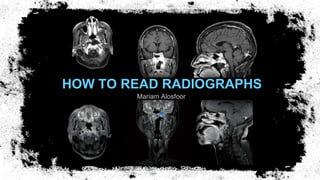
- 1. HOW TO READ RADIOGRAPHS Mariam Alosfoor
- 2. OBJECTIVES:- Steps of correctly reading radiographs Different techniques for taking radiographs Radiological findings in the case
- 3. 1 2 3 4 5 HOW TO READ A RADIOGRAPH? Follow these steps Type of Image Anatomical part Section Technique Lesion description
- 4. TYPE OF IMAGE Magnetic Resonance Image (MRI) •Soft tissue, Brain tumors, Spine injuries, Multiple sclerosis, Brain step lesions •Does not use radiation Computed Tomography (CT scan) •Bone injuries, Chest, Cancer detection, Brain hemorrhage •X-rays Positron Emission Tomography (PET scan) •Functional imaging •Cancer, Epilepsy, Heard disease •Radioactive materials 1
- 5. TECHNIQUE With / Without contrast medium Signal characteristics (MRI): 4 T1 T2 Black Air Bone Air Bone Dark CSF Edema Tissue Brigh t Fat Blood Contrast medium CSF Blood Edema
- 6. LESION DESCRIPTION Anatomy Exact location Three dimensional size Radiological Criteria CT scan: Isodense, hypodense, hyperdense. MRI: Isointense, hypointense, hyperintense. Edema Compression Midline shift Herniation 5
- 8. The Case
- 9. CT SCAN The right sphenoidal sinus and cavernous sinus are filled with tissue. There is a destruction of osseous structures on the caudal and lateral side. Destruction of the skull base.
- 10. MRI Irregular structure in the nasopharyngeal space with destruction of the skull base and ingrowth into the right cavernous sinus and into the pterygopalatinoid sinus.
- 11. PET SCAN
- 12. Any Questions? Feel Free to Ask
Editor's Notes
- The use of artificial contrast media enhances the contrast between different parts of the body. Contrast media is used to help distinguish between parts of the body that have a similar composition to provide a clearer image of how the body is working, or if there is any disease present.