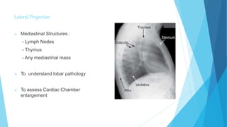
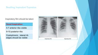
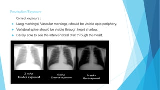
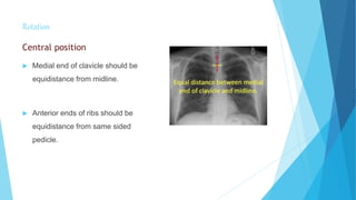
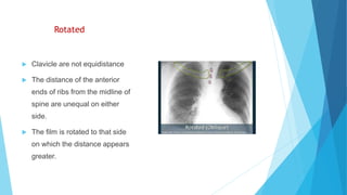
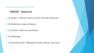
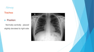
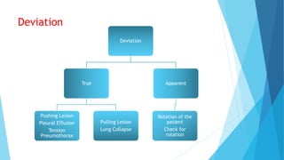
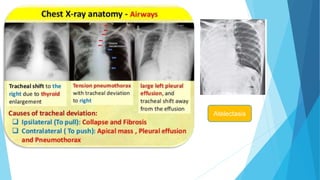
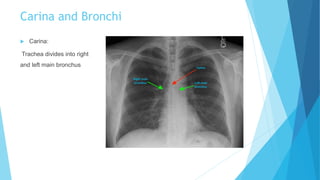
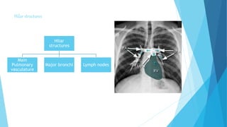
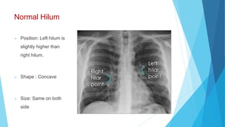
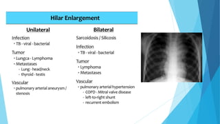
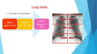
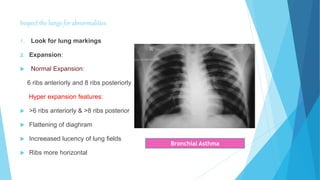
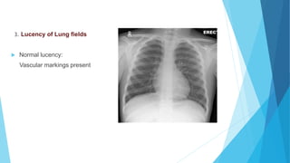
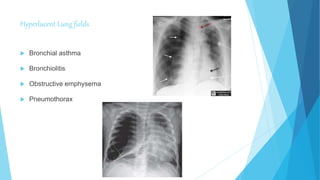
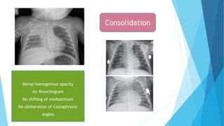
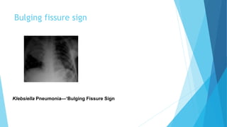
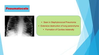
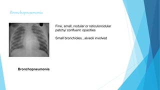
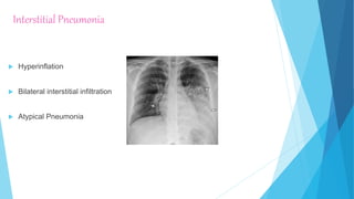
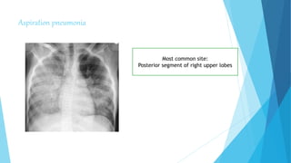
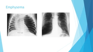
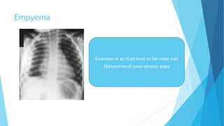
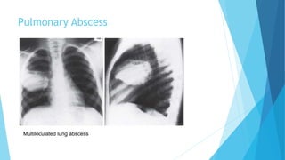
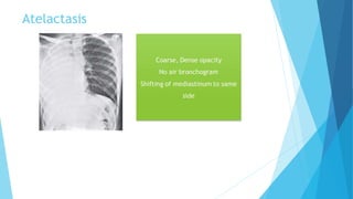
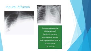
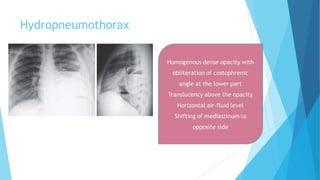
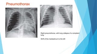
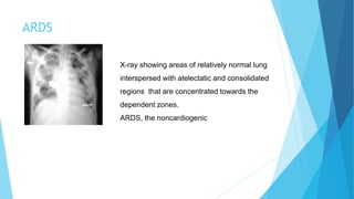
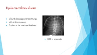
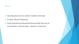
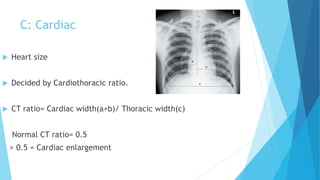
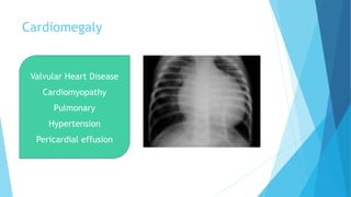
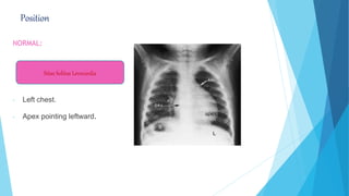
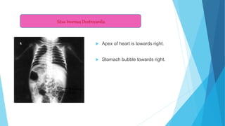
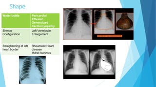
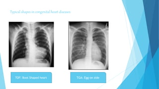
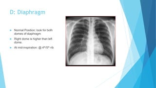
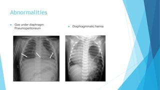
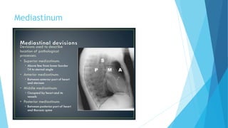
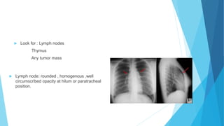
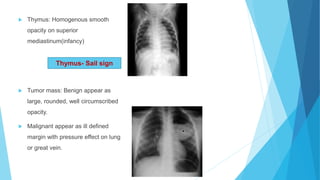
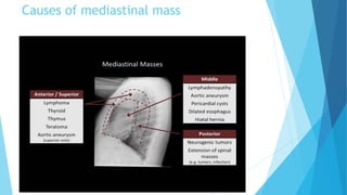
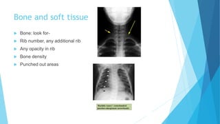
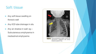
Chest x-ray interpretation
Presenter: Dr Manidipa Barman
MD PAEDIATRICS
AIIMS RISHIKESH
Chest x-ray interpretation begins with following
details-
 Patient details: Name, Age, Gender, Unique identification number.
 Date and time
 Previous imaging(if present)
Assessment of quality of image
 Projection/ View
 Breath : Inspiration/ Expiration
 Position
 Exposure
Projection/ Views
CXR
views
Frontal
Postero-
anterior
Antero-
posterior
Lateral
Frontal Projection
PA view AP view
Scapula Periphery of Thorax Over lung fields
Clavicle Project over lung fields Above the apex of lung fields
Ribs Posterior ribs are distinct Anterior ribs distinct
Horizontally placed
Heart Clear border Larger and indistinct border
Lateral Projection
 Mediastinal Structures :
- Lymph Nodes
- Thymus
- Any mediastinal mass
 To understand lobar pathology
 To assess Cardiac Chamber
enlargement
Breathing: Inspiration/ Expiration
Inspiratory film should be taken
Good Inspiration
5-7 anterior ribs visible
9-10 posterior ribs
Costophrenic , lateral rib
edges should be visible
Penetration/Exposure
Correct exposure :
 Lung markings( Vascular markings) should be visible upto periphery.
 Vertebral spine should be visible through heart shadow.
 Barely able to see the intervertebral disc through the heart.
Rotation
Central position
 Medial end of clavicle should be
equidistance from midline.
 Anterior ends of ribs should be
equidistance from same sided
pedicle.
Rotated
 Clavicle are not equidistance
 The distance of the anterior
ends of ribs from the midline of
spine are unequal on either
side.
 The film is rotated to that side
on which the distance appears
greater.
Structured interpretation of CXR
“ABCDE” Approach
 A: Airway – Trachea, Carina, bronchi and Hilar Structures
 B: Breathing- Lungs and pleura
 C: Cardiac- Heart size and border
 D: Diaphragm
 E: Everything Else- Mediastinal contour, Bones, soft tisues
Airway
Trachea
 Position:
Normally centrally placed/
slightly deviated to right side
Deviation
Deviation
True
Pushing Lesion
Pleural Effusion
Tension
Pneumothorax
Pulling Lesion
Lung Collapse
Apparent
Rotation of the
patient
Check for
rotation
Atelectasis
Carina and Bronchi
 Carina:
Trachea divides into right
and left main bronchus
Hilar structures
Hilar
structures
Main
Pulmonary
vasculature
Major bronchi Lymph nodes
Normal Hilum
 Position: Left hilum is
slightly higher than
right hilum.
 Shape : Concave
 Size: Same on both
side
B: Breathing( Lungs, Pleura)
Lung fields
 Concept of Lung Zones
Upper
Upto 4th rib
Middle
4th – 6th ribs
Lower
Below 6th
ribs
Inspect the lungs for abnormalities:
1. Look for lung markings
2. Expansion:
 Normal Expansion:
6 ribs anteriorly and 8 ribs posteriorly
Hyper expansion features:
 >6 ribs anteriorly & >8 ribs posterior
 Flattening of diaghram
 Increeased lucency of lung fields
 Ribs more horizontal
Bronchial Asthma
3. Lucency of Lung fields
 Normal lucency:
Vascular markings present
Hyperlucent Lung fields
 Bronchial asthma
 Bronchiolitis
 Obstructive emphysema
 Pneumothorax
Opacity of lung fields
 Consolidation
 Collapse
 Collapse with consolidation
 Pleural effusion
 Empyema
 Fibrosis: TB, Bronchiectasis
 Milliary mottling
Dense homogenous opacity
Air Bronchogram
No shifting of mediastinum
No obliteration of Costophrenic
angles
Consolidation
Bulging fissure sign
Klebsiella Pneumonia—‘Bulging Fissure Sign
Pneumatocele
 Seen in Staphylococcal Pneumonia
 Extensive destruction of lung parenchyma
 Formation of Cavities bilaterally
Bronchopneumonia
Bronchopneumonia
Fine, small, nodular or reticulonodular
patchy/ confluent opacities
Small bronchioles , alveoli involved
Interstitial Pneumonia
 Hyperinflation
 Bilateral interstitial infiltration
 Atypical Pneumonia
Aspiration pneumonia
Most common site:
Posterior segment of right upper lobes
Emphysema
Empyema
Extension of air-fluid level to the chest wall
Obliteration of costo-phrenic angle
Pulmonary Abscess
Multiloculated lung abscess
Atelactasis
Pleural effusion
Hydropneumothorax
Pneumothorax
Right pneumothorax, with lung collapse of a compliant
lung.
Shift of the mediastinum to the left
ARDS
X-ray showing areas of relatively normal lung
interspersed with atelectatic and consolidated
regions that are concentrated towards the
dependent zones.
ARDS, the noncardiogenic
Miliary Tuberculosis of the Lungs
The fine, round, millet-like opacities in both lung fields
(military mottling) with right paratracheal
lymphadenopathy.
Milliary Tuberculosis
Hyaline membrane disease
 Ground-glass appearance of lungs
 with air-bronchogram.
 Borders of the heart are ill-defined
Foreign Body: ? Ingested/ Aspirated
Foreign body in Esophagus
Pleura
 Normally pleura are not visible in healthy individuals.
 If visible: Pleural Thickening
 Fluid( Hydrothorax)/ Blood(hemothorax)/ Both fluid and air
accumulation in pleural space : Opacity on chest xray
C: Cardiac
 Heart size
 Decided by Cardiothoracic ratio.
 CT ratio= Cardiac width(a+b)/ Thoracic width(c)
Normal CT ratio= 0.5
> 0.5 = Cardiac enlargement
Cardiomegaly
Valvular Heart Disease
Cardiomyopathy
Pulmonary
Hypertension
Pericardial effusion
Cardiac Silhouette
 Cardiac silhouette refers to the outline of the heart as seen on frontal and
lateral chest x-ray and forms part of the cardiomediastinal contour.
 The size and shape of the cardiac silhouette provide useful clues for
underlying disease.
 From the frontal projection, the cardiac silhouette can be divided into right
and left borders.
 On the lateral projection the cardiac silhouette is formed by anterior and
posterior border.
 Right Border: Right atrium
SVC
 Left Border: Aortic notch
Left Pulmonary artery
Aorto-Pulmonary window
Left atrial appandages
Left ventricle
Heart borders difficult to distinguish
from the lung fields as a result of
pathology which increases the
opacity of overlying lung tissue
Frontal Projection
Lateral projection
 Anterior border: Right ventricle
 Posterior border: Left atrium
Left Ventricle
Inferior vena cava
Size
 The cardiac silhouette is considered enlarged if the CT ratio is
greater than 50% on a PA view of the chest.
Causes of Enlargement:
 Pericardial effusion
 Cardiomegaly
 Anterior mediastinal mass
 AP projection
 Expiratory Radiograph
Position
NORMAL:
- Left chest.
- Apex pointing leftward.
Situs Solitus Levocardia
 Apex of heart is towards right.
 Stomach bubble towards right.
Situs Inversus Dextrocardia
Shape
Water bottle Pericardial
Effusion
Generalized
Cardiomyopathy
Shmoo
Configuration
Left Ventricular
Enlargement
Straightening of left
heart border
Rheumatic Heart
disease
Mitral Stenosis
Typical shapes in congenital heart diseases
TOF: Boot Shaped heart TGA: Egg-on side
TAPVC: Figure of 8
appearance
Ebstein anomaly: Large
box shaped heart
Normal Pulmonary Vasculature
 Decreased Pulmonary blood
flow.
 Due to right to left shunt.
 Decreased vascular markings.
Tetralogy of Fallot
Tricuspid Atresia
Ebstein Anomaly
PULMONARY OLIGEMIA
PULMONARY PLETHORA
ASD
VSD
PDA
TAPVC
 Increased Pulmonary Blood Flow
 Increased Pulmonary vascular markings
D: Diaphragm
 Normal Position: look for both
domes of diaphragm
 Right dome is higher than left
dome.
 At mid-inspiration: @ 4th/5th rib
Abnormalities
 Gas under diaphragm:
Pneumoperitoneum
 Diaphragmmatic hernia
Mediastinum
 Look for : Lymph nodes
Thymus
Any tumor mass
 Lymph node: rounded , homogenous ,well
circumscribed opacity at hilum or paratracheal
position.
 Thymus: Homogenous smooth
opacity on superior
mediastinum(infancy)
 Tumor mass: Benign appear as
large, rounded, well circumscribed
opacity.
 Malignant appear as ill defined
margin with pressure effect on lung
or great vein.
Thymus- Sail sign
Causes of mediastinal mass
Bone and soft tissue
 Bone: look for-
 Rib number, any additional rib
 Any opacity in rib
 Bone density
 Punched out areas
Soft tissue
 Any soft tissue swelling on
thoracic wall
 Any ICD tube drainage in situ
 Any air shadow in wall: eg –
Subcutaneous emphysema in
mediastinal emphysema
Tubes,Lines
 Nasogastric tube placement
 Endotracheal tube placements
 Lines and cables: Cental line,
ECG cable
 All tube and lines will be visible
as radio-opaque lines on CXR
I. Name the x-ray sign
II. Condition
Figure of 3 sign
Coarctation of Aorta
Diagnosis ?
Tracheo-Esophageal Fistula
Diagnosis ?
Pneumomediastinum
Name the sign
Silhouette sign
References
 Nelson Textbook of Paediatrics.
 IAP Colour Atlas of Paediatrics.
 https://geekymedics.com/chest-x-ray interpretation a methodical approach.
 Bedside clinics in Paediatrics: Sibarjun Ghosh.
 https://radiopaedia.org/articles/chest radiograph assessment.

Paediatric Chest X-ray Interpretation