Download as PDF, PPTX


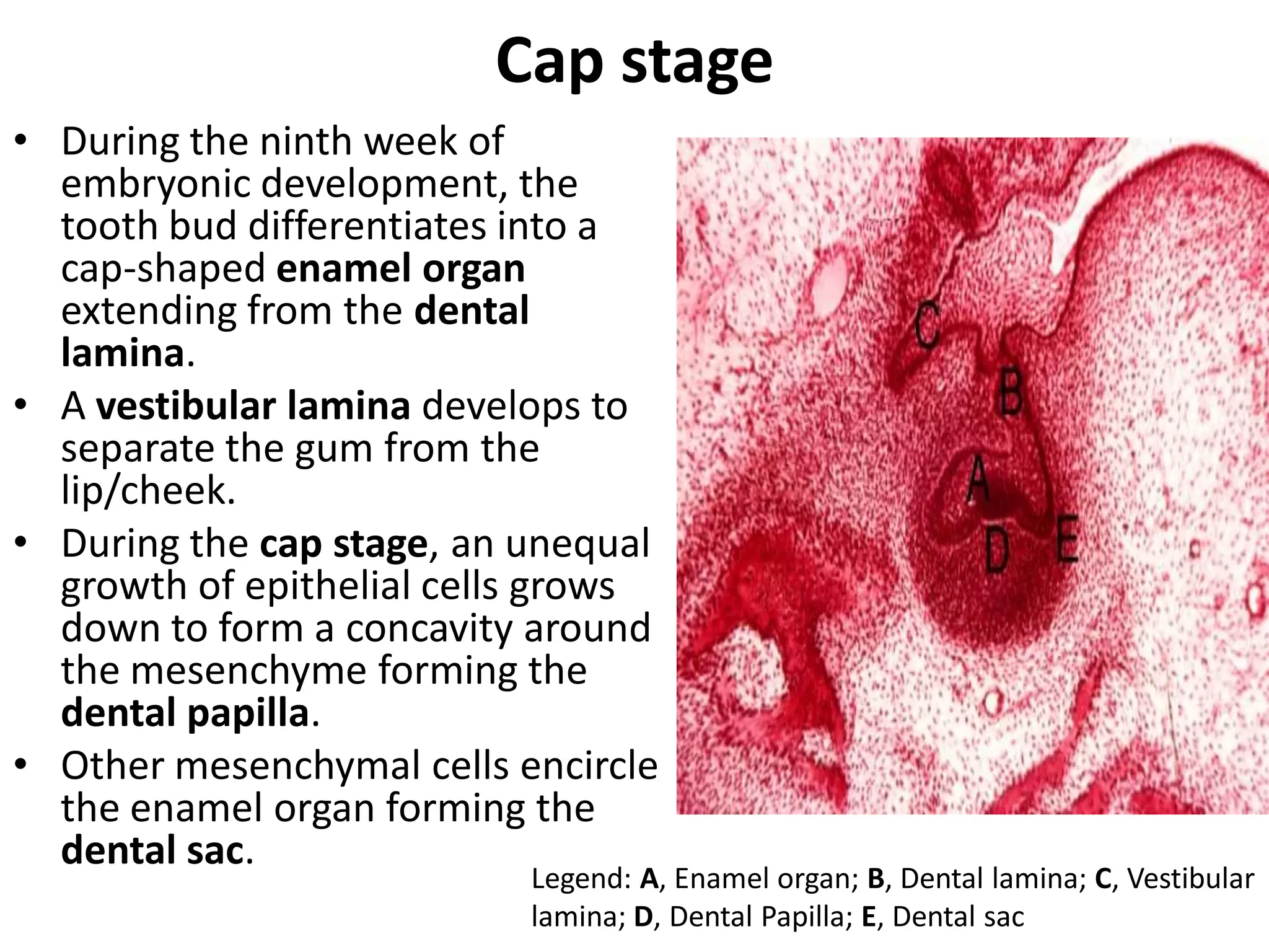


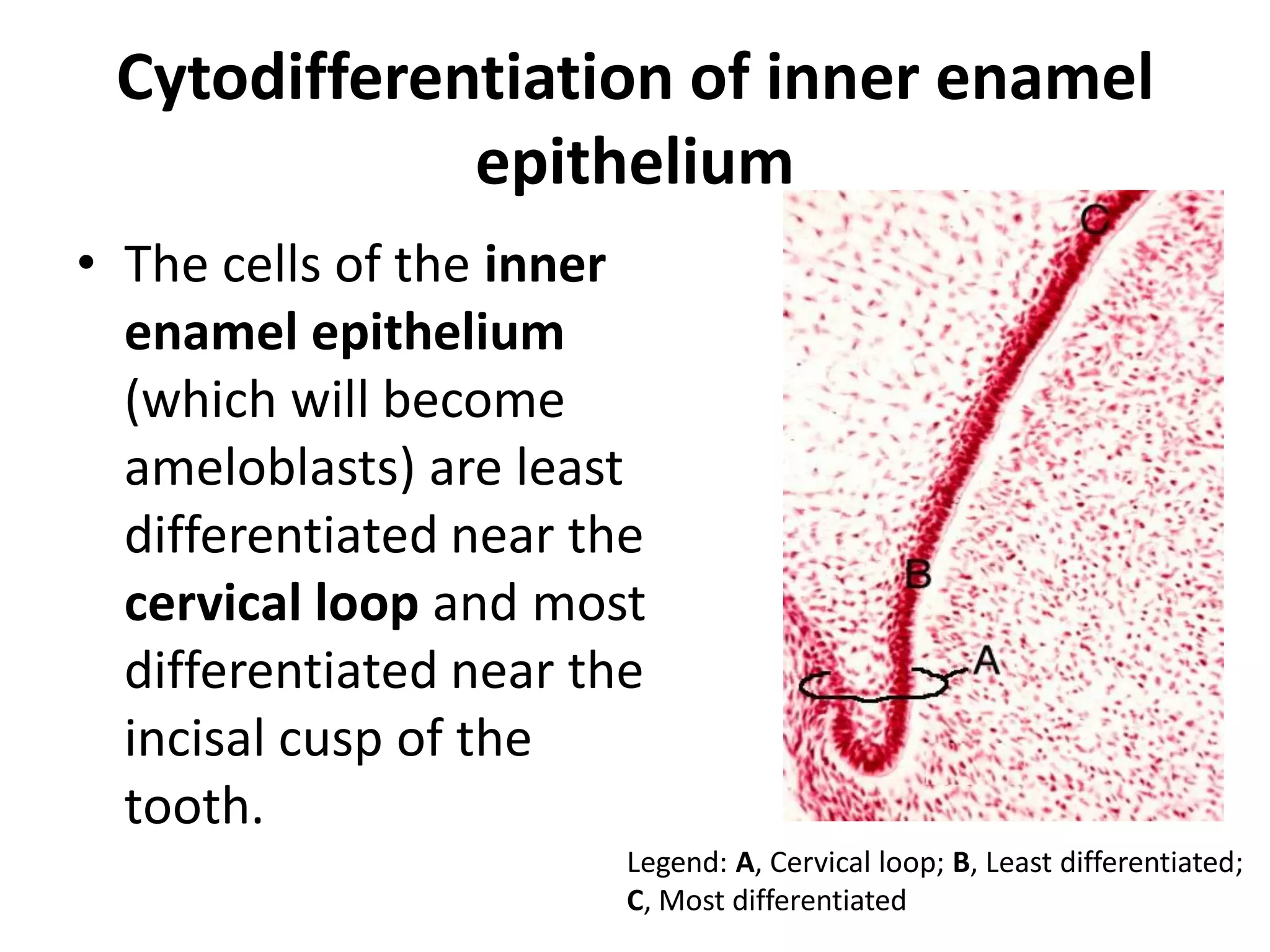


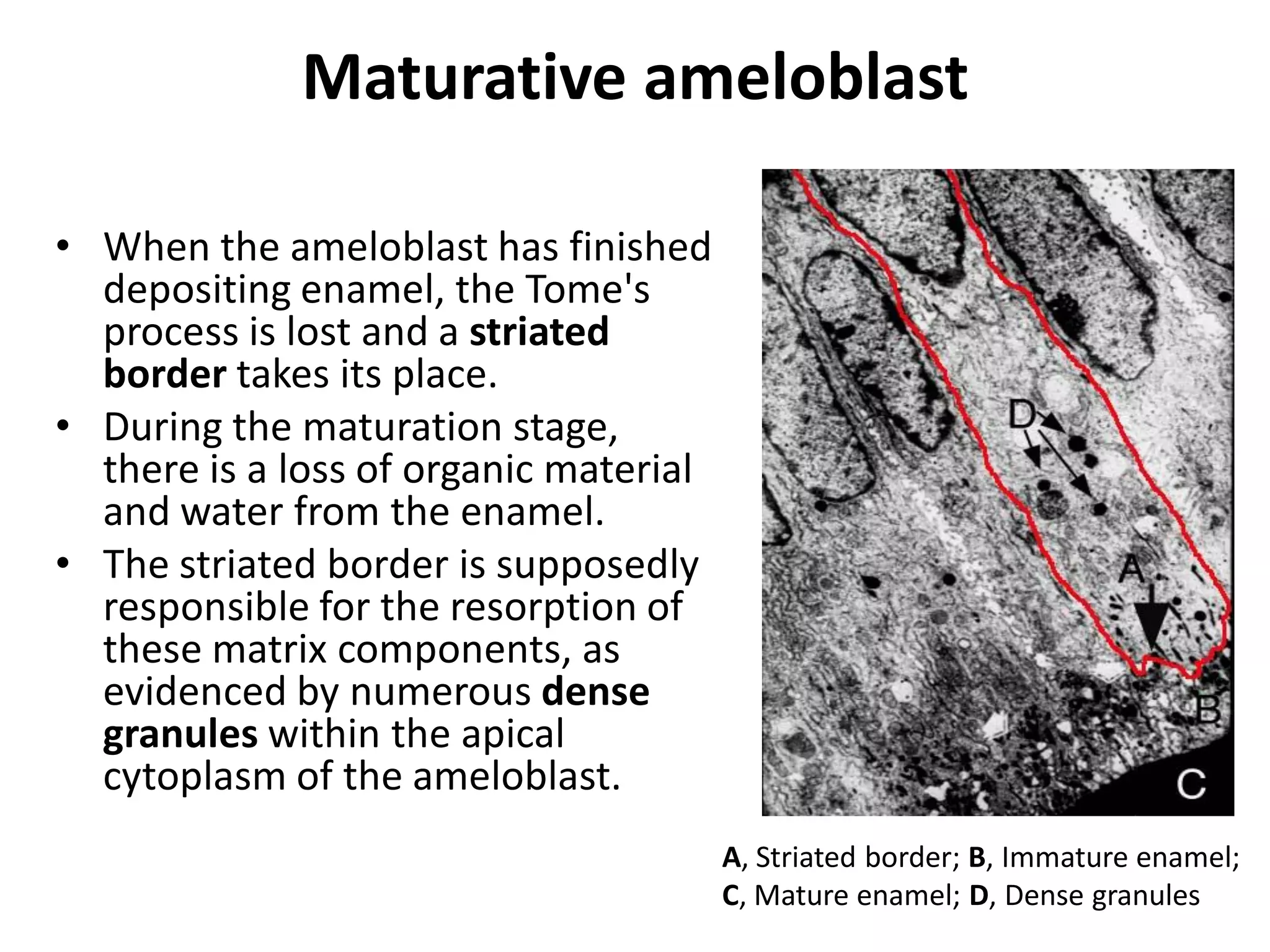

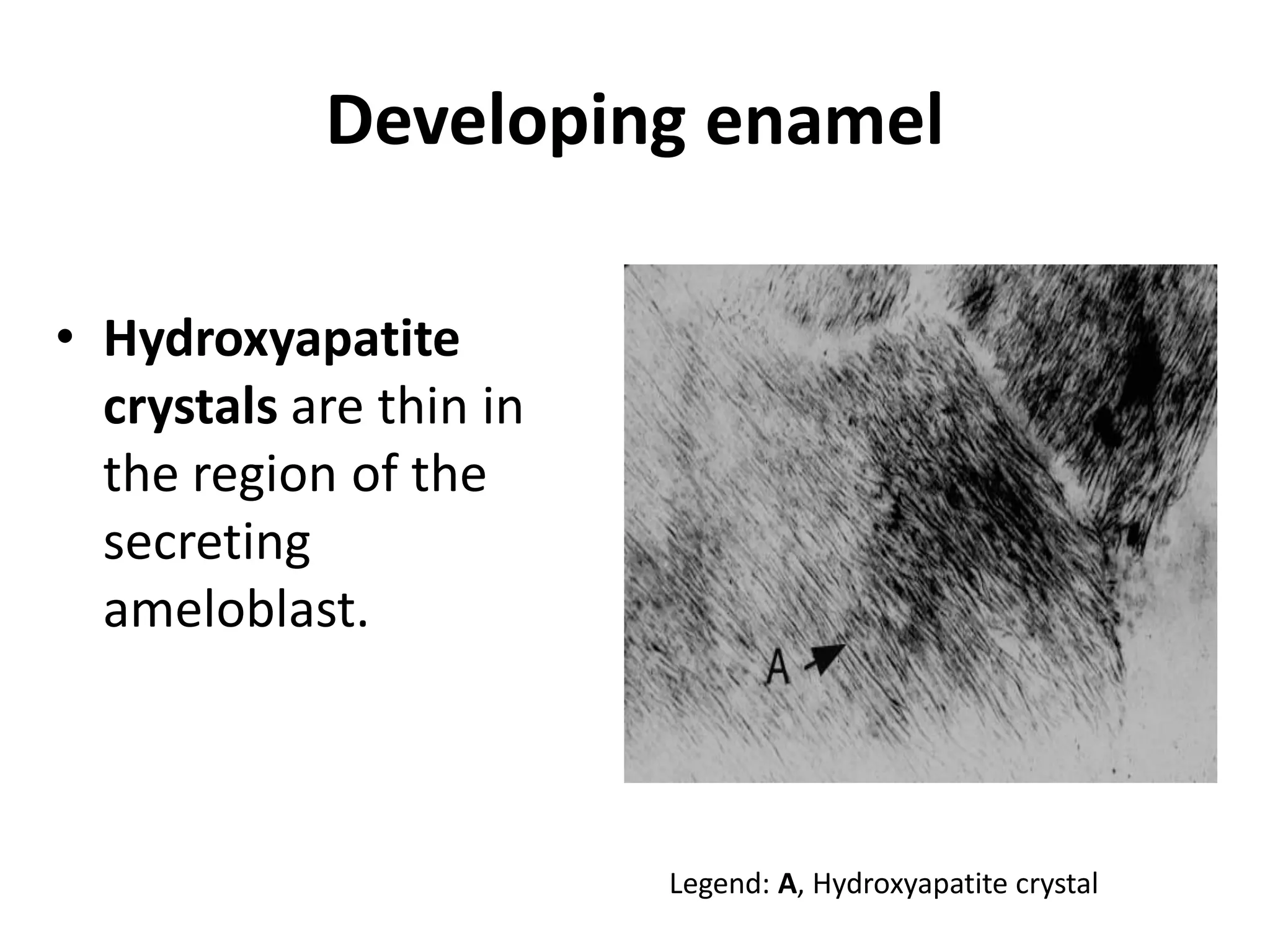
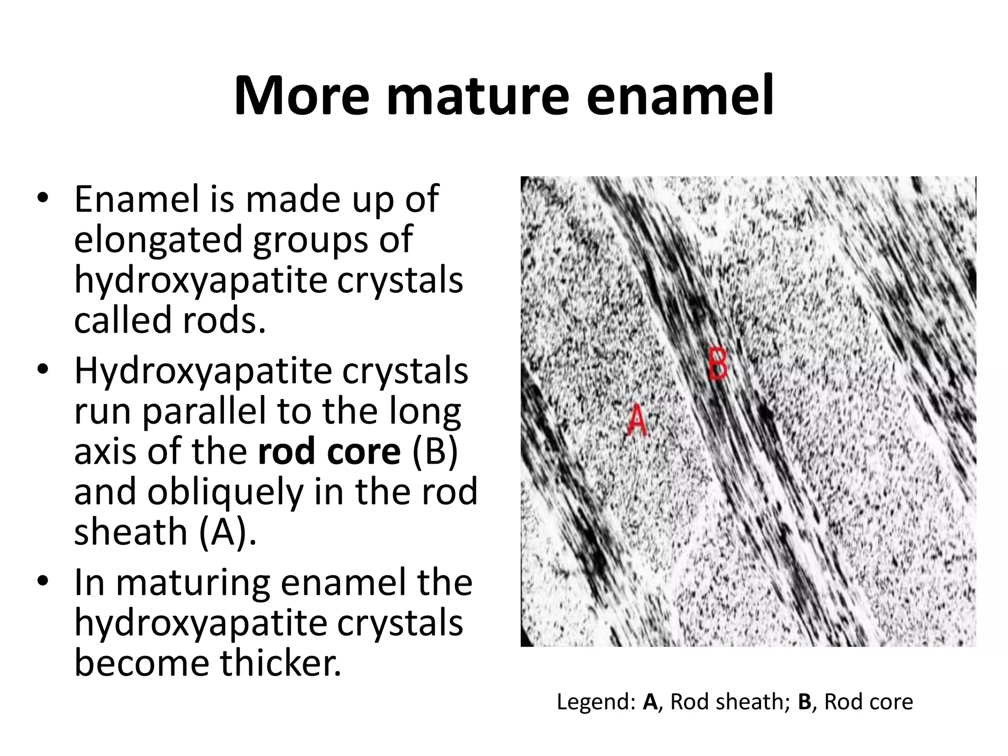

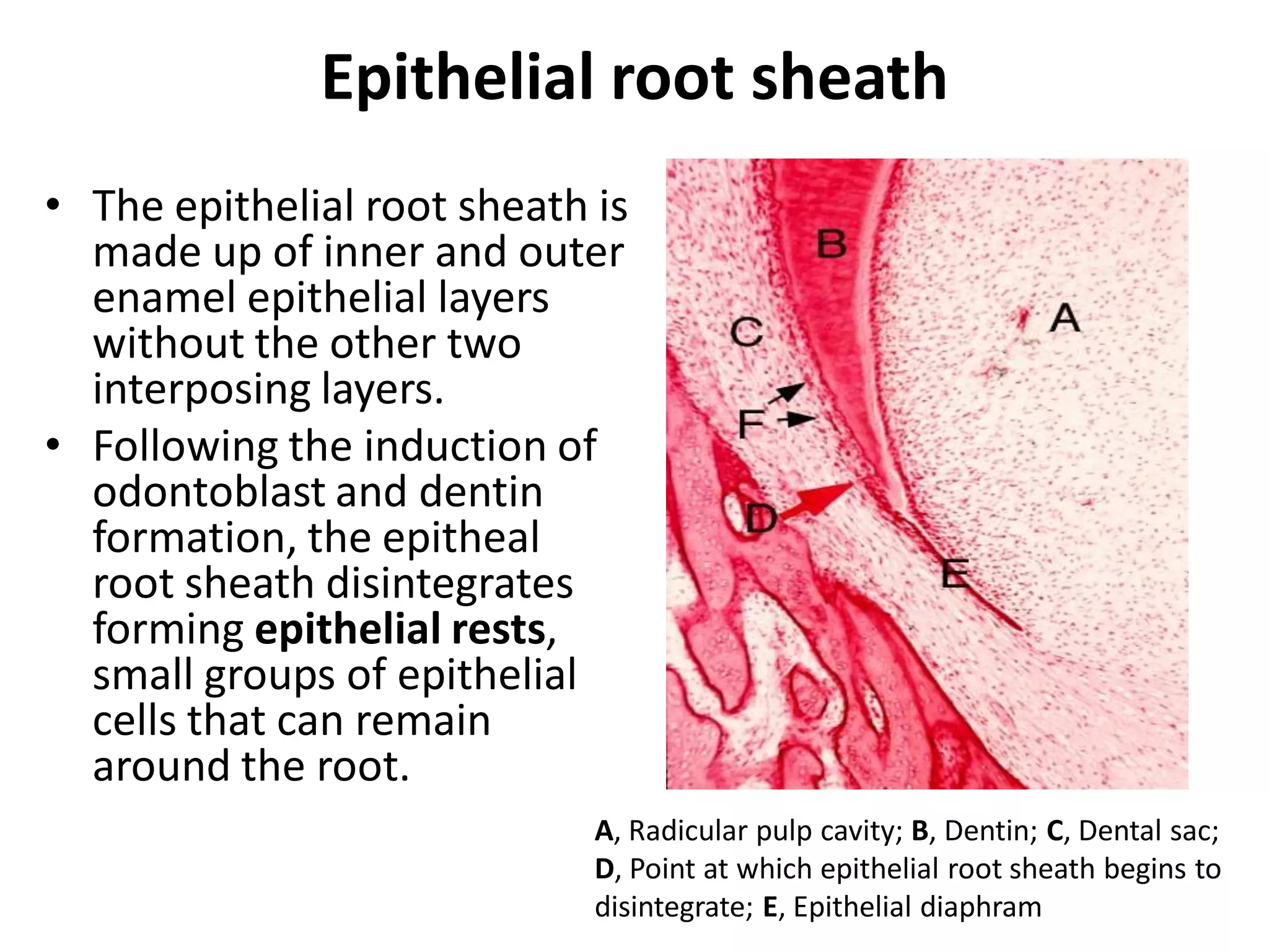
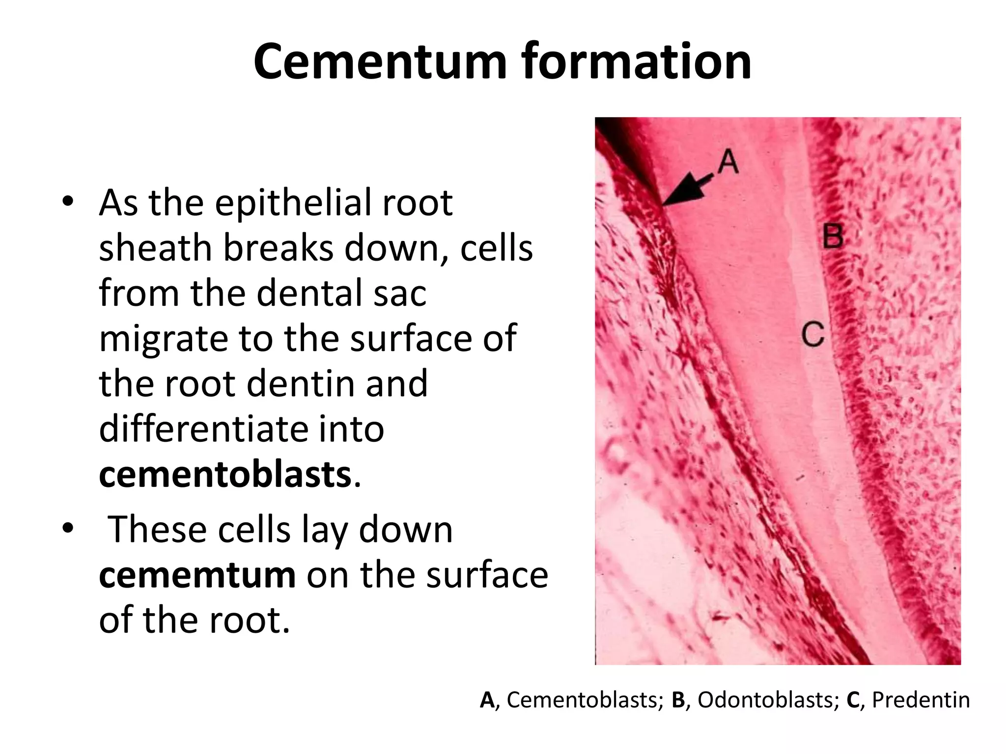


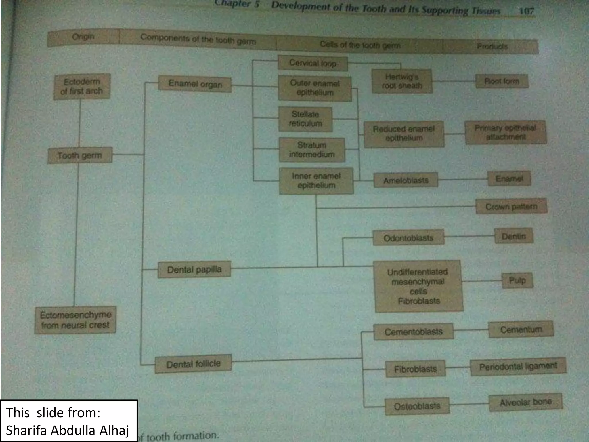


The document summarizes the stages of odontogenesis, or tooth development, from the dental lamina stage through root formation. It describes how during embryonic development, the dental lamina thickens to form tooth buds, which develop through bud, cap, and bell stages. During these stages, the enamel organ and dental papilla form. Odontoblasts then deposit dentin while ameloblasts deposit enamel. After crown formation, the root develops through the formation of the epithelial root sheath. Cementum is later deposited on the root surface by cementoblasts.


![Presentacion formacion dental[1]](https://cdn.slidesharecdn.com/ss_thumbnails/presentacionformaciondental1-110611113239-phpapp01-thumbnail.jpg?width=640&height=640&fit=bounds)


