
Recommended
More Related Content
What's hot
What's hot (19)
Similar to CURC poster presentation 2018
Similar to CURC poster presentation 2018 (20)
Recently uploaded
Recently uploaded (20)
CURC poster presentation 2018
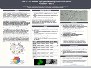
- 1. Role of Clots and Macrophages in the Progression of Idiopathic Pulmonary Fibrosis Jordon Aragon, Cole Mills, Moriah Glennon, Kadi Horn, Catie Pearson, Nikko Garcia, Dr. Alan Schenkel PhD. Colorado State University: Department of Microbiology, Immunology and Pathology Idiopathic Pulmonary Fibrosis (IPF) is a progressive and fatal disease, killing about 30,000 Americans per year. Currently, the only cure is a lung transplant, though there are some new drugs in testing. Currently, it is believed that alveolar epithelial cells die, and the alveolar structure is damaged. Attempts to repair the damage lead to scar tissue (fibrosis). But little is known about what happens between the damage and fibrosis steps. Our hypothesis is that after alveoli structures become damaged, bleeding occurs leaving blood clots in the alveolar spaces. Macrophages in the lung start to eat the red blood cells to clean up the alveoli. They also make repair cytokine signals, like TGF-β. Due to the chronic nature of IPF, the alveoli continue to bleed so the macrophages keep eating the red blood cells and continuously produce the cytokines, IL-10 and TGF-β which leads to scar tissue formation and fibrosis. It is not known how long macrophages live after eating blood clots or what signals they might make. Mouse bone marrow and bronchoalveolar lavage macrophages were cultured with blood clots. We found that macrophages only phagocytosed old blood clots (2-3 days old) and accumulated fluorescent proteins, which may be Ym1, a marker of alternatively activated macrophages. We are using quantitative digital droplet PCR (ddPCR) to examine all the kinetics of several possible repair signals like CD163 (used for hemoglobin phagocytosis), Ym1, IL-10, IL-17A, IL-4, and TGF-β at several time points after incubation with blood clots in different types of macrophages. We are also going to study macrophage function and health after long-term exposure to clots. Abstract This experiment shows that macrophages harvested from vivo, and exposed to RBC’s produce cytokines that promote tissue repair and regeneration. ● TGF-β was seen in cells that were exposed to clot but only on the first day. ● IL10 was also detected only in cells that were exposed to the blood clots as well. ● YM1 was seen in cells only in the 7th day of testing. ● IL4 was seen in day 7 cells with clots. ● Arginase 1 was also only expressed in only the controls of the day 1 and day 7. ● Mouse macrophages were harvested from mouse bone marrow and via bronchoalveolar lavage and cultivated in 2-3 day old clots. ● RNA was extracted from the macrophages and then made into cDNA. ● cDNA was then analyzed with digital droplet PCR. Digital droplet polymerase chain reaction (ddPCR) uses oils to conduct the PCR process. DNA is replicated inside of very small droplets which are then fed through a device that can read fluorescence levels that indicate how much test DNA was being replicated by the macrophages. Methods and Materials From this experiment sever conclusions can be drawn: ● Arg 1 presence in the day 1 and day 7 controls is a little paradoxical due to Arg1 signalling for mediation of inflammation ○ due to Arg1 gene is normally seen as an inflammatory cytokine that is expressed by M2 macrophages. ○ More testing will be necessary ● IL4 is normally seen in macrophages trying to induce an inflammatory response. ○ More testing will be necessary. ● The detection of TGF-β and IL10 shows that we are successfully causing a phenotypic switch. ● The detection of Ym1: ○ Correlated with IPF in mice ○ Is a fluorescent molecule that fluoresces when inside clots. ○ Because it was only detected in the 7 day samples, this supports our hypothesis that YM1 crystals only occur in a chronic environment, where macrophages prefer old clots. Conclusions Results Future Directions Future directions for experimentation are: ● Testing the cytokine response of interstitial macrophages. ● Testing if the fluorescence may be caused by a RBC digestion by product rather than YM1. ● Focus our investigation on whole transcriptome sequencing to see what is upregulated when the macrophages are exposed to RBCs. References and Acknowledgements ● Neil Ahluwalia; Barry S. Shea; Andrew M. Tager; Am J Respir Crit Care Med 190:867-878 (2014)S.Y. Tan & W. Weninger Nature Immunol. 17:1335-1336 (2016). ● Schenkel, Alan R. et al. “Different Susceptibilities of PECAM-Deficient Mouse Strains to Spontaneous Idiopathic Pneumonitis.” Experimental and molecular pathology 81.1 (2006): 23–30. PMC. Web. 29 Mar. 201 Acknowledgements: Dr. Alan Schenkel, Wilusz Lab, John Anderson, Nicholas Garcia, Catie Pearson Possible YM1 Crystals in macrophages Fluorescence in Macrophages