2. URINE
• Urine is a simple type of specimen and it is the most common
test specimen in the clinical laboratory.
• It is a liquid waste produced by our kidneys, a clear,
transparent fluid that normally has an amber colour.
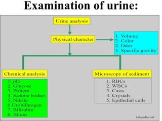
• Urine examination completes into various phases i.e. physical
examination, chemical examination and microscopic
examination.
• Physical examination of urine says about colour, odour,
clarity, volume, and specific gravity while chemical
examination of urine includes the identification of protein,
blood cells, glucose, pH, bilirubin, urobilinogen, ketone
bodies, nitrites, and leukocyte esterase, etc.
• Microscopic examination informs about cells urinary tract,
blood cells, crystals, bacteria, parasites, fungus, etc.
3. Composition of normal urine:
• Urine is a liquid byproduct of the body secreted by
the kidneys through a process called urination and
excreted through the urethra.
• The normal chemical composition of urine is mainly
water content, but it also includes nitrogenous
molecules, such as urea, as well as creatinine and
other metabolic waste components.
• Other substances may be excreted in urine due to
injury or infection of the glomeruli of the kidneys,
which can alter the ability of the nephron to reabsorb
or filter the different components of blood plasma.
4. Normal Chemical Composition of Urine
• Urine is an aqueous solution of greater than 95%
water, with a minimum of these remaining
constituents, in order of decreasing concentration:
• Urea 9.3 g/L.
• Chloride 1.87 g/L.
• Sodium 1.17 g/L.
• Potassium 0.750 g/L.
• Creatinine 0.670 g/L .
• Other dissolved ions, inorganic and organic
compounds (proteins, hormones, metabolites).
5. • Urine is sterile until it reaches the urethra, where
epithelial cells lining the urethra are colonized by
facultatively anaerobic gram-negative rods and
cocci.
• Urea is essentially a processed form of ammonia
that is non-toxic to mammals, unlike ammonia,
which can be highly toxic.
• It is processed from ammonia and carbon dioxide in
the liver.
• There are several conditions that can cause abnormal
components to be excreted in urine or present as
abnormal characteristics of urine.
6. They are mostly referred to by the suffix -uria. Some
of the more common types of abnormal urine include:
• Proteinuria—Protein content in urine, often due to leaky
or damaged glomeruli.
• Oliguria—An abnormally small amount of urine, often
due to shock or kidney damage.
• Polyuria—An abnormally large amount of urine, often
caused by diabetes.
• Dysuria—Painful or uncomfortable urination, often
from urinary tract infections.
• Hematuria—Red blood cells in urine, from infection or
injury.
• Glycosuria—Glucose in urine, due to excess plasma
glucose in diabetes, beyond the amount able to be
reabsorbed in the proximal convoluted tubule.
7. Specimen collection:
• Collection and transportation of urine specimens to
the clinical laboratory are important because
variables such as collection method, container,
transportation, and storage affect the analysis
outcome and consequently diagnostic and
therapeutic decisions based on the results.
• Clinical staff are responsible for patient instruction,
collection and labeling of urine specimens and
timely
transportation of specimens to the Laboratory.
8.
9. Specimen types:
• 1. Random specimen:
• For chemical and microscopic examination, a
voided specimen is usually more suitable.
• A randomly collected specimen may be collected at
unspecified times and is often more convenient for
the patient.
• A random specimen is suitable for most screening
purposes.
10. 2. First morning specimen or 8-hour
specimen:
• The patient should be instructed to collect the
specimen immediately upon rising from a night’s
sleep. Other 8-hour periods may be used to
accommodate insomniacs, night-shift workers, and
in certain pediatric situations.
• The bladder is emptied before lying down and the
specimen is collected on arising so that the urine
collected only reflects the recumbent position.
• Any urine voided during the night should be
collected and pooled with the first morning voided
specimen.
11. 3. Fasting specimen:
• This differs from a first morning specimen by being
the second voided specimen after a period of
fasting.
• 4. 2-Hour postprandial specimen :
• The patient should be instructed to void shortly
before consuming a routine meal and to collect a
specimen 2 hours after eating.
12. 5. 24-hour (or timed) specimen:
• To obtain an accurately timed specimen, it is
necessary to begin and end the collection period
with an empty bladder.
• The following instructions for collecting a 24-hour
specimen can be applied to any timed collection
(consult test requirements to determine if a special
preservative is required): Day 1 - 7 AM: Patient
voids and discards specimen.
• Patient collects all urine for the next 24 hours. Day
2 - 7 AM: Patient voids and adds this urine to the
previously collected urine.
13. 6. Catheterized specimen:
• This specimen is collected under sterile conditions by
passing a hollow tube through the urethra into the
bladder.
• 7. Midstream “clean catch” specimen:
• This specimen provides a safer, less traumatic method
for obtaining urine for bacterial culture.
• It also offers a more representative and less
contaminated specimen for microscopic analysis than
the random specimen.
• Adequate cleansing materials and a sterile container
must be provided for the patient.
• The procedure for the collection of a “clean catch”
urine is described below in section VI of this policy.
14. 8. Suprapubic aspiration:
• Urine may be collected by external introduction of a
needle into the bladder. It is free of extraneous
contamination and may be used for cytologic
examination.
• 9. Pediatric specimens:
• This may be a sterile specimen obtained by
catheterization or by suprapubic aspiration. The random
specimen may be collected by attaching a soft, clear
plastic bag with adhesive to the general area of both
boys and girls.
• B. Transportation of specimens:
• Urine specimens should be delivered to the within 2
hours of collection or refrigerated and transported to the
lab as soon as possible.
15. PRINCIPLE
• Collection and transportation of urine specimens to
the clinical laboratory are important because
variables such as collection method, container,
transportation, and storage affect the analysis
outcome and consequently diagnostic and therapeutic
decisions based on the results.
• Clinical staff are responsible for patient instruction,
collection and labeling of urine specimens and timely
transportation of specimens to the Laboratory.
16. REAGENTS AND SUPPLIES (for collection
of “clean catch” specimens)
• A. Disposable, clean, dry, leak-proof container (sterile
container with lid required for
microbiological cultures)
• B. Screw top specimen tube
• C. Disposable gloves
• D. Betadine swabs (Hibiclens if allergic to betadine)
• E. Dry, clean gauze
• F. Patient’s bedpan or urinal, if patient is unable to go the
bathroom.
• QUALITY CONTROL:
• Identification of the patient must be performed by asking a
conscious patient his or her full name
and birthdate. Verify by checking the identification band if
available.
17. PROCEDURE
• A. Patient preparation:
• For FEMALE patients:
• 1. Wash hands thoroughly before beginning the procedure and put on
disposable
gloves.
• 2. Use betadine swabs or Hibiclens to cleanse the perineal area.
• a. Separate the folds of the labia and wipe the betadine swab or Hibiclens from
front to back (anterior to posterior) on one side, then discard swab or
towelette.
• b. Using a second betadine swab or Hibiclens, wipe the other side from front
to
back, then discard.
• c. Using a third betadine swab or Hibiclens, wipe down the middle from front
to
back, then discard.
• d. Pat dry periurethral area with clean dry gauze to remove excessive betadine
while keeping the labia separated.
18. For MALE patients:
• 1. Wash hands thoroughly before beginning the procedure and put on disposable
gloves.
• 2. If the patient is not circumcised, pull the foreskin back (retract the foreskin) on
the penis to clean and hold it back during urination.
• 3. Using a circular motion, clean the head of the penis with betadine swabs or
Hibiclens. Discard the swab or towelette.
• B. Urination should begin, passing the first portion into the bedpan, urinal, or toilet.
• C. After the flow of urine has started, the urine specimen container should be placed under
the patient collecting the midportion (midstream “clean catch”) without contaminating the
container.
• D. Any excess urine can pass into the bedpan, urinal, or toilet.
• E. Cover the urine container immediately with the lid being careful not to touch the inside of
the container or the inside of the lid.
• F. Transfer urine to specimen tube if tubes are used for transport instead of urine containers.
• G. Attach label to tube or container and place specimen in the transport bag.
• H. Remove gloves and wash hands.
• I. Record date and time of collection and initials of the person collecting (or
submitting) the
specimen on the specimen container. Transport specimen to the Laboratory within 2
hours of collection or refrigerate and transport to the lab as soon as possible.
19. PROCEDURAL NOTES
• Specimens submitted for routine urinalysis should
be collected in clean, dry containers.
• The specimen may be random, first morning,
fasting, 2-hour postprandial, 24-hour (or
timed),catheterized, midstream, clean-catch, or
suprapubic aspiration.
• The specimen should be submitted to the lab in a
plastic screw-top transfer tube or specimen cup.
• Specimens submitted in syringes will not be
accepted by the laboratory.
20. • The specimen containers must be properly labeled with
appropriate patient identification
including: name, medical record number, date of birth/age,
the date and time of collection, and
initials of the person collecting (or submitting) the sample
Specimens should be submitted to the laboratory
immediately.
• A specimen for urinalysis should be examined while fresh.
• Specimens left at room temperature will begin to
decompose resulting in chemical and microscopic changes.
• A minimum of 12 ml of urine should be submitted for
analysis. Smaller sample quantities will be
analyzed but the statement “QNS FOR ACCURACY: < 5
ML SUBMITTED FOR ANALYSIS”
will accompany results of those specimens with volumes <
5 ml., e.g., babies or newborns.
21. SPECIMENS FOR PREGNANCY TESTING
• First morning specimens are the best for pregnancy
testing because the urine is more concentrated.
• SPECIMENS FOR OSMOLALITY
• No special sample preparation is required. Whole blood,
serum, plasma, or urine may be used.
• LIMITATIONS OF PROCEDURE
• A. Specimens submitted in syringes will not be
accepted.
B. Specimens improperly labeled must be discarded and
recollected.
C. Urine osmolality cannot be collected with
preservatives.
D. Urine samples leaking in the collection bag are
unacceptable.
22. Urine Preservation And Type Of Preservatives:
• Role of urine preservatives:
• These preservatives are added to the following:
– Reduce bacterial growth.
– Decrease the decomposition of the chemicals.
– Keep the substance in solute form.
– Decreases the atmospheric oxidation of unstable compounds.
• Refrigeration is the most useful for the collection and its
utility increases with the preservatives.
• There are tablets commercially available are:
– Potassium acid phosphate.
– Sodium benzoate.
– Benzoic acid.
– Methenamine.
– Sodium bicarbonate.
– Red mercuric acid.
23. Advantages and disadvantages of preservatives:
• These tablets act mainly by lowering the pH of urine and
releasing formaldehyde.
• Formalin is used as the preservative, but it will precipitate
urea and inhibit some chemical reactions when it is in a large
amount.
• Acidification of the urine below pH 3 is widely recommended.
This is widely used to preserve urine for 24 hours and is
particularly useful for calcium, steroids, and vanillylmandelic
acid determination. HCL 10 mL, 6 mol/L per 24 hours of
urine is used.
• Sulfamic acid is used at 10 g/L to reduce the pH.
• Boric acid is used at 5 mg/30 mL, but it causes precipitation
of the urates.
• Toluene is only an organic preservative and is still used; this
will not prevent anaerobic microorganisms’ growth.
• Sodium carbonate is used at 5 g/for 24 hours urine sample to
preserve the porphyrins and urobilinogen.
24. Various urine preservatives are:
• Refrigeration:
• When there is a delay in examining urine, refrigerate at 2 C to
8 C.
• Urine can be freezed at -24 C to -16 C.
• If there is a delay of more than 2 hours of urine collection for
the examination, it needs preservatives.
• When urine is refrigerated, then:
– This will not interfere with the urine chemical tests.
– It prevents bacterial growth.
– The disadvantage is there is an increase in the specific gravity.
– Precipitate amorphous phosphates and urates.
• Refrigerate for albumin, aldosterone, amylase, chloride,
cortisol, estradiol, FSH, lipase, oxalate, protein, uric acid.
25. HCl (Hydrochloric acid):
• Add 6 mL of hydrochloric acid (HCL) for Aldosterone,
calcium, creatinine, cystine, homovanillic acid, 17-
ketosteroids, oxalate, and VMA.
• Boric acid:
• It will preserve protein.
• Preserves formed elements.
• It will not interfere with the routine examination except for the
pH.
• This is bacteriostatic.
• It interferes with drugs and hormones.
• The disadvantage is that it may precipitate crystals when used
in large amounts.
• Add boric acid for albumin, cortisol, estrogen, aldosterone,
amino acid, chloride, HCG, citrate, cortisol,
creatine, DHEA, FSH, glucose, phosphate, 17-
Ketosteroids, protein, uric acid, and zinc.
26. Formalin (formaldehyde):
• It is the best for urine sediments.
– It acts as a reducing agent.
– It interferes with chemical tests like glucose, esterase,
blood, and white blood cells.
– Rinse the container with formalin to preserve the cells
and casts.
27. Physical:
• Physical characteristics that can be applied to urine
include color, turbidity (transparency), smell (odor),
pH (acidity – alkalinity) and density. Many of these
characteristics are notable and identifiable by by
vision alone, but some require laboratory testing.
• Color:
• Typically yellow-amber, but varies according to
recent diet and the concentration of the urine.
Drinking more water generally tends to reduce the
concentration of urine, and therefore causes it to
have a lighter color. Dark urine may indicate
dehydration. Red urine indicates red blood cells
within the urine, a sign of kidney damage and
disease.
28. Smell:
• The smell of urine may provide health information.
For example, urine of diabetics may have a sweet or
fruity odor due to the presence of ketones (organic
molecules of a particular structure) or glucose.
• Generally fresh urine has a mild smell but aged
urine has a stronger odor similar to that of ammonia.
• The pH of normal urine is generally in the range 4.6
– 8, with a typical average being around 6.0. Much
of the variation occurs due to diet.
• For example, high protein diets result in more acidic
urine, but vegetarian diets generally result in more
alkaline urine (both within the typical range of 4.6 –
8).
29. Density:
• Density is also known as “specific gravity.” This is the
ratio of the weight of a volume of a substance compared
with the weight of the same volume of distilled water.
The density of normal urine ranges from 0.001 to 0.035.
• Turbidity:
• The turbidity of the urine sample is gauged subjectively
and reported as clear, slightly cloudy, cloudy, opaque or
flocculent. Normally, fresh urine is either clear or very
slightly cloudy. Excess turbidity results from the
presence of suspended particles in the urine, the cause
of which can usually be determined by the results of the
microscopic urine sediment examination. Common
causes of abnormal turbidity include: increased cells,
urinary tract infections or obstructions.
30. • Abnormalities in any of these of physical
characteristics may indicate disease or
metabolic imbalances.
• These problems may seem superficial or
minor on their own, but can actually be the
symptoms for more serious diseases, such as
diabetes mellitus, or a damaged glomerulus.
31. Chemical:
• Normal Chemical Composition of Urine
• Urine is an aqueous solution of greater than 95%
water, with a minimum of these remaining
constituents, in order of decreasing concentration:
• Urea 9.3 g/L.
• Chloride 1.87 g/L.
• Sodium 1.17 g/L.
• Potassium 0.750 g/L.
• Creatinine 0.670 g/L.
• Other dissolved ions, inorganic and organic
compounds (proteins, hormones, metabolites).
32. • Urine is sterile until it reaches the urethra, where
epithelial cells lining the urethra are colonized by
facultatively anaerobic gram-negative rods and cocci.
• Urea is essentially a processed form of ammonia that is
non-toxic to mammals, unlike ammonia, which can be
highly toxic.
• It is processed from ammonia and carbon dioxide in the
liver.
• Abnormal Types of Urine
• There are several conditions that can cause abnormal
components to be excreted in urine or present as
abnormal characteristics of urine. They are mostly
referred to by the suffix -uria. Some of the more
common types of abnormal urine include:
33. Abnormal Types of Urine
• Proteinuria—Protein content in urine, often due to leaky
or damaged glomeruli.
• Oliguria—An abnormally small amount of urine, often
due to shock or kidney damage.
• Polyuria—An abnormally large amount of urine, often
caused by diabetes.
• Dysuria—Painful or uncomfortable urination, often
from urinary tract infections.
• Hematuria—Red blood cells in urine, from infection or
injury.
• Glycosuria— Glucose in urine, due to excess plasma
glucose in diabetes, beyond the amount able to be
reabsorbed in the proximal convoluted tubule.
34. Physical Examination Of Urine:
• Objective:
• At the end of this chapter, the student shall be able to carry out
physical examination of urine such as odour, volume, color,
transparency, foam, specific gravity, pH of uirne and interprete the
result of the investigation so that to identify further the necessary
type of examination ( chemical or microscopic or both).
• Introduction :
• Physical examination of urine is the first part of routine urinalysis.
It is the simplest procedure of all urine examination, but this
simplicity does not mean that any one can do it with out any
background knowledge and experience.
• Physical examination of urine usually gives hint for the subsequent
urinalysis.
• For example, white turbid urine sample may suggest to the
technician the presence of Leukocytes (pus cells) and/or Epithelial
cells in microscopic examination, and in chemical examination,
with positive result of Nitrite.
35. Volume:
• Normally, 600 – 2000 ml of urine is voided per 24
hr. Volume of urine excreted is related to: ƒ
• Individual fluid intake
• Body temperature
• Climate
• Individual’s health status
• Abnormally higher amount (greater than 2000
ml/24) or very low amount i.e. less than 600 ml/24
occur mostly due to some pathological conditions.
37. Test Procedure:
• For the measurement of the volume of urine, the patient should
collect 24 hr urine specimen. The laboratory technician supplies
the urine container, and it should be
• Clean and dry.
• Brown colored to avoid direct sunlight contact with the collected
urine and interaction of sunlight with the chemicals.
• Contains appropriate preservative for the desired urine chemical
test, or that is kept after each urine collection within refrigerator.
• Labeled on the wall, that indicates
• o Name of patient
• o Collection time and date
• o Type of chemical test ordered
• o Preservative used
• * Using graduated cylinder does measurement of urine volume.
The amount is recorded in terms of ml/24 hr.
38. Clinical Significance:
• The Measurement of the volume of urine indicates the evaluation
of fluid balance and kidney function. When an individual excretes
more than 2000 ml of urine/24 hr, consistently (for long period) it
is called Polyuria. It may occur due to:
• Diabetic mellitus
• Diabetic insipidus
• Certain tumors of brain and spinal cord
• Acromegaly(Acromegaly is a rare condition where the body
produces too much growth hormone, causing body tissues and
bones to grow more quickly)
• Myxedema (Myxedema is a term generally used to denote severe
hypothyroidism. Myxedema is also used to describe the
dermatologic changes that occur in hypothyroidism and
occasionally)
• Some type of tubular necrosis( improper function of urine tubules)
39. • Any increased amount of urine volume, even if for
short period, is called Diuresis. It is usually due to
excessive fluid intake. Excretion of constantly small
amount of urine, i.e. below 400 ml of urine/24 hr is
called Oliguria. It may occur due to:
• Dehydration or poor blood supply to kidney that may
be due to prolonged vomiting, diarrhea, etc.
• Obstruction of some area of the urinary tract/system
(mechanical)
• Cardiac insufficiency
• Various renal diseases such as glomerulonephritis, etc.
• Fasting
• Excessive salt intake etc
40. • Complete absence of urine excretion, is called Anuria. It
is less than 100 ml of urine per 24 hr. It may occur due
to:
• Complete urinary tract obstruction
• Acute renal failure
• Acute glomerulonephritis
• Hemolytic transfusion reaction, etc
• Polyuria may result physiologically after consumption
of
• Intravenous glucose or saline
• Coffee, alcohol, tea, caffeine
• Pharmacological agent, such as thiazides and other
diuretics
41. Odor:
• Normally fresh voided urine from healthy individuals
has faint aromatic odor, which comes from volatile
acids, normally found in urine, mostly, ammonia.
• Test Procedure:
• The test is conducted by smelling of urine and the
result is based on the perception of the technician.
• Clinical Significance:
• Abnormal urine odor may result from aging of urine,
disease and diet.
• If the urine specimen is old, i.e. after collection, left on
the bench with out preservative for more than 2 hrs, it
will have ammonical (pungent) odor. The ammonical
odor result is due to break down and conversion of urea
in the urine into ammonia by the action of bacteria.
42. • Cystinuria and homocystinuria (type of amino acids,
voided from abnormal metabolism) have sulfurous odor.
• Oasthouse urine disease has a smell associated with the
smell of a brewery (yeast).
• Tyrosenemia is characterized by cabbage like or “fishy”
urine odor.
• The presence of ketone bodies in the urine, that may be
due to diabetes mellitus, vomiting, starvation, strenuous
exercise, characterized by “sweet fruity” odor.
• Butyric / hexanoic acidemia produce a urine odor
resembling that of sweat.
• Urine of infants, which has inherited amino acid
metabolism disorder, smells like “burnt sugar” or
maple, hence the name, “maple sugar urine disease”.
• Also due to some food stuff such as asparagus,
characteristic, urine odor is produced, which has no
clinical significance.
43. Foam:
• Normally when urine specimen is voided in a container,
it produces small amount of white foam.
• But during certain abnormal physiological and
metabolic conditions, the color and amount of foam
may be changed.
• For example, when there is high bile pigment in the
urine, the amount of foam increases, and the color of
foam becomes yellowish.
• This may indicate the presence of bilirubin in the urine.
But the presence of yellowish foam should not be taken
as a confirmatory test for the presence of bilirubin in
urine.
• Chemical analysis of urine for billirubin should be done
44. Color:
• Normally color of urine may vary within a day; in the
morning it has dark yellow color, while in the afternoon
or evening, the color ranges from light yellow to
colorless.
• Normal urine color varies from straw (light yellow
color) to dark amber (dark yellow).
• Light yellow indicate that the urine is more diluted, and
has low specific gravity. Such exceptional condition
occurs in case of diabetic mellitus. In this condition the
color of urine is mostly light yellow, but because of
having high glucose content, its specific gravity is high.
• On the other hand, dark amber (dark yellow) color
mostly indicates that the urine is concentrated, and has
high specific gravity. This type of urine is seen normally
in the first morning urination.
45. Normal urine color resultes from three
pigments. They are:
• - Urochrome, responsible for yellow color formation.
This pigment is found in high proportion than the other
two.
• - Uroerythrin, – responsible for red color formation.
• - Urobilin, – responsible for the orange-yellow color
formation.
• Thus, normal urine gets its color from a combination of
the above-mentioned three pigments.
• Procedure of the Test Urine color is recorded, after
looking at freshly voided urine specimen.
• If the urine sample color is not recorded within 30
minutes after collection, chemical changes will occur in
it, and so its color will change, and will result in false
report.
46. Clinical Implication:
• By observing the color of freshly voided urine, an
experienced laboratory technician can forecast the
possible findings in the chemical and microscopical
examination of urine.
• Depending up on the constituents of urine, the abnormal
color of urine varies as follows:
• 1. Pale to colorless urine may indicate:
• • Large fluid intake
• • Diabetic mellitus
• • Diabetic insipidus
• • Alcohol consumption
• • Nervousness
47. • Dark yellow or brown red urine may indicate:
• • Concentrated urine
• • Decreased fluid consumption
• • Dehydration
• • Fever
• • Certain urinary tract medication (eg.
phenazophyridine)
• • Yellow brown or “beer brown” color may indicate
the presence of bilirubin.
48. Appearance (Transparency):
• Fresh voided urine specimen is normally clear and
transparent. On long standing, due to chemical
changes that occur in normal constituents of urine
through time, as described in the introduction part of
this lecture note, it becomes turbid.
• Procedure of the Test:
• Appearance (transparency) of urine can be measured
only by observation of fresh voided urine specimen.
• Degree of cloudiness of the urine is described by
using common terms, starting by clear to turbid i.e.
clear, hazy, cloudy, very cloudy and turbid.
49. Clinical Implications:
• Freshly voided urine specimen appearance may
indicate the presence of some abnormal constituents in
it. Causes of turbid urine, as it is freshly voided
includes:
• • White blood cells (pus cells) that occur due to UTI
• • Kidney stones
• • RBC’s
• • Yeast cells,
• • High number of bacteria cells
• • High number of epithelial cells
• • Fat droplets in urine, which give opalescent
appearance (rare condition).
• • Amorphous urates, in case of gout and leukemia.
• • High number of mucus trades.
50. pH :
• A test that determine acidity, neutrality or alkalinity of a
solution.
• pH 7 indicates neutrality.
• pH < 7 indicate acidity.
• pH > 7 indicate alkalinity.
• Normally, freshly voided urine pH range from 5-7 in healthy
individuals, and average is pH 6.
• Persistent acid urine (pH < 6) may be caused by:
• Diarrhea
• Malabsorption syndromes
• Diabetic ketoacidosis
• Dehydration
• Fever
• Starvation
51. Specific Gravity of Urine:
• Specific gravity is defined as the ratio of the weight
of a fixed volume of solution to that of the same
volume of water at a specified temperature, usually
20o C (in some books 25o C).
• The specific gravity of urine has been used for years
as measure of the total amount of material dissolved
in it (total solids), and thus of the concentrating and
excretory power of the kidneys.
52. Chemical Analysis of Urine:
• Chemical analysis of urine is an important procedure,
which is impotant in the detection of many diseases.
Urine contains normal chemical compositions.
• But in abnormal ( pathological ) conditions its
composition varies in kind and quantities. So the
chemical changes of urine can indicate disease at the
early stage.
• The composition of urine varies because it is the
principal route for soluble waste material from body
metabolism. Its composiotion therefore depends greatly
on how much and what specific waste material is to be
excreted. Urea, creatinine, uric acid, ammonium salts,
chlorides, sulphates and phosphates of sodium,
potassium, calcium and magnesiumn are the normal
composition of urine.
53. • They are excreted through the urine as a final body
metabolism. Glucose, protein, ketone bodies,
bilirubin , bile salts etc. are the abnormal
constituents of urine.
• Normaly these substances do not appear in the urine
in detectable amount .
• So their appearance in the urine shows the
pathological condition. For example, glucose does
not appear in the urine in detectable amount .
• But during diabetes mellitus it appears in the urine.
Protein also appears in the urine during renal
disease. Generally the chemical examination of
urine helps to investigate the health condition of
individual.
54. Determination of Urinary Sugar
(Glucose):
• Introduction:
• Glucose, a monosaccharide, is the principal sugar in blood,
serving the tissues as a major metabolic fuel. It is mainly the
end- product of carbohydrate digestion, which provides
energy for life process.
• When body requires energy glucose oxidized to pyruvate and
then to acetylCoA and enter cycle Krebs (tricarboxilic
acid,TCA,cycle).
• Along these metabolic processes it gives energy in the form of
adenosine triphosphate (ATP). ATP is very important energetic
organic compound used for proper body function.
• When glucose is not required for the body’s immediate energy
needs, it is converted to glycogen and stored in liver and
muscles by the metabolic process called glycogenesis.
55. • When there is an excess glucose in the blood (specially
after carbohydrate meal), it can be also converted to
fats. Glucose first oxidized to acetylCoA through
glycolysis. The formed excess acetyl-CoA and then
converted to fats to be stored in the tissue.
• When it is required to maintain the blood glucose level,
particularly during starvation, glycogen is converted to
glucose by glycogenolysis. For maintaining the blood
glucose level, it can be synthesized from non-
carbohydrate precursors like amino acids, glycerol,
lactate and etc. by the metabolic process, which is
called gluconeogensis.
• The blood glucose level is controlled by a hormone,
insulin, which is produced by the beta-islets of
Langerhans of the pancreas.
56. Clinical Significance
• The presence of detectable amount of glucose in the
urine is known as glycosuria. Normally almost all the
glucose, which passes from the blood into the
glomerular filtrate, is reabsorbed back into the
circulation by the kidney tubules ( proximal convoluted
tubules ).
• Usually less than 15 - 20 mg/dl (0.8 mmol) is excreted
in the urine. But this amount cannot be detected by the
routine laboratory tests.
• The term glycosuria is usually used to describe the
presence of more than the normal amount ( 15- 20 mg
/dl ) of glucose in the urine.
• The occurrence of glucose in the urine is not normal if
more than 15 - 20 mg/dl.
57. Causes of Glycosuria
• • Physiological
• • Pathological
• Physiological :
• Sometimes under physiological situations, glycosuria can
occur
• a. After large ingestion of carbohydrates
• b. Anything that stimulates sympathetic nervous system such
as excitement, stress etc.
• c. 15 to 20% cases of pregnancy may be associated with
physiological glycosuria.
• d. Renal Glycosuria: In some persons, glycosuria is found
when blood glucose is in normal range. This is known as renal
glycosuria. This is again due to lowered renal threshold.
Usually this is a benign condition
58. Pathological Glycosuria
• A. Diabetes mellitus The most common condition for
glycosuria is diabetes mellitus, a metabolic disorder due
to deficiencies of insulin. Glucose is not properly
metabolized and blood glucose concentration rises, and
when it is in range of 170 - 180 mg /dl , glucose starts
appearing in urine
• B. Glycosuria due to other endocrine disorders
Deranged function of a number of endocrine disorders
can cause hyperglycemia and this may result in
glycosuria, e.g.
• - Hyperthyroidism
• - Hyperadrenalism
• - Hyperpitutarism
• - Some diseases of pancreas
59. Benedict's Qualitative Test
• Benedict is a very sensetive copper reduction test
and may give positive reactions with non-specific
non-glucose reducing substances normally present in
urine. Since glucose is the reducing agent, it is
oxidized to gluconic acid.
• The positive reaction is indicated by a color change.
It is a qualitative test in which the degree of color
formation is proportional to the amount of reducing
substance present in the specimen and the results are
graded as negative, trace 1+, 2+, 3+, and 4+.
60. Principle:
• When boiled in an alkaline copper sulphate solution,
glucose and other reducing substances reduce
(convert) the blue copper (II) in Benedict's
qualitative reagent to copper (I) oxide (Cu2O),
which is orange to red in color.
• A positive reaction is graded as a change in color
ranging from blue to green, yellow, orange and
finally red.
• The overall reaction is:
61.
62. • The copper (II) ions are supplied in Benedict's
qualitative reagent in the form of copper sulphate
(CuS04).
• In the presence of a strong alkali this is converted to
copper ( I) oxide (Cu2O ). The heat is supplied by
means of a boiling-water (100O C) bath.
• The tubes are brought back to room temperature,
and the results are read when convenient.
63. Procedure:
• 1. Measure 8 to 10 drops or 0.5 ml of well-mixed urine
in a test tube.
• 2. Add 5 ml of Benedict's qualitative reagent. Mix well.
• 3. Place in boiling-water bath for exactly 5 minutes (or
boil in naked flame for exactly 2 minutes.
• 4. Remove from the boiling-water bath and immediately
cool to room temperature in a cold water bath (about 10
minutes).
• 5. Observe the color change. A positive reaction
depends on the presence of a fine yellow, orange, or
brick red precipitate. The test is then graded on the basis
of the color of the mixed solution.
64. Grade results according to the following criteria:
• Negative: No change in the blue color of the reagent or the occurrence
of a white or green precipitate from phosphates in the urine.
• Trace: Slight amount of yellow precipitate with a greenish blue to
bluish green mixed solution. (This represents less than 500mg/dl of
sugar).
• + : Moderate amount of yellow precipitate with green, often referred
to as apple green, mixed solution. (Approximately 500mg/dl of sugar).
• ++: Large amount of yellow precipitate with a yellowish green, often
called muddy green mixed solution. (Appr. 750mg/dl of sugar).
• +++: Large amount of yellow precipitate with green yellow, or
muddy orange, mixed solution. Some blue color remains
insupernatant. (Appr. 1000mg/dl of sugar)
• ++++: Large amount of yellow to red precipitate with reddish yellow
to red mixed solution. No blue remains in the supernatant. (Appr.
2000mg/dl
65.
66. Clinistix Reagent Strip Test
• Principle:
• This is a specific test for glucose based on the use of
the enzyme glucose oxidase, which is impregnated on a
dip strip. In this test glucose oxidase oxidizes glucose to
gluconic acid and at the same time reduces atmospheric
oxygen to hydrogen peroxide.
• The hydrogen peroxide formed , in the presence of the
enzyme peroxidase, oxidizes the reduced form of o-
toluidine( a chromogen ) to oxidized form of the
indicator, wich produces a color change proportional to
the amount of glucose in the urine. Other sugars are not
substrates for the enzyme do not react with this test
67. A positive reaction is seen as a change of color from red to blue,
depending on the amount of glucose present in the urine.
The overall reaction is :
• Glucose + 02(air ) GLUCOSE OXIDASE
Gluconic acid + Hydrogen Peroxide (H2O2)
• H2O2 + o-tolidine(red) PERIOXIDASE
Oxidized o-tolidine( blue ) + H2O
• Contents of the reagent strip:
• The clinistix, reagent strip contains
glucoseoxidase,peroxidase,and 0-toluidine
68. Procedure:
• Follow the directions supplied with the strips.
• 1. Rapidly dip the test end of the strip in the urine.
• 2. Read the results after exactly 10 seconds, looking for
the presence of a purple color.
• 3. Record the results as positive or negative. If the test
area remains red, the result is negative. A positive result
is indicated by the appearance of a purple color in the
test area.
• Sensitivity:
• Clinistix is more sensitive to the presence of glucose
than Benedict's Test or the Clinitest tablets and will
detect 100mg/dl of glucose or less in the urine.
69. Determination of Ketone Bodies:
• Introduction:
• Ketone bodies, also called Ketones, are a group of three
related substances such as, acetone, acetoacetate (acetoacetic
acid or diacetic acid), and β -hydroxybutyrate (β -
hydroxybutyric acid).
• Ketone bodies are normal products of fat metabolism.They
are normally not detectable in the blood or urine.
• In normal metabolism, fat is broken down in the tissues to
glycerol and fatty acids. The free fatty acids are transported by
the plasma albumin to the liver where they are broken down to
acetyl coenzyme A (acetyl Co-A) molecules.
• These condense with oxaloacetate in the Krebs cycle to
produce citrate.
70. • The citrate is then oxidized to produce heat and energy.
Whenever there is inadequate carbohydrate in the diet
or a defect in carbohydratemetabolism or absorption,
the body metabolizes increasing amounts of fatty acids,
which is then converted into excessive amount of
acetyl-CoA.
• The extra acetyl-CoA molecules join up in pairs to form
acetoacetic acid. Most of this is reduced to β-
hydroxybutyric acid while some is decarboxylated to
acetone.
• Acetoacetic and β-hydroxybutyric acids are transported
in the blood to the peripheral tissues to serve as an
alternative fuel for cells. In the peripheral tissues these
ketone bodies are reconverted to acetyl- CoA, and
oxidized by the tricarboxylic acid cycle to give energy.
Acetone is excreted in the urine.
71. Clinical Significance
• When the rate of formation of ketone bodies is
greater than the rate of their use, their levels begin
to rise in the blood, which is called ketonemia, and
eventually in the urine, which is known as
ketonuria.
• These two conditions are seen most often in cases
of starvation and diabetes mellitus. Ketone bodies
can be seen also in the urine during prolonged
vomiting, severe diarrhea, anesthesia, severe liver
damage, high fat intake and low carbohydrate diet.
72. Rothera's Test for Acetone and
Acetoacetate
• Procedure
• 1. To 5 ml of fresh urine, add ammonium sulphate crystals
until saturated (about 1 g.).
• 2. Add 2 drops of sodium nitroprusside reagent and mix
thoroughly.
• 3. Overlay with ammonium hydroxide solution (28% full
strength).
• 4. If acetone or acetoacetate is present, a red to purple color
will develop at the line of contact. The color may not appear
for 10-15 minutes. Disregard any brown or orange colors.
• 5. Report the test as positive or negative.
• Note: Urine collected after a big meal may give a purplish
color within 30 seconds but it fades within 3-4 minutes. This
is not a positive test.
73. Determination of Urinary Protein:
• Introduction:
• Protein is a macromolecule, composed of one or
more polypeptide chains, each possessing a
characteristic amino acid sequence and molecular
weight. It has many biologically important
functions. Someof the functions are acting as
enzyme(e.g trypsin), transport protein ( e.g
hemoglobin, myoglobin ) nutrient and storage
protein (e.g ovalbumin (egg), casein (milk),
contractile or motile protein (e.g actin, myosin )
structural protein ( e.g keratin, fibroin, collagen ),
defense protein (e.g antibodies, fibrinogen ), and
regulatory protein (e.g insulin, growth hormone ).
74. Clinical Significance:
• The presence of protein in the urine is called
Proteinuria. It is one of the most important indicatior
of renal disease. Its presence in the urine depends on
the nature of the clinical and pathological disorder
and the severity of the specific disease.
• Causes of Proteinuria
• 1. Increased permeability of the glomerulus
Normally, the glomerular membrane, the initial
stage in the formation of urine, is not permeable for
protein molecules. If the glomerular membrane is
damaged these large protein molecules can pass
through, and end up in the urine.
75. • 2. A decrease in normal reabsorption in the tubules
Under normal conditions, the small amount of
protein (with lower molecular weight), which does
filter through the glomerulus, is reabsorbed back
into the blood stream. Normal urine, therefore,
contains only traces of protein, insufficient for
detection by routine laboratory tests.
• Tests for Urinary Protein
• Precipitation or Turbidimetric Tests:
76. Precipitation or Turbidimetric Tests:
• Principle:
• The general principle of these tests is that protein is
either precipitated out of the urine specimen by
means of a chemical, which is usually a strong acid,
or it is coagulated out of solution with heat. These
tests include:
• - Robert's test
• - Heller's test
• - Sulphosalicylic Acid Test
• - Heat and Acetic Acid Test
77. Heat and Acetic Acid Test:
• Principle:
• The test is based on the precipitation of protein by heat.
• Procedure
• 1. Fill a test tube three-fourth full of clear urine, and gently heat the
upper portion of urine for 2 minutes to boil, being careful not to
shake the tube more than necessary. The lower portion of urine is
not heated so that it can be used as a control for comparing. Note:
Rotate the tube to prevent cracking.
• 2. Now turbidity ( a white cloud ) can arise due either of
phosphates, carbonates, or protein.
• 3. Add 3 drops of 10% acetic acid drop by drop, boiling between
each drop.
• 4. A white cloud that disappeared is due to phosphates or
carbonates. Persistence or development of turbidity implies
proteinuria.
• 5. Read the test and record results according to the chart for
nonring precipition test.
78. Result and Interpretation:
• Grade the turbidity as follows:
• Negative : No cloudiness
• Trace: Barely visible cloudiness.
• 1+ : definite cloud without granular flocculation
• 2+ : heavy and granular cloud without granular
flocculation
• 3+ : densed cloud with marked flocculation.
• 4+ : thick curdy precipitation and coagulation
79. Determination of Bilirubin:
• Introduction:
• Bilirubin is a waste product that must be eliminated from the
body. It is formed by the breakdown of hemoglobin in the
reticuloendothelial cells of the spleen and bone marrow, and
then transported to the liver.
• On its way to the liver it is not water-soluble, and is carried
through the blood stream linked to plasma albumin. This
water insoluble form of bilirubin is often referred to as free
bilirubin or unconjugated bilirubin or indirect bilirubin. Since
this albumin - bound form is insoluble in water; it does not
appear in the urine.
• In the liver bilirubin is converted to a watersoluble product by
conjugation with glucuronic acid to form bilirubin
glucuronide.
• The water-soluble form is called cunjugated bilirubin. It is
also called direct bilirubin.
80. • The liver cells that form the conjugated bilirubin
excrete it into the bile and it is then excreted into the
intestinal tract through the bile duct.
• In the small intestine this conjugated bilirubin is
converted by intestinal bacteria to urobilinogen or
stercobilinogen.
• Even though normally the level of conjugated
bilirubin in the blood is not high enough to cause
significant amounts to appear in the urine, this water
soluble and conjugated bilirubin can be excreted by
the kidneys.
• Normal Value: approximately up to 0.02 mg/dl (This
amount is not detected by routine qualitative or semi
quantitative techniques).
81. Clinical Significance:
• Tests for urinary bilirubin and urobilingen were
normally performed only indicated by abnormal color
of the urine or when liver disease or a hemolytic
condition was suspected from the patient's history.
• The presence of bilirubin and urobilinogen in the urine
is an early sign of liver cell disease (hepatocellular
disease) and obstruction of the bile flow from the liver
(Obstructive or post - hepatic jaundice).
• Urine containing bilirubin will typically have been
brown color and produce a yellow foam when shaken.
82. • Tests for Bilirubin:
• Tests for bilirubin are based on the oxidation of
bilirubin to biliverdin. Specimen: Freshly passed
urine is required.
• Urine containing bilirubin should be analyzed
immediately after collection (with in 2 hrs of
voiding).
• If bilirubin exposed to sunlight, it will oxidize to
biliverdin, which cannot be detected by the reagents
used in any of the tests.
• The following tests are used to detect bilirubin in the
urine.
83. Harrison's (Fouchet's) Test
• Principle This test depends on precipitation of bilirubin
with barium chloride, which is then oxidized to biliverdin
with Fouchet's reagent. The formation of biliverdin gives a
green color, which constitutes a positive reaction.
• Procedure:
• 1. Add 5 ml of a 10% solution of barium chloride to 10 ml
of urine. Mix, and let stand a Few minutes.
• 2. Filter through a small filter paper.
• 3. Spread the filter paper on a dry piece of filter paper and
place one or two drops of Fouchet's reagent.
• 4. A blue to green color indicates a positive reaction. 5.
Report as positive or negative.
• Prparation of Fouchet's Reagent
84. Determination of Bile salt:
• Bile salts are made of four types of acids. Bile salts help in the
digestion and absorption of food. The presence of bile salts in
urine is an indicator of liver problems.
• Bile is a greenish yellowish liquid that is made and released
by the liver but stored by the gallbladder which is a small
pouch located below the liver.
• Bile mainly consists of water in which many constituents such
as bile salts, bilirubin, phospholipids, cholesterol, enzymes,
and vitamins are dissolved.
• There are five main constituents of bile:
• Bile acids and bile salts
• Cholesterol
• Phospholipid
• Bile pigments
• Electrolytes and water
85. Bile Salt Test
• This test is done to find the presence of bile
salts/pigments in urine.
• Bile salts/pigments present in urine in diseased
conditions. This test can be done to find certain
diseases such as jaundice and other liver diseases.
• To detect the presence of bile salts in the urine, a
smith's test is done.
• Gmelin's test is done to find the presence of bile
pigments in urine.
86. Smith's test
• Procedure
• Take a sterile and dried test tube
• Using a measuring cylinder add 1 ml of smith's reagent into
the test tube
• Take the urine sample in the dropper and hold the test tube in
an inclined position and slowly add urine to the sides or walls
of the test tube.
• Observe the change and note the color of the solution
• Observation and Conclusion
• If the green color ring is formed at the intersection of both
layers, this indicates the presence of bile salts in the urine.
• Observation and Conclusion
• If the green color ring is formed at the intersection of both
layers, this indicates the presence of bile salts in the urine.
87. Hay’s suphur test
• Principle:
• Bile salts lower the surface tension allowing the sulphur
powder to sink.
• Procedure:
• Sprinkle a little dry sulphur powder on the surface of
fresh urine in at test tube taking distilled water as
control .Sulphur powder sinks in the presence of bile
salt
• Result:
• In the control, sulphur powder remains immiscible with
the underlying liquid in the hay’s sulphur test the
sulphur powder sinks to the bottom
• Interpretation: Bile salts and bile pigment are present in
urine in obstructive jaundice
88.
89. Determination of Ketone Bodies:
• Introduction:
• Ketone bodies, also called Ketones, are a group of
three related substances such as, acetone,
acetoacetate (acetoacetic acid or diacetic acid), and β
-hydroxybutyrate (β -hydroxybutyric acid).
• Ketone bodies are normal products of fat
metabolism.
• They are normally not detectable in the blood or
urine. In normal metabolism, fat is broken down in
the tissues to glycerol and fatty acids.
• The free fatty acids are transported by the plasma
albumin to the liver where they are broken down to
acetyl coenzyme A (acetyl Co-A) molecules. These
condense with oxaloacetate in the Krebs cycle to
produce citrate.
90. • The citrate is then oxidized to produce heat and energy.
Whenever there is inadequate carbohydrate in the diet
or a defect in carbohydrate metabolism or absorption,
the body metabolizes increasing amounts of fatty acids,
which is then converted into excessive amount of
acetyl-CoA.
• The extra acetyl-CoA molecules join up in pairs to
form acetoacetic acid. Most of this is reduced to β-
hydroxybutyric acid while some is decarboxylated to
acetone.
• Acetoacetic and β-hydroxybutyric acids are transported
in the blood to the peripheral tissues to serve as an
alternative fuel for cells. In the peripheral tissues these
ketone bodies are reconverted to acetyl- CoA, and
oxidized by the tricarboxylic acid cycle to give energy.
Acetone is excreted in the urine.
91. Clinical Significance :
• When the rate of formation of ketone bodies is greater than
the rate of their use, their levels begin to rise in the blood,
which is called ketonemia, and eventually in the urine, which
is known as ketonuria.
• These two conditions are seen most often in cases of
starvation and diabetes mellitus. Ketone bodies can be seen
also in the urine during prolonged vomiting, severe diarrhea,
anesthesia, severe liver damage, high fat intake and low
carbohydrate diet.
• The excessive production and accumulation of ketone bodies
may lead to ketosis. Its physiological effect is serious
because acetoacetic acid and βhydroxybutyric acid contribute
excess hydrogen ions to the blood, resulting in acidosis - a
condition that tends to lower the blood pH. If not corrected in
time this may result in death
92. • Another physiological effect of ketone accumulation
concerns the substance acetone and acetoacetic acid.
Both have been found to be toxic to brain tissue when
present in increased amounts in the blood.
• So this condition can result in permanent brain damage.
When ketones accumulate in the blood and urine, they
do not occur in equal concentrations.
• β-hydroxybutric acid is present in the greatest
concentration and acetone in the smallest
concentrations. However most of the tests for ketonuria
are most sensitive to the presence of acetoacetate.
• There are no simple laboratory tests for β-
hydroxybutyric acid. Most tests react with acetone and
acetoacetate or both.
93. Types of Tests for Ketone Bodies:
• A test for ketone bodies should be done routinely on
any urine that is positive for glucose because they
appear in the urine of diabetics. Test for ketones
should be done with in 2 hours after collection Some
of the commonly used tests for ketone bodies are the
following:-
• - Acetest tablet test,
• - Acetone powder test,
• - Reagent strip tests (Ex. Ketostix),
• - Lang's test,
• - Rothera's test
94. Principle of the Tests:
• Both acetone and acetoacetate give a purple color with alkaline sodium
nitroprusside. This is the general principle for the tests mentioned above.
• Results - Report the test as positive or negative
– Rothera's Test for Acetone and Acetoacetate:
• Procedure
• 1. To 5 ml of fresh urine, add ammonium sulphate crystals until saturated
(about 1 g.).
• 2. Add 2 drops of sodium nitroprusside reagent and mix thoroughly.
• 3. Overlay with ammonium hydroxide solution (28% full strength).
• 4. If acetone or acetoacetate is present, a red to purple color will develop at
the line of contact. The color may not appear for 10-15 minutes. Disregard
any brown or orange colors.
• 5. Report the test as positive or negative.
• Note: Urine collected after a big meal may give a purplish color within 30
seconds but it fades within 3-4 minutes. This is not a positive test.
Preparation of Sodium Nitroprusside Reagent
95. Result :
Immediate formation of purple permanganate colored
ring at the interface: Ketone bodies present (Positive)
No formation of purple permanganate colored ring at
the interface: ketone bodies absent (Negative)
96. Determination of Urobilinogen:
• Introduction:
• In the intestine, most of the bilirubin is converted to
urobilinogen or stercobilinogen by the action of certain
bacteria that make up the intestinal flora.
• Approximately half of the urobilinogen formed in the intestine
is absorbed into the portal blood circulation and returned to
the liver. In the liver most of the urobilinogen is excreted into
the bile once again and returned to the intestine.
• A very small amount of urobilinogen about 1 percent of the
formed urobilinogen is excreted from the body in the urine as
urobilinogen or can be also converted into urobilin, which
gives the urine its characteristic color with the other color
pigments (urochroms).
• Urobilinogen is also converted into urobilin when exposed to
air.
97. • Stercobilinogen in the intestine is either eliminated from the
body unchanged or oxidized to the colored compound
stercobilin, which gives the faces its characteristic color. Thus,
urine normally contains only a very small amount of
urobilinogen and no bilirubin.
• Both are abnormal urinary constituents. However, there are
several serious conditions in which either one or both of these
substances are found in the urine.
• When testing for urobilinogen the urine specimen must be
fresh, since it is usually unstable and it is rapidly oxidized to
urobilin.
• This oxidation takes place so readily that most urine
specimens that contain urobilinogen will show an abnormal
color caused by partial oxidation of urobilin. The presence of
urobilinogen and that of urobilin have the same clinical
significance, however, they take part in different chemical
reactions, and urine is more frequently tested for urobilinogen.
• Normal value: Normally 1-4 mg of urobilinogen is excreted in
the urine each day.
98. Clinical Significance:
• Urine is often tested for increases in urobilinogen
when investigating hemolytic jaundice or liver
disorder in which liver function is impaired.
– Qualitative Ehrlich's Test for Urobilinogen:
• Principle:
• The test depends upon the reaction between
urobilinogen and paradimethylaminobenzaldhyde to
form a cherry (deep) red.
99. Procedure:
• 1. Place 10 ml urine in a test tube. Allow warming to
room temperature.
• 2. Add 1 ml Ehrlich's reagent and mix.
• 3. Let stand 3 to 5 minutes
• 4. Normal amounts of urobilinogen present in the
urine sample will change the solution to pink.
Abnormally high amounts of urobilinogen will
change the solution to a Cherry red color. This must
be reported positive for urobilinogen.
• Disregard any pink or light red coloration. This test
is of no value in infections of the Urinary tract
because some bacteria produce nitrites, which give
false positive reaction. Formaline interferes with the
test and should not be used as a preservative.
100. • Results
• The urine urobilinogen test is read by comparing
the color of the test strip to a color chart provided by
the manufacturer. The color of the test strip will
change depending on the level of urobilinogen in the
urine sample.
• False positive results:
• Highly colored pigments and their metabolites.
• Any other Ehrlich reagent
• False negative results:
• Formaldehyde (>200 mg/dL), a urine preservative.
• Improper storage, leading to oxidation of
urobilinogen to urobilin.
101.
102. Chyle :
• Chyluria is a condition in which fats and lymph (chyle)
are in your pee. A note from Cleveland Clinic. Chyluria
is a condition in which chyle is present in your pee.
Chyle turns your pee milky white, which can sometimes
cause problems such as vitamin deficiencies and
malnutrition.
• Chyluria, also called chylous urine, is a medical
condition involving the presence of chyle in
the urine stream, which results in urine appearing milky
white. The condition is usually classified as being either
parasitic or non parasitic. It is a condition that is more
prevalent among people of Africa and the Indian
subcontinent.
• Chyluria appearance is irregular and intermittent. It may
last several days, weeks or even months. There are
several factors that trigger Chyluria recurrence.
103. Signs and symptoms
• Once the lymph channels are blocked, one may open
into the kidney hilum or ureter or sometimes into
the bladder and chyle can leak into the urinary tract
resulting in milky white urine. Blood sometimes
mixes with the urine resulting in haemato-chyluria.
• Usually the condition is self-limiting and can
sometimes lead to complications. If left untreated,
chronic chyluria can lead to malnutrition and fat-
soluble vitamin deficiency.
104. Causes
• Chyluria is often caused by filariasis due to the
parasite Wuchereria bancrofti, a thready nematode which
lodges the lymph channels.
• The parasitic infection can lead to obstruction of peripheral
lymphatic vessels and increased pressure within the vessels
causing collateral flow of the lymph, redirecting the lymph
flow from the intestinal lymphatic vessels into the lymphatic
vessels of the kidney and ureter.
• Because of obstruction, subsequent local inflammation of
the area leads to dilation of the lymph vessels and the
development of a urinary fistulae due to rupture of the
lymphatic vessel, which allows for the passage of white
blood cells, fat, and fat-soluble vitamins into the urine
105. causes
Chyluria has parasitic and non-parasitic causes.
• Parasitic causes:
• The roundworm Wuchereria bancrofti causes 95% of parasitic cases (filariasis). Causes
of the other 5% of cases include:
• Taenia echinococcus.
• Taenia nana.
• Ankylostomiasis.
• Trichinosis.
• Malaria.
• Non-parasitic causes:
• Non-parasitic causes of chyluria include:
• Injury to your abdomen (abdominal trauma).
• Surgery, including partial nephrectomy and scoliosis
• Infections.
• Abdominal lymph node enlargement.
• Tumors.
• Radiation.
• Abscesses.
• Lymphangioma of your bladder or kidney.
• Narrowing (stenosis) of your thoracic duct.
• Pregnancy.
106. Benzidine test :
• Principle:
• The peroxidase activity of hemoglobin decomposes hydrogen
peroxide releasing nascent oxygen which in turn oxidizes
benzidine to give blue colour
• Reagent: Saturated solution of benzidine in glacial acetic acid,
hydrogen peroxide
• Reagent: Saturated solution of benzidine in glacial acetic acid,
hydrogen peroxide
• Procedure:
• Add 2 ml of urine in test tube.
• Add 2 ml of 1% benzidine solution in acetic acid. Shake well
• Add 2 ml of hydrogen peroxide
• Mix and observe for a change in colour
• Result:
• Positive result: Green or blue (hematuria)
108. Bence Jones (B-J) protein
• Bence Jones protein is the light chain of
immunoglobulins. It is observable in urine in the
case of plasma cell cancer (multiple myeloma).
• Introduction of Bence Jones Protein
• Bence Jones protein consists of dimers of either
kappa or lambda (K/λ) light chains from
immunoglobulins (IgS). The molecular weight is
very small about 44,000 Dalton, hence it is usually
filtered through the normal glomerulus . It was first
described by Henry Bence Jones in 1847.
109. Principle of Bence Jones Protein
• Bence Jones protein has usually solubility
properties. It precipitates when boiled to 40-60°C,
but becomes soluble when boiled. It reappears after
cooling .
• There is malignant proliferation of plasma cells in
multiple myeloma ( a type of cancer) , usually in
the bone marrow.
• This disease is associated with Bence Jones
proteins. Nearly 50-80% patient with multiple
myeloma will have Bence Jones proteins in their
urine.
110. Test procedure for Bence Jones Protein
• Take 5 drops urine in a clean tube.
• Heat up to 60°C and observe for cloudiness.
• If cloudiness exists, it may be due to protein or carbonate or
phosphate or nucleoprotein or mucin.
• Add 2-3 drops acetic acid.
• If cloudiness disappears, it may be due to either carbonate or
phosphate.
• When cloudiness does not disappear, add 2-3 drops nitric acid.
• If cloudiness disappears, it may be due to nucleoprotein or
mucin.
• When cloudiness persists, protein present.
• Now, boil up to 100°C.
• If cloudiness disappears, cool to 60°C.
• cloudiness reappears, which is Bence Jones protein.
111. Clinical Significance of the Bence Jones
Protein
• This test is useful when you have some of these
symptoms like-
• Bone pain or breaks, especially in the back, hips, or
skull
• High level of blood calcium
• Low levels of red blood cells (anemia) or white blood
cells (leucopenia) or platelets (thrombocytopenia)
• Nervous system problems e.g. pain, numbness, or
weakness
• Stroke-like symptoms such as confusion and dizziness
• Weakness and swelling of the legs
• Variety of infections
112. Confirmatory test for Bence Jones
Protein
• Serum protein electrophoresis is the confirmatory
test for Bence Jones protein
113. Determination of Hemoglobin:
• Introduction :
• Hemoglobin is a respiratory pigment in red blood cells
composed of an iron- containing group (heme) and a complex
protein (globin).
• In combination as hemoglobin it has the property of forming
a reversible combination with oxygen. So, it serves as a
transporter of oxygen in the blood from the lung to
metabolically active tissues.
• It also transports carbon dioxide and hydrogen ions to the lung
from metabolically active tissues.
• Hemoglobin appears in the urine when there is extensive or
rapid destruction (hemolysis) of circulating erythrocytes that
the reticuloendothelial system cannot metabolize or store the
excessive amounts of free hemoglobin. Normal Value: The
renal threshold for hemoglobin is 1.0 - 1.4 g/1.
114. Clinical significance :
• The presence of free Hemoglobin in the urine is referred to as
hemoglobinuria.
• Hemoglobinuria is usually related to hematuria- a condition
when intact red blood cells are present in the urine. Hematuria
is used to indicate bleeding somewhere in the urinary
tract.Usually both red blood cells and hemoglobin mark this
disorder.
• Therefore, hematuria can be distinguished from
hemoglobinuria by a microscopic examination of the sediment
from a fresh urine specimen.The presence of hemoglobin and
the absence of red cells in the urine does not necessarily mean
that the hemoglobin was originally free urinary hemoglobin.
• Red cells rapidly lysis in urine, especially when it has a
specific gravity of 1.006 or less or is alkaline. For this reason
urine should be absolutely fresh when examined for the
presence of red cells.
115. • Hemoglobinuria may occur with severe intravascular
hemolysis when the amount of hemoglobin being released into
the plasma is more than can be taken up by haptoglobin (the
plasma protein that binds free hemoglobin to prevent it being
lost from the body). This results from a variety of conditions
and disease states. It may be the result of hemolysis in the
blood stream, in a particular organ, in the kidney of lower
urinary tract or in the sample itself.
• Causes of Intravascular Hemolysis Intravascular Hemolysis is
associated with:
• - Hemolytic anemia.
• - Severe infectious disease such as Falciparum Malaria,
Yellow Fever, Small Pox and Typhoid Fever.
• - Glucose - 6- phosphate dehydrogenase (G6PD) deficiency.
• - The ingestion of certain drugs.
• - Escherichia coli septicaemic.
• - Incompatible blood transfusion.
116. Specimen :
• Urine containing hemoglobin appears brown or brown-
gray on color and is usually cloudy. It should be tested
as soon as possible after it has been passed.
• Factors that Affect Hemoglobin Determination
• False negative
• - High specific gravity such as heavy proteinuria (over 5
g/1). This prevent lysis of RBCs and may reduce the
color reaction.
• - Low to false negative results are obtained if the urine
contains large amounts of ascorbic acid.
• - Nitrite delays test reaction.
• - Formaline used as preservative, fix the cell and
prevent hemolysis.
117. False positive
• - Low specific gravity < 1.010 enhances lysis and
produces color reaction.
• - Microbial peroxidase produces false the color
reaction.
• - False positive reactions can result from the presence
of contaminating oxidizing detergents on the urine such
as bleach.
• Occultest (tablet method):
• Principle:
• When the tablet is moistened with water tartaric acid
and calcium acetate react with strontium peroxide to
form hydrogen peroxide. The hemoglobin in the urine
catalytically decomposes hydrogen peroxide liberating
oxygen, which oxidizes orthotolidine to a blue
derivative.
118. Procedure:
• 1. Place a piece of filter paper, which comes with the reagent
on a clean surface.
• 2. Place one drop of well-mixed urine in the middle of filter
paper.
• 3. Place occultest tablet in middle of the moist area.
• 4. Flow two drops of water over the tablet.
• 5. Observe color of filter paper around tablet exactly two
minutes later.
• 6. The test is positive, if a blue color appears on the filter
paper around the tablet.
• 7. Report as positive or negative.
• Content of the tablet :
• The tablet consists of orthotolidine, tartaric acid, strontium
peroxide, sodium bicarbonate, calcium acetate, and red dye.
120. Microscopic and Microbiological investigations
• Objective:
• It is expected that using the information presented in
this chapter the students will be able to describe normal
and abnormal urine sediments with their diagnostic
features.
• Introduction:
• Microscopic examination of urine is one of the routine
tests of urinalysis. As mentioned in the introductory part
of this lecture note, urine contains many substances in
addition to water.
• The amounts of solid substances, which are found in
the urine, may indicate an individual's health status, i.e.
whether one is healthy or sick.
121. • Normally small amount of solid substances is found in
the urine. But when their concentration become high, it
may indicate the existence of abnormal physiological
function of our body.
• Microscopic examination of urine to some extent can be
considered as “renal biopsy” because it reveals more
about the function of the kidneys.
• Repeated evaluation of urine sediment is frequently
valuable in following the course and management of
urinary tract disorders, because the appearance of
cellular elements, and casts in the urine is a reflection of
changes that take place in the kidney.
• Urine sediments can grossly be categorized into
organized and nonorganized sediments based on the
substances they are composed of.
122. Procedure for Microscopic Examination of Urine:
• Assemble all necessary materials used for the collection,
centrifugation and examination. This include:
• Clean dry plastic or Glass containers, which enable
to collect at least up to 15 ml of urine for routine
urinalysis.
• Hand (manual), or electrical centrifuge.
• Conical centrifuge tubes, or regular test tubes.
• Pasture pipette with rubber fit or automatic pipettes
if possible.
• Slides and cover slides 20 x 20 mm.
• Electrical or solar microscope, which has 10x and
40 x objectives.
123. Preparation of patient:
• Explain the purpose of the test by using simple
language. Do not use medical terms or try to explain
details of the procedure.
• Advise the patient how to collect the specimen. The
first morning urine or mid-stream urine specimen is
more preferable, because it is more concentrated.
• If the patient is female, advice her to wash her genital
organ before giving the specimen. This is because
bacteria that are normally found on the genital tract may
contaminate the sample and affect the result.
• Advise the patient to collect at least 15 ml of urine in to
the clean, sterilize and dry urine cup that is supplied
from the laboratory.
124. The collected urine sample should arrive at a
diagnostic laboratory as soon as possible.
• If the urine sample is delayed by more than 2 hours,
without preservation, urine sediment appearance and
constituent may be changed and false results may be
obtained and reported.
• If it is difficult to deliver within 2 hrs, it is better to
preserve specimen in the refrigerator at the
temperature between 2-6 0 C or use chemical
preservatives.
125. Centrifugation of the urine specimen:
• a) Mix the urine specimen
• b) Transfer about 10 ml of urine in the centrifuge tube.
Balance tubes in the centrifuge.
• c) Centrifuge the specimen at a medium speed (from
1500 – 2000 rpm) for 3-5 minutes
• d) Discard the supernatant by quick inversion of the
tube
• e) Re suspend the sediment that is at the bottom of the
tube, by tapping the tube by your fingers
• f) Take the sediment by Pasteur pipette from the tube
and transfer a drop into the clean, sterilized and dry
slide. If Pasteur pipette is not available, gently incline
the tube and place drop of sediment into the clean,
sterilized and dry slide.
126. • g) Apply cover slide on the urine sediment that is on the slide. This
will make specimen to be spread on the slide on one cell thickness.
• h) Put the slide on the stage of microscope and tie it by clips on the
stage.
• i) Lower the condenser, close the diaphragm and look under 10x
objective of the microscope. Casts tend to concentrate near the
edge of cover slide.
• j) Then after looking through at least 20 fields of the low power
objective, change the objective in to 40x objective. Do notforget to
raise the condenser and opening of the diaphragm when you
change the objective in to the high power (40x). Under high power
objective also you should have to look for a minimum of 10-15
fields).
• k) Then report what you get under10 x (low power) and 40 x (high
power) on the laboratory request form of the patient. For
determination of cellular elements, casts, etc, the number of
elements seen under at least 10 fields should be counted and the
average of this number is used for report value. Other elements
such as parasites are usually reported as well.
127. Urinary Sediments:
• Organized Urinary Sediments
• RED BLOOD CELLS Appearance: Normally RBCs
appear in the fresh sample as intact, small and faint
yellowish discs, darker at the edges
• - Measure 7-8 μm
• - In concentrated urine may be crenated, and their size
became small (5-6 μm)
• - In diluted urine, RBCs may be turgid and increase in
size (9-10 μm)
• - In alkaline urine, they may be small or entirely
destroyed forming massive of brownish granules
• How to report result:
• • After looking RBCs under the 40x objective, they can
be reported by mentioning the average number of
RBCs/HPF
128.
129. Clinical Implications:
• When the number of RBCs is found more than their
normal range, usually greater than 5 RBCs/HPF it may
indicate:Presence of disease conditions in the urinary
tract, such as:
• • Acute and chronic glomerulonephritis
• • Renal stone
• • Cystitis
• • Prostates
• • Trauma of the kidney
• • Presence of parasites, such as: schistosoma.
• • Presence of bacterial infection, such as: renal
tuberculosis
130. LEUKOCYTES (WBCs) :
• Normal range: 0-4 WBC/HPF.
• Appearance: normally, clear granular disc shaped,
• Measure 10-15 μm, the nuclei may be visible. ƒ
• In alkaline urine, they may increase their size and
become irregular. ƒ
• Predominantly, polymorph nuclear neutrophils are seen.
• ƒ
Sometimes because of predominance of neutrophils
and the occurrence of bacterial cell together with
polymorphonuclear cells, WBCs are called pus cells.
• WBCs (pus cells) may be seen in clumps.
• It is also possible to see single irregular nuclei and
small round lobed nuclei in the WBCs, that are seen in
the urine sediment
131. Clinical implication: increased number
of leukocyte urine are seen in case of:
• Urinary tract infection
• All renal disease
• Bladder tumor
• Cystitis
• Prostates
• Acute or chronic bacterial infection such as renal tuberculosis,
temporarily increased number of leukocytes are also seen during:
• - Fever, and
• - After strenuous exercise
• How to report the result:
• - After observing the distribution of leukocytes under 40x
objective, at least 10 fields of microscope, it is possible to report
as : 0-5 leukocytes / HPF, 20-39 leukocytes / HPF etc, that is by
counting the total leukocytes in 10 HPF and divide by 10.
132. • Or, When 0-5 leukocytes / HPF are seen.............
normal
• 5-10 leukocytes / HPF are seen...................... few
leukocytes / HPF
• 10-20 leukocytes/HPF are seen…...........moderate
leukocytes/ HPF
• 20-30 leukocytes /HPF are seen …............... many
leukocytes / HPF
• Above 30 leukocytes / HPF / are seen ………
full/field
133.
134. EPITHELIAL CELLS :
• • Normally few epithelial cells (0-2 / HPF) can be
found
• • Appearance: ƒ Their size differs depending on the site
from which they originated.
• a. Those coming from renal cells
• - Size is small as compared to other epithelial cells
• - It measures 10μ to 18 μm in length, i.e., slightly larger
than leukocytes
• - Very granular
• - Have refractive and clearly visible nucleus
• - Usually seen in association with proteins or casts ( in
renal disease).
135. • b. Cells from pelvis and urethra of the kidney
• - Size is larger than renal epithelia’s
• - Those from pelvis area are granular with sort of tail,
while those from urethra are oval in shape
• - Most of the time urethral epithelia is seen with
together of leukocytes and filaments (mucus trades and
large in number)
• - Pelvic epithelia’s seen usually with no leukocyte and
mucus trade, and are few in number
• c. Bladder cells
• - Are squamious epithelial cells?
• - Very large in size.
• - Shape seems rectangular and often with irregular
border.
• - Have single nucleus.
136. Reporting of the result:
• Reporting of the result:
• • Epithelial cells distribution reported after looking under 10x
(low power objective) of the microscope.
• • Usually they are reported semi quantitatively by saying
• - Occasional epithelial cells /LPF ……1-3 epithelial cells seen in
the whole LPF
• - Few epithelial cells / LPF...........……......... 2-4 epithelial / LPF
• - Moderate epithelial cells / LPF..……........ 6-14 epithelial / LPF
• - Many epithelial cells / LPF.............……... 15-25 epithelial/ LPF
• - Full of epithelial cells / LPF..............……...when the whole
field of 10 x objective covered by epithelial cells.
• Interfering factors
• • Squameous epithelial cells from female patients that shade from
vaginal area (together with vaginal discharge) may give false
result of high epithelial cells.
137.
138. CASTS:
• • Formed by precipitation of proteins, and
aggregation of cells within the renal tubules. Most of
them dissociate in alkaline urine, and diluted urine
(specific gravity ≤ 1.010) even in the presence of
proteinurea.
• Most of them are transparent. Thus to look them
clearly, it is important to lower the condenser and
close (partially) the diaphragm.
• Look them under 10 x (low power objective) of the
microscope. There are different kinds of casts based
on their shape and content (morphologically) may be
grouped in to the following.
139. Hyaline Casts
• • Normal range: 0-2/HPF
• • Appearance
• - Transparent (clear), cylindrical shape
• - Have parallels side with slightly round ends
• - Their appearance in urine depends on rate of urine
flow, i.e. many hyaline casts are seen when the flow rate
is slow, and are not seen in alkaline urine mostly; and as
the degree of proteinurea is high, there concentration
also increase.
• Clinical Implication:
• • Nephritis
• • Meningitis
• • Chronic renal disease
• • Congenital heart failure
140. Granular Casts
• • More similar in appearance with hyaline casts and in which
homogenous, course granules are seen. More dense (opaque)
than hyaline cast, thus can be more easily seen than hyaline
casts.
• They are also shorter and broader than hyaline casts. May
represent the first stage of epithelial cell cast degeneration.
Some other studies also suggest that, they are formed
independently from cellular cast degeneration, and stated that
they result from aggregation of serum proteins into cast matrix
of mucoproteins
• • Based on the amount and type of granules, they can be
further divided into fine, and course granular casts.
• Clinical implication Granular casts may be seen in:
• • Acute tubular necrosis
• • Advanced granulonephritis
• • Pyelonephrites
141. Waxy Casts (Renal Failure Casts) Normal value
• • Not seen in normal individuals.
• Appearance :
• • Shorter and broader than hyaline casts.
• • Composed of homogeneous, yellowish materials.
• • Broad waxy casts are from two to six times the width
of ordinary
• • casts and appear waxy and granular.
• • Have high retractile index
• . • May occur from cells (WBC, RBC, or Epithelial)
casts, hyaline casts.
• Clinical significance Waxy casts are found in
• • Chronic renal disease.
• • Tubular inflammation and degeneration.
• • Localized nephron obstruction.
142. Cellular Casts
• Cellular casts are casts, which contain
• • Epithelial cells
• • White blood cells
• • Red blood cells
• Normal range: normally not seen in normal individual
• Appearance
• • These are casts in which cellular elements are seen.
• • Formed usually after accumulation of cellular eleme
• Clinical Significance:
• • Epithelial / renal / casts mostly seen in tubular degeneration.
• • Red cell cast usually seen in acute glomerulonephritis cases.
• • White blood cell casts seen mostly during pyelonephrites
conditions nt in the renal tubules
• Reporting of Laboratory Result:
• • Casts are examined under 10x objective of the microscope
143. PARASITES :
• Parasites that can be seen in urine microscopy are:
• Trichomonas vaginalis
• Schistosoma haematobium
• Wuchereria bancroftie
• * Other parasites also may occur due to
contamination of the urine with stool.
144. YEAST CELL :
• Yeast cells are fungi that are not normally seen in health
individuals.
• Appearance
• - Variable in size
• - Colorless. - Oval in shape, and usually form budding.
• - Have high refractive index.
• - Usually confused with Red Blood Cells. The way in which
one can differentiate yeast cells from RBC is discussed in
detail under Red Blood Cells.
• Clinical Significance
• • They are usually of candida species (candida albicans) and
are common in patients with
• - Urinary tract infection
• - Vaginites
• - Diabetic mellitus
145. BACTERIA:
• Bacteria are the most common cause of UTI and aerobic
gram-negative bacilli, particularly, members of the
enterobacteriacea, are the most dominant agents. The
Gram-positives account for proportionately large
number of infections in hospital inpatients.
• Normally, bacteria are not seen in the healthy
individual’s urine. To check the presence or absence of
bacteria a technician can either check for Nitrate that
was formed in the urine after breakdown of nitrite into
nitrate by the metabolic action of bacteria.
• Hence, dipstick test can give indirect clue. Or one can
use urine microscopy test to check the presence of pus
cells within the drop of urine or its sediment. Further
the observed bacterial cell can be identified by
bacteriological culture.
146. Appearance :
• - Bacteria that are seen in the microscopic
examination of the drop of urine sample. Their
shape varies with the type of bacteria observed..
• - Depending on the type of bacteria they can be
either motile or non motile organisms.
• - They can be observed when examined under less
than 40 x (high power) objective of the microscope.
• Clinical Significance :
• - Presence of bacteria may indicate the presence of
UTI or contamination by genital or intestinal
microflora.
147. Report of the Result:
• The bacteria concentration before or without
performing culture and identification of the bacteria,
can be reported as:
• • Occasional bacteria HPF
• • Few bacteria / HPF
• • Moderate bacteria / HPF
• • Many bacteria / HPF
• • Full of bacteria / HPF.
148. Non-organized Elements (Urine Crystals):
• Appear usually after the specimen (urine) collected and left with
out examination. Mostly occur during metabolic abnormalities and
excessive consumption of certain foodstuffs. May be classified into
acidic, basic, and both acidic and basic based on:
• • pH of urine in which they are usually seen.
• • Solubility characters
• Identification of particular urine crystals from patient urine-
sediment mainly serves as
• • Guide to diagnose most likely type of calculus present.
• • Mode of therapy of calculus by adjusting of urine, and by
avoiding the intake of certain calculus precursors.
• • Occurrence of certain abnormal urine crystals, such as cystine.
Leucine, and Tyrosine, indicate the patient is in certain metabolic
disorders andSome drug crystals in the urine include,
sulfonamides, aspirin, caffeine, used to follow the treatment
condition.
149. I. Acidic Urine Crystals:
• a. Amorphous Urates (Anhydrous uric acid)
• - Normally present in urine in different quantity.
• - Have pink to “brick red” color.
• - From very small granules and seen in cluster.
• - Dissolve in urine when the sample is gently heated.
• - When urine is left in the refrigerator, it shows
heavy precipitation of urates.
150. b. Uric Acid Crystals:
• - Polymorphs (different in shape) i.e. square, prism,
hexagonal, rostelles etc.
• - Yellow to yellow brown in color.
• - Size is 30-150 μm
• - Small quantity found in normal urine, but increases
in association with:
• - Increased Purine metabolism in case of gout.
• - Increased Nucleic Acid turn over, such as
leukemia.
151. c. Cystine Crystals :
• - Rarely found.
• - Flat, hexagonal plates with well defined edges.
• - Colorless, and highly retractile.
• - Size is 30-60 μm.
• - Found only in fresh urine, because if there is
delay, they are soluble and not seen.
• - Appeared during cystinosis, which is a hereditary
disease (Wilson disease), or during transient acute
phase of pyelonephritis. Its appearance in the urine
is called cystinuria.
152.
153. II. Acidic, Neutral, or Basic Urine Crystals
• Calcium Oxalate Crystal
• - Are colorless and refractive.
• - Have octahedral, envelope, shape.
• - Size 10-12 μm.
• - Normally seen in small amount.
• - After consumption of high calcium, or oxalate rich
foods, such as milk, tomatoes, asparagus, and orange,
normally the crystals may be seen.
• - In dehydration condition, such as, in hot weather
where there is high perspiration and only small amount
of water is consumed per day Calcium oxalate crystals
may be seen.
• - Pathologically in large quantity may be seen in (severe
chronic renal disease, and urinary calculus).
154.
155. Urine Crystals Triple Phosphates
• - Colorless and refractive.
• - Have “coffin lids” 3 to 4 to 6 – sided prism.
• - Shape, or fern leaf or star shape.
• - Size 13 0- 150μm.
• - Seen in urine stasis (obstructive uropathy), or in
urinary tract infections
• - Their presence is frequently indicative of bacterial
infection by proteus marbles.
156. Amorphous Phosphates
• a. Amorphous Phosphates
• - Normally seen in alkaline urine.
• - Small, whitish granules usually seen scattered.
• - Soluble in 100g/1 acetic acid.
157. b. Calcium Carbonate
• - Less commonly seen.
• - Colorless.
• - Have needle, spherical or dumbbells shape.
• - Have very small crystals.
• - If 100g/1 , i.e. 10% acetic acid is added, they
dissolve, give off bubbles of gas.
158. Calcium Phosphates
• - Seen in small amount in normal individual urine,
and when they are in large amount, may indicate
chronic cystitis, or prosthetic hypertrophy. . Have
star or needle shape.
• - Colorless.
• d. Ammonium Bruits (Urates)
• - Normally seen in alkaline urine.
• - Have bundle of needles or “thorn apple” sphere
shape.
• - Size is about 20 μm.
• - Often found together with phosphates.
• - Yellowish or brown refractive color.
159.
160. MISCELLANEOUS:
• I. Spermatozoa:
• - Are small structures consisting of a head and tail,
connected by a short middle piece (neck).
• - Easily recognized especially if they are motile. -
Frequently seen in the urine of males.
• - They may see in the urine of females, when the
urine collected after coitus usually not reported,
unless the physician has special interest in it.