SlideShare a Scribd company logo
1 of 25
Effusions pleural
Effusions pleural:
• Pleural effusion, also called water on the lung, happens
when fluid builds up in the space between your lungs
and chest cavity. You may have symptoms, such as
chest pain, depending on the cause.
• Thin membranes, called pleura, cover the outside of the
lungs and the inside of the chest cavity.
• There’s always a small amount of liquid within this
lining to help lubricate the lungs as they expand within
the chest during breathing.
• However, if too much fluid builds up, for example,
because of a medical condition, problems can arise.
Doctors call this pleural effusion.
• Various conditions can lead to pleural effusion,
but congestive heart failure is the most commonTrusted
Source cause.
Symptoms:
• Some people with pleural effusion don’t have any symptoms.
They may have symptoms of an underlying disease, such as a
cough or fever.
• You may find out you have pleural effusion through a chest X-
ray or physical examination done for another reason.
• When a doctor examines you, they may notice expansion on
one side of the chest and a dull sound when they tap on that
side.
• Depending on the cause, a person with pleural effusion may
also have:
• chest pain
• cough
• fever
• shortness of breath
• See your doctor immediately if you have symptoms of pleural
effusion.
Causes and types:
• Pleural effusion happens when fluid accumulates in
the chest cavity outside the lung.
• It can be either transudative or exudative.
• Transudative pleural effusion happens when
increased pressure on the small and large vessels of
various organs causes them to be leaky, resulting in
protein-filled fluid collections. It occurs with
coronary heart disease or cirrhosis.
• Exudative pleural effusion happens when there is
irritation, inflammation, or infection. These can
result in extra fluid production, reduced drainage, or
both.
Causes of pleural
effusion includeTrusted Source:
• congestive heart failure
• cirrhosis or poor liver function
• pulmonary embolism, which is caused by a blood
clot and is a blockage in the lung arteries
• complications from open-heart surgery
• pneumonia
• severe kidney disease
• autoimmune diseases, such as lupus and rheumatoid
arthritis
• the use of some medications, such
as methotrexate, phenytoin, or amiodarone
• radiation therapy
• rupture of the esophagus
• ovarian hyperstimulation
syndrome
• certain types of cancer for
instance, lung cancer and breast
cancer
• COVID-19
Types:
• Pleural effusion can be classifiedTrusted Source as
transudative or exudative.
• Transudative pleural effusions:
• This type is caused by fluid leaking into the pleural
space due to increased pressure in the blood vessels.
• It can happen if you:
• have congestive heart failure
• have cirrhosis
• have kidney disease
• have just started peritoneal dialysis
• have malnutrition due to low albumin levels in the
blood
Exudative effusions:
• This happens when a buildup of fluid occurs as the result of:
• inflammation
• infection
• tumors
• a lung injury
• Conditions that could result in this type of pleural infusion include:
• tuberculosis
• cancer
• inflammatory conditions, such as pancreatitis, lupus, or rheumatoid arthritis
• complications from heart surgery
• chylothorax, which results from an obstruction in the lymph vessels
• pneumonia
• hemothorax, when blood collects in the pleural cavity
• Some conditions, such as pulmonary embolism, can lead to either transudate
or exudate pleural effusion.
Pleural effusions and cancer:
• Pleural effusions can occur when cancer cells spread to the
pleura or block the flow of normal fluid within the pleura.
Fluid may also build up due to certain cancer treatments, such
as radiation therapy or chemotherapy.
• Certain cancers are more likely to cause pleural effusions than
others, including:
• lung cancer
• breast cancer
• ovarian cancer
• leukemia
• melanoma
• cervical cancer
• uterine cancer
• mesothelioma, which results from exposure to asbestos
Signs and symptoms often include:
• shortness of breath
• cough
• chest pain
• weight loss
• The doctor may drain the fluid or carry out pleurodesis if
you’re likely to need repeated drainage. This involves
insering a shunt that redirects the fluid away from the chest.
• They may prescribe antibiotics if you have or are susceptible
to infection. Steroids or other anti-inflammatory medications
may reduce pain and inflammation. They will also discuss
other treatment options for cancer.
• People who are undergoing treatment for cancer may also
have compromised immune systems, making them more
prone to infections or other complications.
Treatment:
• The treatment and outcome will depend on the cause of the
pleural effusion.
• Draining fluid:
• One way to treat pleural effusion is by draining the fluid from
the chest cavity, either with a needle or by inserting a small
tube into the chest.
• You’ll receive a local anesthetic before this procedure, which
will make the treatment more comfortable. You may feel
some pain or discomfort at the incision site after the
anesthetic wears off. Most doctors will prescribe medication
to help relieve pain.
• You may need this treatment more than once if fluid builds up
again.
• Other treatments may be necessary to manage fluid buildup if
cancer is the cause of the pleural effusion.
Antibiotics:
• If you have a bacterial infection, the doctor will likely
prescribe antibiotics or administer them intravenously.
They will usually do this alongside drainage.
• Pleurodesis:
• Pleurodesis is a treatment that creates mild
inflammation between the lung and chest cavity pleura.
After drawing the excess fluid out of the chest cavity, a
doctor injects a drug into the area. This drug causes the
two layers of the pleura to stick together, which
prevents the future buildup of fluid between them.
• A doctor may decide to do this if pleural effusion is due
to cancer. It reduces the need for frequent drainage.
Surgery:
• If symptoms don’t improve with drainage and
antibiotics, the doctor may recommend thoracoscopic
decortication or thorascopic debridement. They will
insert a thoracoscope into the pleural space then either
remove any tissue that is causing a problem
(decortication) or surgically clean a wound to enable it
heal (debridement). A doctor may call thisTrusted
Source a medical thorocoscopy or a pleuroscopy.
• In some cases, a doctor surgically inserts a shunt, or
small tube, into the chest cavity. This helps redirect the
fluid from the chest into the abdomen, where it can be
more easily removed by the body. This may be an
option for those who don’t respond to other treatments.
• Pleurectomy, in which the surgeon removes part of the
pleural lining, may also be an option in certain cases.
Diagnosis:
• Your doctor will perform a physical examination and listen to your
lungs with a stethoscope. They may also order a chest X-ray to
help diagnose pleural effusion.
• Other possible tests includeTrusted Source:
• chest ultrasound
• CT scan
• thoracentesis, where the doctor removes some pleural fluid for
analysis
• bronchoscopy
• pleural biopsy
• Thoracentesis involves removing fluid from the pleural membrane
area by inserting a needle into the chest cavity and suctioning the
fluid into a syringe. The doctor will use ultrasound to guide the
needle. At the same time, they may drain the excess fluid from the
chest cavity. The fluid will then be tested to determine the cause.
• Your doctor may also choose to perform a pleural
biopsy, which involves taking a tissue sample from the
pleura. They do this by inserting a small needle from
outside the chest wall into the chest cavity.
• If they discover you have a pleural effusion but are
unable to diagnose which type, your doctor may
schedule a thoracoscopy. This is a surgical procedure
that lets the doctor see inside the chest cavity using a
fiber-optic camera.
• For this procedure, your doctor will make a few small
incisions in the chest area while you’re under general
anesthesia. Then they’ll insert the camera through one
incision and the surgical tool through the other to
extract a small amount of fluid or tissue for analysis.
Risks and complications:
• Pleural effusions can be complicated or uncomplicated.
Uncomplicated pleural effusions contain fluid without signs of
infection or inflammation. They’re less likely to cause
permanent lung problems.
• Complicated pleural effusions, however, contain fluid with
significant infection or inflammation. They require prompt
treatment that frequently includes chest drainage.
• Pleural effusion can be a sign of severe symptoms with some
diseases. In 2021, some scientistsTrusted Source found that
people with COVID-19 who developed pleural effusion were
more likely to have severe inflammation and complications,
which could affect their chances of recovery.
• Treatment can also lead to complications.
• Minor complications from more invasive treatments can
include slight pain and discomfort, which often go away with
time. More serious complications will depend on the severity
of the condition, the cause, and the treatment used.
Serious complications can include:
• pulmonary edema or fluid in the lungs, which can result
from draining fluid too quickly during thoracentesis
• partial collapsed lung
• infection or bleeding
• empyema, when there is pus in the pleural space
• trapped lung, when a layer formsTrusted Source around
the lung that prevents it from expanding
• These complications, while serious, are rare. Your
doctor will help determine the most effective treatment
option and discuss the benefits and risks of each
procedure.
Diagnostic investigations
1st investigations
to order
Investigations to
consider
Emerging tests
•posteroanterior and
lateral chest x-ray
•pleural ultrasound
•LDH and protein in
pleural fluid and serum
•red blood cell count in
pleural fluid
More 1st investigations
to order
•pleural fluid
cholesterol level
•thoracic CT scan
•thoracic MRI
•helical CT scan
•More investigations to
consider
•tumor markers in
pleural fluid
•procalcitonin
Sugar:
• Pleural fluid analysis is a test that examines a sample of fluid that has been
collected in the pleural space.
• This is the space between the lining of the outside of the lungs (pleura) and
the wall of the chest.
• When fluid collects in the pleural space, the condition is called pleural
effusion.
• Thoracentesis should be performed for new and unexplained pleural
effusions when sufficient fluid is present to allow a safe procedure.
• Observation of pleural effusion is reasonable when benign etiologies are
likely, as in the setting of overt congestive heart failure, viral pleurisy, or
recent thoracic or abdominal surgery.
• Laboratory testing helps to distinguish pleural fluid transudates from
exudates. However, certain types of exudative pleural effusions might
be suspected simply by observing the gross characteristics of the fluid
obtained during thoracentesis.
Note the following:
• Frankly purulent fluid indicates an empyema
• A putrid odor suggests an anaerobic empyema
• A milky, opalescent fluid suggests a chylothorax, resulting in
most often from lymphatic obstruction by malignancy or
thoracic duct injury by trauma or surgical procedure
• Grossly bloody fluid may result from trauma, malignancy,
postpericardiotomy syndrome, or asbestos-related effusion
and indicates the need for a spun hematocrit test of the
sample. A pleural fluid hematocrit level of more than 50% of
the peripheral hematocrit level defines a hemothorax, which
often requires tube thoracostomy
• Black pleural fluid suggests a limited number of diseases,
including infection with Aspergillus niger or Rhizopus oryzae,
malignant melanoma, non-small cell lung cancer or ruptured
pancreatic pseudocyst, or charcoal-containing empyema
Normal pleural fluid
• Normal pleural fluid has the following
characteristics:
• Clear ultrafiltrate of plasma that originates from the
parietal pleura
• A pH of 7.60-7.64
• Protein content of less than 2% (1-2 g/dL)
• Fewer than 1000 white blood cells (WBCs) per
cubic millimeter
• Glucose content similar to that of plasma
• Lactate dehydrogenase (LDH) less than 50% of
plasma
Pleural fluid laboratory findings
• Lights criteria (High protein and LDH = exudate),
determines presence of exudate with protein and LDH
levels
– Pleural fluid protein to serum protein ratio >0.5
– Pleural fluid LDH to serum LDH ratio >0.6
– Pleural fluid level >2/3 of upper value for serum LDH
• Additional criteria – Confirm exudate if results
equivocal
– Serum albumin – pleural fluid albumin <1.2g/dL
•
• Glucose
– Low
• Common: Infection (pneumonia) and malignancy
• Rare: TB, haemothorax, Churg-Strauss
Protein:
• LDH level – This is classically high in exudates
– Repeated testing confirms continuation or cessation of
process
• Increasing LDH (ongoing inflammation)
• Decreasing LDH (cessation of process)
• Pleural fluid pH (Low glucose and pH = infection or
malignancy)
– Taken if suspect pneumonic or malignant process (Low
glucose)
– <7.20 with pneumonia…Drain the fluid
– <7.20 with malignancy …Life expectancy 30 days
• Amylase
– Useful if suspect pancreatitis as cause
Cell count:
• If exudate is confirmed, further testing required to
evaluate cause of exudate
• Differential cell count (predominance of white cells)
– Neutrophils – PTE, pancreatitis, pneumonia, empyema
– Lymphocytes – Cancer, TB pleuritis
– Eosinophila – Pneumothorax, haemothorax,
asbestosis, Churg-Strauss
– Mononuclear cells – Chronic inflammatory process
• Gram stain and culture and cytology
– Use blood culture bottles and specimen jars – especially if
chronic illness or suspect TB or fungus
– Cytology useful in cases of suspected malignancy
•

More Related Content

Similar to Effusions pleural.pptx

pleural effusion
pleural effusionpleural effusion
pleural effusion
RxSlides PPT
 
INTUSSUSCEPTION.pptx, introduction to the pathologies of the git
INTUSSUSCEPTION.pptx, introduction to the pathologies of the gitINTUSSUSCEPTION.pptx, introduction to the pathologies of the git
INTUSSUSCEPTION.pptx, introduction to the pathologies of the git
SweetPotatoe1
 
Session 3_ Care of a Patient with Pleural Effusion.ppt
Session 3_  Care of a Patient with Pleural Effusion.pptSession 3_  Care of a Patient with Pleural Effusion.ppt
Session 3_ Care of a Patient with Pleural Effusion.ppt
Juma675663
 

Similar to Effusions pleural.pptx (20)

Hemoptysis , definition, classification, causes and management
Hemoptysis , definition, classification, causes and managementHemoptysis , definition, classification, causes and management
Hemoptysis , definition, classification, causes and management
 
Pleural disease
Pleural disease Pleural disease
Pleural disease
 
pleural effusion
pleural effusionpleural effusion
pleural effusion
 
Pulmonary embolism
Pulmonary embolismPulmonary embolism
Pulmonary embolism
 
Pleural effusion
Pleural effusionPleural effusion
Pleural effusion
 
PLEURAL EFFUSION, TYPES AND MANAGEMENT.pdf
PLEURAL EFFUSION, TYPES AND MANAGEMENT.pdfPLEURAL EFFUSION, TYPES AND MANAGEMENT.pdf
PLEURAL EFFUSION, TYPES AND MANAGEMENT.pdf
 
PLEURAL EFFUSION.pptx
PLEURAL EFFUSION.pptxPLEURAL EFFUSION.pptx
PLEURAL EFFUSION.pptx
 
Suppurative lung diseases
Suppurative lung diseasesSuppurative lung diseases
Suppurative lung diseases
 
Respiratory system 1
Respiratory system 1Respiratory system 1
Respiratory system 1
 
GROUP 2 RMHN PULMONARY HEART DISEASE.pptx
GROUP 2 RMHN PULMONARY HEART DISEASE.pptxGROUP 2 RMHN PULMONARY HEART DISEASE.pptx
GROUP 2 RMHN PULMONARY HEART DISEASE.pptx
 
dvt-good.ppt
dvt-good.pptdvt-good.ppt
dvt-good.ppt
 
17.pleurisy.pptx
17.pleurisy.pptx17.pleurisy.pptx
17.pleurisy.pptx
 
INTUSSUSCEPTION.pptx, introduction to the pathologies of the git
INTUSSUSCEPTION.pptx, introduction to the pathologies of the gitINTUSSUSCEPTION.pptx, introduction to the pathologies of the git
INTUSSUSCEPTION.pptx, introduction to the pathologies of the git
 
Plural effusion, PE & lung abscess, pneumothorax_014402.pptx
Plural effusion, PE & lung abscess, pneumothorax_014402.pptxPlural effusion, PE & lung abscess, pneumothorax_014402.pptx
Plural effusion, PE & lung abscess, pneumothorax_014402.pptx
 
Pneumothorax : Cause and management
Pneumothorax : Cause and managementPneumothorax : Cause and management
Pneumothorax : Cause and management
 
Effusions peritoneal fluids.pptx
Effusions peritoneal fluids.pptxEffusions peritoneal fluids.pptx
Effusions peritoneal fluids.pptx
 
Pleural effusion
Pleural effusionPleural effusion
Pleural effusion
 
Lung cancer-medical discussion | study
Lung cancer-medical discussion | study Lung cancer-medical discussion | study
Lung cancer-medical discussion | study
 
Malignant pleural effusion
Malignant pleural effusion Malignant pleural effusion
Malignant pleural effusion
 
Session 3_ Care of a Patient with Pleural Effusion.ppt
Session 3_  Care of a Patient with Pleural Effusion.pptSession 3_  Care of a Patient with Pleural Effusion.ppt
Session 3_ Care of a Patient with Pleural Effusion.ppt
 

More from Jyoti Balmiki

More from Jyoti Balmiki (20)

Hema II Chapter 4_OFT.ppt
Hema II Chapter 4_OFT.pptHema II Chapter 4_OFT.ppt
Hema II Chapter 4_OFT.ppt
 
Cytological tests.pptx
Cytological tests.pptxCytological tests.pptx
Cytological tests.pptx
 
Staining methods.pptx
Staining methods.pptxStaining methods.pptx
Staining methods.pptx
 
UNIT 1 biochem and micro.pptx
UNIT 1 biochem and micro.pptxUNIT 1 biochem and micro.pptx
UNIT 1 biochem and micro.pptx
 
Viruses.pptx
Viruses.pptxViruses.pptx
Viruses.pptx
 
Role of minerals, ions and water in.pptx
Role of minerals, ions and water in.pptxRole of minerals, ions and water in.pptx
Role of minerals, ions and water in.pptx
 
vitamins.pptx
vitamins.pptxvitamins.pptx
vitamins.pptx
 
National Health Policy 2019_DoHS Annual Report_Public Health Update.pdf
National Health Policy 2019_DoHS Annual Report_Public Health Update.pdfNational Health Policy 2019_DoHS Annual Report_Public Health Update.pdf
National Health Policy 2019_DoHS Annual Report_Public Health Update.pdf
 
Viruses.pptx
Viruses.pptxViruses.pptx
Viruses.pptx
 
Etiology of cancer (carcinogenic agents).pptx
Etiology of cancer (carcinogenic agents).pptxEtiology of cancer (carcinogenic agents).pptx
Etiology of cancer (carcinogenic agents).pptx
 
Effusions pericardial.pptx
Effusions pericardial.pptxEffusions pericardial.pptx
Effusions pericardial.pptx
 
Examination of urine.pptx
Examination of urine.pptxExamination of urine.pptx
Examination of urine.pptx
 
Clinical Pathology unit -1.pptx
Clinical Pathology unit -1.pptxClinical Pathology unit -1.pptx
Clinical Pathology unit -1.pptx
 
Depression.pptx
Depression.pptxDepression.pptx
Depression.pptx
 
Laboratory diagnosis of cancer.pptx
Laboratory diagnosis of cancer.pptxLaboratory diagnosis of cancer.pptx
Laboratory diagnosis of cancer.pptx
 
Cyto-pathology.pptx
Cyto-pathology.pptxCyto-pathology.pptx
Cyto-pathology.pptx
 
Cytological tests
Cytological testsCytological tests
Cytological tests
 
Unit -4 pathology.pptx
Unit -4 pathology.pptxUnit -4 pathology.pptx
Unit -4 pathology.pptx
 
Causes and Types of cell injury.pptx
Causes and Types of cell injury.pptxCauses and Types of cell injury.pptx
Causes and Types of cell injury.pptx
 
Causes and Types of cell injury.pptx
Causes and Types of cell injury.pptxCauses and Types of cell injury.pptx
Causes and Types of cell injury.pptx
 

Recently uploaded

An Overview of Mutual Funds Bcom Project.pdf
An Overview of Mutual Funds Bcom Project.pdfAn Overview of Mutual Funds Bcom Project.pdf
An Overview of Mutual Funds Bcom Project.pdf
SanaAli374401
 
The basics of sentences session 2pptx copy.pptx
The basics of sentences session 2pptx copy.pptxThe basics of sentences session 2pptx copy.pptx
The basics of sentences session 2pptx copy.pptx
heathfieldcps1
 
Making and Justifying Mathematical Decisions.pdf
Making and Justifying Mathematical Decisions.pdfMaking and Justifying Mathematical Decisions.pdf
Making and Justifying Mathematical Decisions.pdf
Chris Hunter
 

Recently uploaded (20)

Unit-IV- Pharma. Marketing Channels.pptx
Unit-IV- Pharma. Marketing Channels.pptxUnit-IV- Pharma. Marketing Channels.pptx
Unit-IV- Pharma. Marketing Channels.pptx
 
Nutritional Needs Presentation - HLTH 104
Nutritional Needs Presentation - HLTH 104Nutritional Needs Presentation - HLTH 104
Nutritional Needs Presentation - HLTH 104
 
SOCIAL AND HISTORICAL CONTEXT - LFTVD.pptx
SOCIAL AND HISTORICAL CONTEXT - LFTVD.pptxSOCIAL AND HISTORICAL CONTEXT - LFTVD.pptx
SOCIAL AND HISTORICAL CONTEXT - LFTVD.pptx
 
An Overview of Mutual Funds Bcom Project.pdf
An Overview of Mutual Funds Bcom Project.pdfAn Overview of Mutual Funds Bcom Project.pdf
An Overview of Mutual Funds Bcom Project.pdf
 
The basics of sentences session 2pptx copy.pptx
The basics of sentences session 2pptx copy.pptxThe basics of sentences session 2pptx copy.pptx
The basics of sentences session 2pptx copy.pptx
 
psychiatric nursing HISTORY COLLECTION .docx
psychiatric  nursing HISTORY  COLLECTION  .docxpsychiatric  nursing HISTORY  COLLECTION  .docx
psychiatric nursing HISTORY COLLECTION .docx
 
Ecological Succession. ( ECOSYSTEM, B. Pharmacy, 1st Year, Sem-II, Environmen...
Ecological Succession. ( ECOSYSTEM, B. Pharmacy, 1st Year, Sem-II, Environmen...Ecological Succession. ( ECOSYSTEM, B. Pharmacy, 1st Year, Sem-II, Environmen...
Ecological Succession. ( ECOSYSTEM, B. Pharmacy, 1st Year, Sem-II, Environmen...
 
Key note speaker Neum_Admir Softic_ENG.pdf
Key note speaker Neum_Admir Softic_ENG.pdfKey note speaker Neum_Admir Softic_ENG.pdf
Key note speaker Neum_Admir Softic_ENG.pdf
 
APM Welcome, APM North West Network Conference, Synergies Across Sectors
APM Welcome, APM North West Network Conference, Synergies Across SectorsAPM Welcome, APM North West Network Conference, Synergies Across Sectors
APM Welcome, APM North West Network Conference, Synergies Across Sectors
 
Unit-IV; Professional Sales Representative (PSR).pptx
Unit-IV; Professional Sales Representative (PSR).pptxUnit-IV; Professional Sales Representative (PSR).pptx
Unit-IV; Professional Sales Representative (PSR).pptx
 
PROCESS RECORDING FORMAT.docx
PROCESS      RECORDING        FORMAT.docxPROCESS      RECORDING        FORMAT.docx
PROCESS RECORDING FORMAT.docx
 
Making and Justifying Mathematical Decisions.pdf
Making and Justifying Mathematical Decisions.pdfMaking and Justifying Mathematical Decisions.pdf
Making and Justifying Mathematical Decisions.pdf
 
ICT Role in 21st Century Education & its Challenges.pptx
ICT Role in 21st Century Education & its Challenges.pptxICT Role in 21st Century Education & its Challenges.pptx
ICT Role in 21st Century Education & its Challenges.pptx
 
INDIA QUIZ 2024 RLAC DELHI UNIVERSITY.pptx
INDIA QUIZ 2024 RLAC DELHI UNIVERSITY.pptxINDIA QUIZ 2024 RLAC DELHI UNIVERSITY.pptx
INDIA QUIZ 2024 RLAC DELHI UNIVERSITY.pptx
 
Mattingly "AI & Prompt Design: Structured Data, Assistants, & RAG"
Mattingly "AI & Prompt Design: Structured Data, Assistants, & RAG"Mattingly "AI & Prompt Design: Structured Data, Assistants, & RAG"
Mattingly "AI & Prompt Design: Structured Data, Assistants, & RAG"
 
Explore beautiful and ugly buildings. Mathematics helps us create beautiful d...
Explore beautiful and ugly buildings. Mathematics helps us create beautiful d...Explore beautiful and ugly buildings. Mathematics helps us create beautiful d...
Explore beautiful and ugly buildings. Mathematics helps us create beautiful d...
 
Basic Civil Engineering first year Notes- Chapter 4 Building.pptx
Basic Civil Engineering first year Notes- Chapter 4 Building.pptxBasic Civil Engineering first year Notes- Chapter 4 Building.pptx
Basic Civil Engineering first year Notes- Chapter 4 Building.pptx
 
Advanced Views - Calendar View in Odoo 17
Advanced Views - Calendar View in Odoo 17Advanced Views - Calendar View in Odoo 17
Advanced Views - Calendar View in Odoo 17
 
Código Creativo y Arte de Software | Unidad 1
Código Creativo y Arte de Software | Unidad 1Código Creativo y Arte de Software | Unidad 1
Código Creativo y Arte de Software | Unidad 1
 
Presentation by Andreas Schleicher Tackling the School Absenteeism Crisis 30 ...
Presentation by Andreas Schleicher Tackling the School Absenteeism Crisis 30 ...Presentation by Andreas Schleicher Tackling the School Absenteeism Crisis 30 ...
Presentation by Andreas Schleicher Tackling the School Absenteeism Crisis 30 ...
 

Effusions pleural.pptx

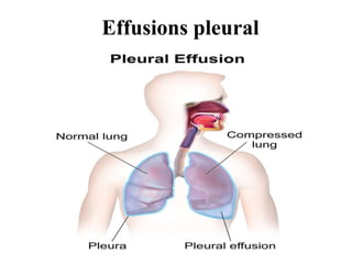
  • 2. Effusions pleural: • Pleural effusion, also called water on the lung, happens when fluid builds up in the space between your lungs and chest cavity. You may have symptoms, such as chest pain, depending on the cause. • Thin membranes, called pleura, cover the outside of the lungs and the inside of the chest cavity. • There’s always a small amount of liquid within this lining to help lubricate the lungs as they expand within the chest during breathing. • However, if too much fluid builds up, for example, because of a medical condition, problems can arise. Doctors call this pleural effusion. • Various conditions can lead to pleural effusion, but congestive heart failure is the most commonTrusted Source cause.
  • 3. Symptoms: • Some people with pleural effusion don’t have any symptoms. They may have symptoms of an underlying disease, such as a cough or fever. • You may find out you have pleural effusion through a chest X- ray or physical examination done for another reason. • When a doctor examines you, they may notice expansion on one side of the chest and a dull sound when they tap on that side. • Depending on the cause, a person with pleural effusion may also have: • chest pain • cough • fever • shortness of breath • See your doctor immediately if you have symptoms of pleural effusion.
  • 4. Causes and types: • Pleural effusion happens when fluid accumulates in the chest cavity outside the lung. • It can be either transudative or exudative. • Transudative pleural effusion happens when increased pressure on the small and large vessels of various organs causes them to be leaky, resulting in protein-filled fluid collections. It occurs with coronary heart disease or cirrhosis. • Exudative pleural effusion happens when there is irritation, inflammation, or infection. These can result in extra fluid production, reduced drainage, or both.
  • 5. Causes of pleural effusion includeTrusted Source: • congestive heart failure • cirrhosis or poor liver function • pulmonary embolism, which is caused by a blood clot and is a blockage in the lung arteries • complications from open-heart surgery • pneumonia • severe kidney disease • autoimmune diseases, such as lupus and rheumatoid arthritis • the use of some medications, such as methotrexate, phenytoin, or amiodarone
  • 6. • radiation therapy • rupture of the esophagus • ovarian hyperstimulation syndrome • certain types of cancer for instance, lung cancer and breast cancer • COVID-19
  • 7. Types: • Pleural effusion can be classifiedTrusted Source as transudative or exudative. • Transudative pleural effusions: • This type is caused by fluid leaking into the pleural space due to increased pressure in the blood vessels. • It can happen if you: • have congestive heart failure • have cirrhosis • have kidney disease • have just started peritoneal dialysis • have malnutrition due to low albumin levels in the blood
  • 8. Exudative effusions: • This happens when a buildup of fluid occurs as the result of: • inflammation • infection • tumors • a lung injury • Conditions that could result in this type of pleural infusion include: • tuberculosis • cancer • inflammatory conditions, such as pancreatitis, lupus, or rheumatoid arthritis • complications from heart surgery • chylothorax, which results from an obstruction in the lymph vessels • pneumonia • hemothorax, when blood collects in the pleural cavity • Some conditions, such as pulmonary embolism, can lead to either transudate or exudate pleural effusion.
  • 9. Pleural effusions and cancer: • Pleural effusions can occur when cancer cells spread to the pleura or block the flow of normal fluid within the pleura. Fluid may also build up due to certain cancer treatments, such as radiation therapy or chemotherapy. • Certain cancers are more likely to cause pleural effusions than others, including: • lung cancer • breast cancer • ovarian cancer • leukemia • melanoma • cervical cancer • uterine cancer • mesothelioma, which results from exposure to asbestos
  • 10.
  • 11. Signs and symptoms often include: • shortness of breath • cough • chest pain • weight loss • The doctor may drain the fluid or carry out pleurodesis if you’re likely to need repeated drainage. This involves insering a shunt that redirects the fluid away from the chest. • They may prescribe antibiotics if you have or are susceptible to infection. Steroids or other anti-inflammatory medications may reduce pain and inflammation. They will also discuss other treatment options for cancer. • People who are undergoing treatment for cancer may also have compromised immune systems, making them more prone to infections or other complications.
  • 12. Treatment: • The treatment and outcome will depend on the cause of the pleural effusion. • Draining fluid: • One way to treat pleural effusion is by draining the fluid from the chest cavity, either with a needle or by inserting a small tube into the chest. • You’ll receive a local anesthetic before this procedure, which will make the treatment more comfortable. You may feel some pain or discomfort at the incision site after the anesthetic wears off. Most doctors will prescribe medication to help relieve pain. • You may need this treatment more than once if fluid builds up again. • Other treatments may be necessary to manage fluid buildup if cancer is the cause of the pleural effusion.
  • 13. Antibiotics: • If you have a bacterial infection, the doctor will likely prescribe antibiotics or administer them intravenously. They will usually do this alongside drainage. • Pleurodesis: • Pleurodesis is a treatment that creates mild inflammation between the lung and chest cavity pleura. After drawing the excess fluid out of the chest cavity, a doctor injects a drug into the area. This drug causes the two layers of the pleura to stick together, which prevents the future buildup of fluid between them. • A doctor may decide to do this if pleural effusion is due to cancer. It reduces the need for frequent drainage.
  • 14. Surgery: • If symptoms don’t improve with drainage and antibiotics, the doctor may recommend thoracoscopic decortication or thorascopic debridement. They will insert a thoracoscope into the pleural space then either remove any tissue that is causing a problem (decortication) or surgically clean a wound to enable it heal (debridement). A doctor may call thisTrusted Source a medical thorocoscopy or a pleuroscopy. • In some cases, a doctor surgically inserts a shunt, or small tube, into the chest cavity. This helps redirect the fluid from the chest into the abdomen, where it can be more easily removed by the body. This may be an option for those who don’t respond to other treatments. • Pleurectomy, in which the surgeon removes part of the pleural lining, may also be an option in certain cases.
  • 15. Diagnosis: • Your doctor will perform a physical examination and listen to your lungs with a stethoscope. They may also order a chest X-ray to help diagnose pleural effusion. • Other possible tests includeTrusted Source: • chest ultrasound • CT scan • thoracentesis, where the doctor removes some pleural fluid for analysis • bronchoscopy • pleural biopsy • Thoracentesis involves removing fluid from the pleural membrane area by inserting a needle into the chest cavity and suctioning the fluid into a syringe. The doctor will use ultrasound to guide the needle. At the same time, they may drain the excess fluid from the chest cavity. The fluid will then be tested to determine the cause.
  • 16. • Your doctor may also choose to perform a pleural biopsy, which involves taking a tissue sample from the pleura. They do this by inserting a small needle from outside the chest wall into the chest cavity. • If they discover you have a pleural effusion but are unable to diagnose which type, your doctor may schedule a thoracoscopy. This is a surgical procedure that lets the doctor see inside the chest cavity using a fiber-optic camera. • For this procedure, your doctor will make a few small incisions in the chest area while you’re under general anesthesia. Then they’ll insert the camera through one incision and the surgical tool through the other to extract a small amount of fluid or tissue for analysis.
  • 17. Risks and complications: • Pleural effusions can be complicated or uncomplicated. Uncomplicated pleural effusions contain fluid without signs of infection or inflammation. They’re less likely to cause permanent lung problems. • Complicated pleural effusions, however, contain fluid with significant infection or inflammation. They require prompt treatment that frequently includes chest drainage. • Pleural effusion can be a sign of severe symptoms with some diseases. In 2021, some scientistsTrusted Source found that people with COVID-19 who developed pleural effusion were more likely to have severe inflammation and complications, which could affect their chances of recovery. • Treatment can also lead to complications. • Minor complications from more invasive treatments can include slight pain and discomfort, which often go away with time. More serious complications will depend on the severity of the condition, the cause, and the treatment used.
  • 18. Serious complications can include: • pulmonary edema or fluid in the lungs, which can result from draining fluid too quickly during thoracentesis • partial collapsed lung • infection or bleeding • empyema, when there is pus in the pleural space • trapped lung, when a layer formsTrusted Source around the lung that prevents it from expanding • These complications, while serious, are rare. Your doctor will help determine the most effective treatment option and discuss the benefits and risks of each procedure.
  • 19. Diagnostic investigations 1st investigations to order Investigations to consider Emerging tests •posteroanterior and lateral chest x-ray •pleural ultrasound •LDH and protein in pleural fluid and serum •red blood cell count in pleural fluid More 1st investigations to order •pleural fluid cholesterol level •thoracic CT scan •thoracic MRI •helical CT scan •More investigations to consider •tumor markers in pleural fluid •procalcitonin
  • 20. Sugar: • Pleural fluid analysis is a test that examines a sample of fluid that has been collected in the pleural space. • This is the space between the lining of the outside of the lungs (pleura) and the wall of the chest. • When fluid collects in the pleural space, the condition is called pleural effusion. • Thoracentesis should be performed for new and unexplained pleural effusions when sufficient fluid is present to allow a safe procedure. • Observation of pleural effusion is reasonable when benign etiologies are likely, as in the setting of overt congestive heart failure, viral pleurisy, or recent thoracic or abdominal surgery. • Laboratory testing helps to distinguish pleural fluid transudates from exudates. However, certain types of exudative pleural effusions might be suspected simply by observing the gross characteristics of the fluid obtained during thoracentesis.
  • 21. Note the following: • Frankly purulent fluid indicates an empyema • A putrid odor suggests an anaerobic empyema • A milky, opalescent fluid suggests a chylothorax, resulting in most often from lymphatic obstruction by malignancy or thoracic duct injury by trauma or surgical procedure • Grossly bloody fluid may result from trauma, malignancy, postpericardiotomy syndrome, or asbestos-related effusion and indicates the need for a spun hematocrit test of the sample. A pleural fluid hematocrit level of more than 50% of the peripheral hematocrit level defines a hemothorax, which often requires tube thoracostomy • Black pleural fluid suggests a limited number of diseases, including infection with Aspergillus niger or Rhizopus oryzae, malignant melanoma, non-small cell lung cancer or ruptured pancreatic pseudocyst, or charcoal-containing empyema
  • 22. Normal pleural fluid • Normal pleural fluid has the following characteristics: • Clear ultrafiltrate of plasma that originates from the parietal pleura • A pH of 7.60-7.64 • Protein content of less than 2% (1-2 g/dL) • Fewer than 1000 white blood cells (WBCs) per cubic millimeter • Glucose content similar to that of plasma • Lactate dehydrogenase (LDH) less than 50% of plasma
  • 23. Pleural fluid laboratory findings • Lights criteria (High protein and LDH = exudate), determines presence of exudate with protein and LDH levels – Pleural fluid protein to serum protein ratio >0.5 – Pleural fluid LDH to serum LDH ratio >0.6 – Pleural fluid level >2/3 of upper value for serum LDH • Additional criteria – Confirm exudate if results equivocal – Serum albumin – pleural fluid albumin <1.2g/dL • • Glucose – Low • Common: Infection (pneumonia) and malignancy • Rare: TB, haemothorax, Churg-Strauss
  • 24. Protein: • LDH level – This is classically high in exudates – Repeated testing confirms continuation or cessation of process • Increasing LDH (ongoing inflammation) • Decreasing LDH (cessation of process) • Pleural fluid pH (Low glucose and pH = infection or malignancy) – Taken if suspect pneumonic or malignant process (Low glucose) – <7.20 with pneumonia…Drain the fluid – <7.20 with malignancy …Life expectancy 30 days • Amylase – Useful if suspect pancreatitis as cause
  • 25. Cell count: • If exudate is confirmed, further testing required to evaluate cause of exudate • Differential cell count (predominance of white cells) – Neutrophils – PTE, pancreatitis, pneumonia, empyema – Lymphocytes – Cancer, TB pleuritis – Eosinophila – Pneumothorax, haemothorax, asbestosis, Churg-Strauss – Mononuclear cells – Chronic inflammatory process • Gram stain and culture and cytology – Use blood culture bottles and specimen jars – especially if chronic illness or suspect TB or fungus – Cytology useful in cases of suspected malignancy •