2. Cell Structure
In 1655, the English scientist Robert Hooke coined
the term “cellulae” for the small box-like structures
he saw while examining a thin slice of cork under a
microscope.
3. TWO TYPES OF CELLS
Prokaryotes
•single compartment and surrounded
by the plasma membrane
•lacks a defined nucleus
•simple internal organization
•ex. bacteria, blue-green algae
(cyanobacteria)
Eukaryotes
•
•contain a defined membrane-bound nucleus
• extensive internal membranes that enclose
compartments
main parts: plasma membrane, cytoplasm and nucleus
• ex. all members of the plant and animal kingdom, fungi
(molds* and yeasts**), protozoan*
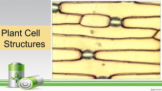
5. The Structure of Eukaryotic Plant Cell
• A living plant cell is composed of three main parts
namely: cell wall, protoplast and inclusions
6. 1. The Cell
Wall
• The outermost part of the plant cell
• Non-living structure with main chemical
component cellullose (a polysaccharide)
• Other substances which may form part of the
cell wall are lignin, suberin and cutin
• Between neighboring cell walls is a
cementing intercellular layer composed of
pectin substance
• The nonliving layer is often referred to as the
middle lamella
7.
8. PROTOPLASM
a mass of proteins, lipids, nucleic acids,
and water within a cell; except for the
wall, everything in the
cell is protoplasm
• Portions of the protoplast is organized into
protoplasmic bodies with specific functions and
are generally referred to as ORGANELLES
• The protoplast is composed of two main regions,
namely: an outer region called as the
CYTOPLASM and an inner region as the
NUCLEUS
9.
10. • Is bounded by a protoplasmic membrane
called the plasma membrane,
plasmalemma or cell membrane
• The plasma membrane is a selectively
permeable protoplasmic membrane which
regulates the entry and exit of materials in
a cell
11. Other main cytoplasmic structures of plant cells:
1. Mitochondria
2. Ribosomes
3. Endoplasmic Reticulum (ER)
4. Golgi bodies or dictyosomes
5. Lysosomes
6. Plastids
7. Microtubules
12. Mitochondria
• Mitochondria are found in PLANT and animal cells.
• Sites of cellular respiration, ATP synthesis
• Bound by a double membrane surrounding fluid-filled matrix.
• The inner membranes of mitochondria are cristae
• The matrix contains enzymes that break down carbohydrates and
the cristae house protein complexes that produce ATP
THE POWER
HOUSE OF THE
CELL
ATP
13. Ribosomes
• Granular structures visible under electron
microscope
• Protein synthesis
• Ribosomes are composed of a large subunit
and a small subunit.
• Ribosomes can be found alone in the
cytoplasm, in groups called polyribosomes, or
attached to the endoplasmic reticulum.
• Ribosomes are RNA-protein complexes
composed of two subunits that join and
attach to messenger RNA.
– site of protein synthesis
– assembled in nucleoli
14. Endomembrane System
• Compartmentalizes cell, channeling
passage of molecules through cell’s interior.
– Endoplasmic reticulum
• Rough ER - studded with ribosomes
• Smooth ER - few ribosomes
15. Rough ER
• Rough ER is especially abundant in cells that secrete
proteins.
– As a polypeptide is synthesized on a ribosome
attached to rough ER, it is threaded into the
cisternal space through a pore formed by a protein
complex in the ER membrane.
– As it enters the cisternal space, the new protein
folds into its native conformation.
– Most secretory polypeptides are glycoproteins,
proteins to which a carbohydrate is attached.
– Secretory proteins are packaged in transport
vesicles that carry them to their next stage.
16. Smooth ER
• The smooth ER is rich in enzymes and plays a role in a variety of
metabolic processes.
17. The Golgi bodies/ dictyosomes
• The Golgi body is the shipping and receiving center for cell
products.
– Many transport vesicles from the ER travel to the Golgi apparatus
for modification of their contents.
– The Golgi is a center of manufacturing, warehousing, sorting, and
shipping.
– The Golgi apparatus consists of flattened membranous sacs—
cisternae—looking like a stack of pita bread.
– The Golgi sorts and packages materials into transport vesicles.
18. Functions Of The Golgi Bodies
TEM of Golgi apparatus
cis face
(“receiving” side of
Golgi apparatus)
Vesicles move
from ER to GolgiVesicles also
transport certain
proteins back to ER
Vesicles coalesce to
form new cis Golgi cisternae
Cisternal
maturation:
Golgi cisternae
move in a cis-
to-trans
direction
Vesicles form and
leave Golgi, carrying
specific proteins to
other locations or to
the plasma mem-
brane for secretion
Vesicles transport specific
proteins backward to newer
Golgi cisternae
Cisternae
trans face
(“shipping” side of
Golgi apparatus)
0.1 0 µm1
6
5
2
3
4
Golgi
apparatus
19. Lysosomes and Peroxisomes
• Lysosomes – vesicle containing hydrolytic or
digestive enzymes that break down food/foreign
particles
• Peroxisomes - contain enzymes that catalyze
the removal of electrons and associated
hydrogen atoms
20. Lysosome
• Rounded or iregularly-
shaped organelles
bounded by a single
membrane
• Said to play important
role in the destruction
of worn-out or
defective parts of the
cell
• Probably belong to a
group of structures like
microbodies (a) Phagocytosis: lysosome digesting food
1 µm
Lysosome contains
active hydrolytic
enzymes
Food vacuole
fuses with
lysosome
Hydrolytic
enzymes digest
food particles
Digestion
Food vacuole
Plasma membrane
Lysosome
Digestive
enzymes
Lysosome
Nucleus
21. Plastids
• Rounded, oval or irregularly-shaped
protoplasmic bodies of three main
parts:
• Leucoplast
• Chroloplast
• Chromoplast
22. Leucoplast
• Colorless plastids
• Some involved in food storage
• If associated with the starch storage, they
are referred to as amyloplasts
• if associated with oil storage= elaioplasts
• With protein storage = aleurone-plasts
24. Chloroplasts
• A chloroplast is bounded by two membranes enclosing a fluid-filled
stroma that contains enzymes.
• Membranes inside the stroma are organized into thylakoids that house
chlorophyll.
• Chlorophyll absorbs solar energy and carbohydrates are made in the
stroma.
27. • the thickest fibers, are hollow rods about 25
microns in diameter.
– are constructed of the globular protein,
tubulin, and they grow or shrink as more
tubulin molecules are added or removed.
• They move chromosomes
during cell division.
Fig. 7.21b
Microtubules
28. • long, hollow cylinders and made-up
of protein tubulin
• outer diameter of 25nm
• much more rigid
• long and straight and typically have
one end attached to a single
microtubule-organizing center
(MTOC) called a centrosome
29. The Nucleus
• Repository for genetic material
• Chromatin: DNA and proteins
• Nucleolus: Chromatin and ribosomal
subunits - region of intensive ribosomal RNA
synthesis. darkly staining rounded bodies rich
in ribosomal RNA, the type of RNA used in
the formation of ribosomes
• Nuclear envelope: Surface of nucleus bound
by two phospholipid bilayer membranes -
Double membrane with pores
• Nucleoplasm: semifluid medium inside the
nucleus
31. Nucleus
– Nickname: “The Control Center”
– Directs cell activities
• Separated from cytoplasm by nuclear
membrane
• Contains genetic material - DNA
33. Chromosomes
• DNA of eukaryotes is divided into linear chromosomes.
– Exist as strands of chromatin, except during cell division
– Histones associated packaging proteins
34. 3. The Inclusions
• Refer to the nonprotoplasmic structures found
within the protoplast
• Among these are the VACUOLES which are
fluid-filled structures
• The fluid vacuole commonly referred to as the
cell sap
• Contains various dissolved substances such
as anthocyanins (water-soluble pigments) and
various metabolites (e.g. Sugars, inorganic
salts, organic acids, alkaloids)
35. Cont...
• Other inclusions are waste products which may be in
a form of crystals
• The crystals may be contained in vacoules and in the
cytoplasm
• Plant cell may start with many but small vacuoles
• Upon maturity, the small vacuoles may coalesce to
form bigger but fewer vacuoles
• Eventually, a plant cell may have but a SINGLE
LARGE CENTRAL VACUOLE
• Some biologist would consider vacuole as an
oganelle. As such the membrane-bounded structure
containing the cell sap.
36. Crystals in Plant Cells
• Many plant cells contain crystals
which are a product of metabolism
• There are many forms
• Most common are composed of
calcium oxide and calcium carbonate
37. Calcium oxalate crystals
• Generally found in the vacuoles and are as follows:
1. Raphides – needle like crystals which occur singly
or in groups or bundles as in gabi and other succulent plants
2. Prismatic – prism-like or pyramid-like crystals found
in leaves of begonia and bangka-bangkaan
(Rhoeo discolor)
3. Rosette – aggregate of crystals which has
flower-like apperance in santan (Ixora sp.) and
stem of Kutsarita plant
41. Calcium carbonate crystals
• Cystotith- grape-like as seen in a hypodermal cell of the
leaf of Indian rubber tree or ampalaya-like plant.
42. BASIC TYPES OF CELLS
• Despite the diversity of types of stems that have
originated by natural selection, all share a basic, rather
simple organization.
• The same is true for leaves and roots.
• Although we might suspect that numerous types of cells
are present within a plant, actually the various kinds of
plant cells are customarily grouped into three classes
based on the nature of their walls:
Parenchyma, Collenchyma, and Sclerenchyma.
43. 1. PARENCHYMA
• Parenchyma cells have only primary walls that remain
thin.
• Parenchyma tissue is a mass of parenchyma cells. This is
the most common type of cell and tissue, constituting all soft
parts of a plant.
• Parenchyma cells are active metabolically and usually remain
alive once they mature.
• Numerous subtypes are specialized for particular tasks:
44. Parenchyma cells of Geranium
spp.; their walls (green) are thin,
and their vacuoles are large and
full of watery contents that did not
stain. Nuclei were present in all
cells,
45. Subtypes of Parenchyma Cells
A. Chlorenchyma cells
B. Glandular cells
C.Transfer cells
D.(Aerenchyma cells)
46. A. Chlorenchyma cells
• Chlorenchyma cells are parenchyma cells involved in
photosynthesis
• they have an abundance of chloroplasts, and the thinness
of the wall is advantageous for allowing light and carbon
dioxide to pass through to the chloroplasts.
• Other types of pigmented cells, as in flower petals and
fruits, also must be parenchyma cells with thin walls that
permit the pigments in the protoplasm to be seen.
47. Chlorenchyma cells from a
leaf of privet- Ligustrum
vulgare L.
-Intercellular spaces, permit
CO2 rapid diffusion in the
leaf
49. B. Glandular cells
• Glandular cells that secrete nectar, fragrances, mucilage,
resins, and oils are also parenchyma cells; they typically
contain few chloroplasts but have elevated amounts of
dictyosomes and endoplasmic reticulum.
• They must transport large quantities of sugar and
minerals into themselves, transform them metabolically,
then transport the product out.
51. C. Transfer cells
• Transfer cells are parenchyma cells that mediate the short-
distance transport of material by means of a large, extensive
plasma membrane capable of holding numerous molecular
pumps.
• Unlike animal cells, plant cells cannot form folds or
projections of their plasma membranes; instead, transfer
cells increase their surface area by having extensive knobs,
ridges, and other ingrowths on the inner surface of their walls
• Because the plasma membrane follows the contour of all
these, it is extensive and capable of large-scale molecular
pumping.
52. Transfer cells in the salt gland of
Frankenia grandifolia.
The wall ingrowths increase the
surface area of the cell membrane,
providing
more room for salt-pumping proteins in
the membrane.
W. W. Thomson
and R. Balsamo, University of
California, Riverside
53. D. (Aerenchyma cells)
• Parenchyma tissue with extensive connected air
spaces.
• refers to spaces or air channels in the leaves, stems
and roots of some plants, which allows exchange of
gases between the shoot and the root.
• The channels of air-filled cavities provide a low-
resistance internal pathway for the exchange of
gases such as oxygen and ethylene between the
plant above the water and the submerged tissues.
• Aerenchyma is widespread in aquatic and wetland
plants which must grow in hypoxic soils
54.
55. Stem cross-section of Peperomia. The inner
part of the stem is mostly parenchyma cells.
56. • Some parenchyma cells function by dying at maturity.
• Structures such as stamens and some fruits must open
and release pollen or seeds; the opening may be
formed by parenchyma cells that die and break down or
are torn apart.
• Large spaces may be necessary inside the plant body;
some of these are formed when the middle lamella
decomposes and cells are released from their
neighbors.
• In other cases, the space is formed by the degeneration
of parenchyma cells.
• In a few species, such as milkweeds, as parenchyma
cells die, their protoplasm is converted metabolically
into mucilage or a milky latex.
57. • Parenchyma tissue that conducts nutrients over long distances
is phloem
• Parenchyma cells are relatively inexpensive to build because
little glucose is expended in constructing the wall's cellulose
and hemicellulose.
• Each molecule incorporated into a wall polymer cannot be
used for other purposes such as the generation of ATP or the
synthesis of proteins.
• Consequently, it is disadvantageous to use a cell with thick
walls any time one with thin walls would be just as functional.
58. 2. COLLENCHYMA CELLS
• Collenchyma cells have a primary wall that
remains thin in some areas but becomes thickened
in other areas, most often in the corners.
• The nature of this wall is important in understanding
why it exists and how it functions in the plant.
• Like clay, the wall of collenchyma exhibits plasticity,
the ability to be deformed by pressure or tension and
to retain the new shape even if the pressure or tension
ceases.
59. • Collenchyma is present in elongating shoot tips
that must be long and flexible, such as those of
vining plants like grapes, as a layer just under the
epidermis or as bands located next to vascular
bundles, making the tips stronger and more resistant
to breaking.
• But the tips are still capable of elongating because
collenchyma can be stretched.
• In species whose shoot tips are composed only of
weak parenchyma, the tips are flexible and delicate
and often can be damaged by wind; the elongating
portion must be very short or it simply buckles under
its own weight.
60. Stem cross-section of Peperomia. Masses of collenchyma cells
often occur in the outer parts of stems and leaf stalks. The
collenchyma forms a band about 8 to 12 cells thick.
61. • It is important to think about the method by which
collenchyma provides support.
• If a vine or other collenchyma-rich tissue is cut off from
its water supply, it wilts and droops; the collenchyma is
unable to hold up the stem.
• Parenchyma cells are needed in the inner tissues for
support. Collenchyma and turgid parenchyma work
together like air pressure and a tire: The tire or inner
tube is extremely strong but is useless for support
without air pressure.
• Similarly, air pressure is useless unless it is confined by
a container.
• In stems, the tendency for parenchyma to expand is
counterbalanced by the resistance of the collenchyma,
and the stem becomes rigid.
62. The shoot tips
of long vines
need the plastic
support of
collenchyma
while their
stems are
elongating
63. • Because the walls of collenchyma cells are thick, they
require more glucose for their production.
• Collenchyma is usually produced only in shoot tips and
young petioles, where the need for extra strength justifies
the metabolic cost.
• Subterranean shoots and roots do not need collenchyma
because soil provides support, but the aerial roots of
epiphytes such as orchids and philodendrons have a
thick layer of collenchyma
64. 3. SCLERENCHYMA CELLS
• The third basic type of cell and tissue, sclerenchyma,
has both a primary wall and a thick secondary wall that
is almost always lignified.
• These walls have the property of elasticity: They can be
deformed, but they snap back to their original size and
shape when the pressure or tension is released.
• Sclerenchyma cells develop mainly in mature organs that
have stopped growing and have achieved their proper
size and shape.
65.
66. A stem of bamboo was treated with a mixture of nitric acid and chromic acid to
67. These are sclereids; they are more or less cuboidal, definitely not long like fibers.
These have remained alive at maturity, and nuclei & cytoplasm are visible in
68. astrosclereids (star-shaped sclereids), shines brightly because its cellulose molecules
are packed in a tight, crystalline form, giving the wall extra strength .
69. • Deforming forces such as wind, animals, or snow would
probably be detrimental.
• If mature organs had collenchyma for support, they would be
reshaped constantly by storms or animals, which of course
would not be optimal.
• For example, while growing and elongating, a young leaf
must be supported by collenchyma if it is to continue to grow.
• But once it has achieved its mature size and shape, some
cells of the leaf can mature into sclerenchyma and provide
elastic support that maintains the leaf's shape.
• Unlike collenchyma, sclerenchyma supports the plant by its
strength alone; if sclerenchyma-rich stems are allowed to wilt,
they remain upright and do not droop.
70. • Parenchyma and collenchyma cells can absorb water so
powerfully that they swell and stretch the wall, thereby
growing; sclerenchyma cell walls are strong enough to
prevent the protoplast from expanding.
• The rigidity of sclerenchyma makes it unusable for growing
• shoot tips because it would prevent further shoot elongation.
• Sclerenchyma cells are of two types—conducting
sclerenchyma and mechanical sclerenchyma.
• The mechanical sclerenchyma is subdivided into long fibers
and short sclereids , both of which have thick secondary
walls.
• Because fibers are long, they are flexible and are most often
found in areas where strength and elasticity are important.
71. • The wood of most flowering plants contains abundant
fibers, and their strength supports the tree while their
elasticity allows the trunk and branches to sway in the
wind without breaking (usually) or becoming permanently
bent .
• The fiber-rich bark is important not in holding up the tree
but in resisting insects, fungi, and other pests.
• Sclereids are short and more or less
isodiametric(cuboidal).
• Because sclereids have strong walls oriented in all three
dimensions, sclerenchyma tissue composed of sclereids
is brittle and inflexible.
74. Parenchyma
This tissue is composed of large,
thin walled cells having a single
large vacuole.
This tissue is found in the soft parts
of the plant like the cortex(outer
region) and pith(inner region) of the
roots and stems.
The cells present in these tissues
are living.
These cells store food for the plant,
and thus they also provide
temporary support for the plants.
When parenchymatous cells are
present in leaves, they sometimes
contain chlorophyll, and thus are
green in colour. This tissue is then
referred to as chlorenchyma.
Potatoes are made up of mainly
Parenchyma tissue cells.
75. Collenchyma
This tissue is made up of
cells from the parenchyma
tissue which become
elongated and have thick cell
walls at the corners. This
diagram illustrates the
difference between
parenchyma and
collenchyma cells. The
collenchyma cells are
elongated and thick at the
corners.
This tissue is found mainly in
the leaf stalks and below the
epidermis of stems.
It gives support for the parts
of a plant like leaves, stems,
branches.
76. Sclerenchyma
Sclerenchyma is made up of
thin, narrow and long cells
having very thick cell walls
due to deposition of lignin.
These cells are thin because
they are all dead cells, having
no functions to perform and so
no organelles inside the cell
require space.
They are thick at the cell walls
and thus these cells provide
rigid support for the plant as
they are hard and supportive.
That is why the tissue is
named "Sclerenchyma", as
scleros means hard.
Sclerenchyma tissue is
present in stems of plants and
the veins of leaves in plants. It
provides strength to plant
parts.