
Recommended
Recommended
More Related Content
What's hot
What's hot (20)
Similar to Two new species of Myxobolus (Myxozoa: Myxosporea) parasites of Barbus callipterus Boulenger, 1907 (Cyprinidae) and Oreochromis niloticus Linnaeus, 1758 (Cichlidae) in Cameroon
Similar to Two new species of Myxobolus (Myxozoa: Myxosporea) parasites of Barbus callipterus Boulenger, 1907 (Cyprinidae) and Oreochromis niloticus Linnaeus, 1758 (Cichlidae) in Cameroon (20)
More from Journal of Research in Biology
More from Journal of Research in Biology (20)
Recently uploaded
Recently uploaded (20)
Two new species of Myxobolus (Myxozoa: Myxosporea) parasites of Barbus callipterus Boulenger, 1907 (Cyprinidae) and Oreochromis niloticus Linnaeus, 1758 (Cichlidae) in Cameroon
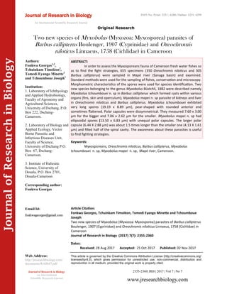
- 1. Article Citation: Fonkwa Georges, Tchuinkam Timoléon, Tomedi Eyango Minette and Tchoumboue Joseph Two new species of Myxobolus (Myxozoa: Myxosporea) parasites of Barbus callipterus Boulenger, 1907 (Cyprinidae) and Oreochromis niloticus Linnaeus, 1758 (Cichlidae) in Cameroon Journal of Research in Biology (2017) 7(7): 2355-2360 JournalofResearchinBiology Two new species of Myxobolus (Myxozoa: Myxosporea) parasites of Barbus callipterus Boulenger, 1907 (Cyprinidae) and Oreochromis niloticus Linnaeus, 1758 (Cichlidae) in Cameroon Keywords: Myxosporeans, Oreochromis niloticus, Barbus callipterus, Myxobolus tchoumbouei n. sp, Myxobolus mapei n. sp, Mapé river, Cameroon. ABSTRACT: In order to assess the Myxosporeans fauna of Cameroon fresh water fishes so as to find the fight strategies, 655 specimens (350 Oreochromis niloticus and 305 Barbus callipterus) were sampled in Mapé river (Sanaga basin) and examined. Standard methods were used for the sampling of fishes, conservation and microscopy. Morphometric characteristics of the spores were used for species identification. Two new species belonging to the genus Myxobolus Büstchli, 1882 were described namely Myxobolus tchoumbouei n. sp in Barbus callipterus which formed cysts within various organs (fins, skin and operculum); Myxobolus mapei n. sp parasite of kidneys and liver in Oreochromis niloticus and Barbus callipterus. Myxobolus tchoumbouei exhibited very long spores (19.19 x 8.89 µm), pear-shaped with rounded anterior end sometimes flattened. Polar capsules were dissymmetrical. They measured 7.60 x 3.00 µm for the bigger and 7.06 x 2.62 µm for the smaller. Myxobolus mapei n. sp had ellipsoidal spores (13.50 x 6.83 µm) with unequal polar capsules. The larger polar capsule (6.44 X 2.88 µm) was about 1.5 times longer than the smaller one (4.13 X 1.61 µm) and filled half of the spiral cavity. The awareness about these parasites is useful to find fighting strategies. 2355-2360| JRB | 2017 | Vol 7 | No 7 This article is governed by the Creative Commons Attribution License (http://creativecommons.org/ licenses/by/4.0), which gives permission for unrestricted use, non-commercial, distribution and reproduction in all medium, provided the original work is properly cited. www.jresearchbiology.com Journal of Research in Biology An International Scientific Research Journal Authors: Fonkwa Georges1,2 , Tchuinkam Timoléon2 , Tomedi Eyango Minette3 and Tchoumboue Joseph1 Institution: 1. Laboratory of Ichthyology and Applied Hydrobiology, Faculty of Agronomy and Agricultural Sciences, University of Dschang, P.O. Box 222, Dschang- Cameroon. 2. Laboratory of Biology and Applied Ecology, Vector Borne Parasitic and Infectious Diseases Unit, Faculty of Science, University of Dschang P.O. Box 67, Dschang- Cameroon. 3. Institute of Halieutic Science, University of Douala, P.O. Box 2701, Douala-Cameroon Corresponding author: Fonkwa Georges Email Id: Web Address: http://jresearchbiology.com/ documents/RA0647.pdf Dates: Received: 28 Aug 2017 Accepted: 25 Oct 2017 Published: 02 Nov 2017 Journal of Research in Biology An International Scientific Research Journal Original Research ISSN No: Print: 2231 –6280; Online: 2231- 6299
- 2. INTRODUCTION Fish is an important source of animal proteins (FAO, 2016). Many factors influence fish productions among which are parasites (Renault and Guichard, 2007). Myxosporeans are primarily fish parasites (Fomena et al., 2010; Eiras et al., 2010). They affect fish growth (Longshaw et al., 2010), their reproduction (Obiekezie and Okaeme, 1990) and are involved in epizooties responsible for massive fish deaths (Gbankoto et al., 2001; Feist and Longshaw, 2005). The world of Myxosporean fauna is composed of about 2180 species gathered within 62 genera among which the genus Myxobolus Bütschli, 1882 is the most abundant with 792 species (Lom et Diková, 2006). The Myxosporean fauna of Africa fresh water fishes comprises about 240 species (Lekeufack, 2010). Moreover, in Cameroon particularly, 74 species were described. Those species belong to the genera Myxobolus, Henneguya, Myxidium, Thelohanellus, Sphaerospora, Chloromyxum and Hoferellus. The objective of this study is to describe two new species of Myxosporean, parasites of economical and dietary important fishes in Cameroon namely Myxobolus tchoumbouei n.sp. and Myxobolus mapei n.sp. MATERIALS AND METHODS Study site Fishes were sampled in Mapé river (tributary of Mbam river), Bankim subdivision (6°00’- 6°20’ NL and 11°20’-11°40’ EL, Adamawa – Cameroon Region, Central Africa). The average altitude is about 724 m. The soil is a mixture of clay and sand. The climate is of tropical Soudano-Guinean type with two seasons: a long rainy season running from March to November and a short dry season from November to March. The annual average temperature is about 23°C and the rainfall varies between 1500 and 2000 mm (Olivry, 1986). Fish sampling and conservation A total of 655 specimens of fishes (305 Barbus callipterus and 350 Oreochromis niloticus) were bought monthly from fishermen during the study period i.e. May 2016 to May 2017. They were captured both at the day and night using fish nets and fishing canes. On the field, specimens were immediately stored at 10% formalin solution and transported to the laboratory for examination. Identification of myxosporeans In the laboratory, fishes were identified according to Stiassny et al. (2007) and examined according to the method used by Abakar (2006). So, standard and total lengths were measured to the closest millimeter using a slide caliper of stainless brand. Fishes were weighed using Sartorius electronic scale of 0.01g accuracy and were sex determined after dissection. External organs (fins, skin, scales and eyes) and internal organs (gills, spleen, kidneys, intestines, gall bladder, stomach and gonads) were examined with naked eyes, then with Motic stereoscopic microscope at 10X to look for the macroscopic cysts. Smears of kidneys, spleen and gonads were made and examined at a total magnification of 1000 X with a light microscope in order to look for spores. Cysts were crushed between slide and cover glass in a drop of distilled water and their contents were identified with the light microscope at 1000X. Spores were fixed using methanol, stained with May- Grünwald-Giemsa and snapped with digital camera, Canon Ixus brand. Species were identified according to Lom and Arthur (1989). The measurements are given in the form of mean (minimum - maximum) values. RESULTS Myxobolus tchoumbouei n. sp Vegetative stage This parasite forms whitish cysts which are ovoid, polysporous and generally macroscopic (1 to 2 mm in diameter). They are embedded in various organs (fins, skin and operculum). A single host can harbor 1 to 7 cysts. Fonkwa et al., 2017 2356 Journal of Research in Biology (2017) 7(7): 2355-2360
- 3. Spores They are big (19.19 x 8.89 µm), pear-shaped with rounded (Figures 1-6) and sometimes with flattened anterior end. The inter capsular appendix is lacking. Polar capsules are slightly dissymmetrical and of unequal dimensions. The number of polar filament coils ranges from 7 to 10. The sporoplasm is very well developed and occupies two third of spiral cavity. Measurements: Length (L): 19.19 (17.50 – 25.50) µm; width (l): 8.89 (7.50 – 10) µm; ratio (L/l): 2.15; length of large polar capsule (L’): 7.60 (6 – 8.75) µm; width of large polar capsule (l’): 3 (2.50 – 3.75) µm; ratio (L’/l’): 2.52; length of small polar capsule (L’’): 7.06 (5.63 – 8.75) µm; width of small polar capsule (l’’): 2.62 (1.88 – 4.50) µm; ratio (L’’/l’’): 2.71 Host type: Barbus callipterus Boulenger, 1907 Location: Fins, operculum, skin, kidneys, liver Prevalence: 11.15% (34 / 305) Myxobolus mapei n .sp (Figures 7-10) Vegetative stage: This myxosporean did not form cysts. Pansporoblasts were found in the kidneys. Spores: From frontal view, spores are ellipsoidal. The polar capsules are dissymmetrical. The larger polar capsule is well developed (6.44 X 2.88 µm) and 1.5 times longer than the smaller one and lies in the half of sporal cavity. The sporoplasm is binucleated and the intercapsular appendix is absent. Measurements : Length (L): 13.35 (10.50 – 16.50) µm; width (l): 6.83 (6 - 9) µm; ratio (L/l): 1.97 length of large polar capsule (L’): 6.44 (6 – 8.25) µm; width of large polar capsule (l’): 2.85 (2.25 – 4.50) µm; ratio (L’/l’): 2.28; length of small polar capsule (L’’): 4.19 (3 – 5.25) µm; width of small polar capsule (l’’): 1.61 (1.50 – 2.25) µm; ratio (L’’/l’’): 2.80. Hosts: Oreochromis niloticus Linnaeus, 1758 and Barbus callipterus Boulenger, 1907 Location: liver and kidneys Prevalence: 3. 43 % for Oreochromis niloticus (12/350); 9.18 % for Barbus callipterus (28 / 305). DISCUSSION Myxobolus tchoumbouei n.sp. There are relevant differences between Myxobolus tchoumbouei n. sp. and four myxosporeans species of the genus Myxobolus described throughout the world (Table 1). In fact, Lom et al. (1992) described Myxobolus aureatus as the fins parasite of Notropis anogenus, Pimephales notatus and P. promelas in Canada, but that species is shorter (14 -16.6 µm) than our specimen (17.50 – 25.50 µm) and possesses polar Journal of Research in Biology (2017) 7(7): 2355-2360 2357 Fonkwa et al., 2017 Parasite species Hosts Infested organs Countries ReferencesS. No Myxobolus aureatus Ward (1919) Notropis anogenus Pimephales notatus P. promelas Fins Canada Lom et al. (1992)1 Myxobolus angustus Kudo (1886) Pimephales vigilax Gills Canada Kudo (1886)2 Myxobolus pseudokoi Li and Desser (1985) Luxilus cornutus Gills Canada Li et Desser (1985) 3 Myxobolus bilobus Cone et al. (2005) Notemigonus crysoleucas Gills Canada Cone et al. (2005)4 Myxobolus mbailaoi Fomena et al. (2004) Citharinus citharus Skin, operculum, intestines Cameroon Fomena et al. (2004) 5 Myxobolus tchoumbouei n. sp Barbus callipterus Fins, operculum, skin, kidneys, liver Cameroon Present study6 Myxobolus mapei n. sp. Barbus callipterus Oreochromis niloticus Kidneys, liver Cameroon Present study7 Table 1. Comparison of Myxobolus tchoumbouei n.sp. and Myxobolus mapei n. sp with five similar species described in Cyprinidae across the world
- 4. capsules which are not only symmetrical but also longer (6.20 -9.4µm). Myxobolus angustus described by Kudo (1886) on the gills of Pimephales vigilax in Canada is far different from Myxobolus tchoumbouei n. sp. by many features: length (14 -15µm), symmetrical polar capsules (8-9.5 X 2.5 -3 µm), less developed polar capsules with iodinophilous vacuole. Moreover, the anterior end of Myxobolus angustus is narrow. Li and Desser (1985) described Myxobolus pseudokoi, gills parasite of Luxilus cornutus in Canada. This species differs from M. tchoumbouei n. sp. by its size (11.50 – 14.50 X 6 - 7 µm), symmetrical polar capsules (6 – 7.5 X 2 -3 µm) and the presence of an iodinophilous vacuole. The species resembling remarkably Myxobolus tchoumbouei n. sp. is Fonkwa et al., 2017 2358 Journal of Research in Biology (2017) 7(7): 2355-2360 Figures 1-10: Spores and cysts micrographs and lines drawings of different Myxobolus species described 1-6: Myxobolus tchoumbouei n.sp. 1: Fresh spore (scale bar: 5.5 µm), dissymmetric polar capsules and well developed sporoplasm 2: Stained spore with May-Grünwald- Giemsa (scale bar: 5.5 µm), rounded anterior end (arrow) 3: Stained spores with May-Grünwald- Giemsa (scale bar: 5.5 µm), flattened anterior end (arrow) 4: Line drawing, scale bar: 10 µm 5: Cyst implanted in the operculum (arrow, white spot) x5 6: Cyst implanted in the skin (arrow, white spot) x5 7-10: Myxobolus mapei n. sp. 7: Stained spore with May-Grünwald- Giemsa, two nuclei (arrows), scale bar: 3.3 µm 8: Fresh spore, the bigger size of the large polar capsule compared to the smaller polar capsule (arrows), scale bar: 3.3 µm 9: Line drawing, scale bar: 10 µm 10: Disporal pansporoblast (arrows), scale bar: 6.7µm 5 7 8 9 10 1 2 43 6 5.5 μm 5.5 μm 10μm 5X5X 3.3 μm 3.3μm 10μm 6.7 μm 5.5μm
- 5. Myxobolus bilobus, a gill myxosporean of Notemigonus crysoleucas, Cyprinidae from Brewer and Opeongo lakes and Algonquin park (Canada). However, there are several diverging characters. Myxobolus bilobus cysts are bilobed (where the name originates) in contrast to those of M. tchoumbouei n. sp. Moreover, both parasites species infest Cyprinidae fishes belonging to the different genera. Myxobolus bilobus seems to be specific to the gills of Notemigonus crysoleucas whereas M. tchoumbouei n. sp. targets several organs (fins, operculum, skin, kidneys, liver) in Barbus callipterus. Myxobolus bilobus spores are shorter (20 – 22.10 µm) and less wide (7. 50 – 9.30 µm). Although its polar capsules are dissymmetrical and of unequal sizes like those of M. tchoumbouei n. sp, they are however longer, hence, reducing the sporoplasm volume. The lengths of the larger and smaller polar capsule are 9.10 -12 µm and 9.10 -11 µm respectively in Myxobolus bilobus against 6 – 8.75 µm and 5.63 – 8.75 µm for Myxobolus tchoumbouei n. sp. The rounded and sometimes flattened anterior end of Myxobolus tchoumbouei n. sp. makes it very different from M. bilobus which rather has a narrower anterior end. In a nutshell, the parasite of Barbus callipterus is probably new and we propose to name it Myxobolus tchoumbouei n. sp. in honor to Tchoumboué Joseph, Emeritus Professor in the University of Dschang- Cameroon. Myxobolus mapei n. sp In Africa, several species of Myxobolus are described. Fomena et al. (2004) described Myxobolus mbailaoi in Cameroon. This parasite infesting skin, operculum and intestines of Citharinus citharus differs from Myxobolus mapei n. sp. which does not form cysts and parasitizes two taxonomically distant hosts namely Oreochromis niloticus and Barbus callipterus. Myxobolus mbailaoi spores are less long and wider (11.57 X 7.7 µm) compared to Myxobolus mapei n. sp. (13.35 X 6.83 µm). Although having unequal polar capsules, they are less developed than those of Myxobolus mapei n. sp. In addition, its sporoplasm has an iodinophilous vacuole lacking in our parasite. Nchoutpouen and Fomena (2011) described Myxobolus nchoutnounensis from Labeo parvus in Cameroon. Although this parasite has two dissymmetrical polar capsules, it possesses an intercapsular triangle and iodinophilous vacuole, features missing in Myxobolus mapei n. sp. Moreover, the latter does not form cysts. We think that the parasite being described is new and will be called Myxobolus mapei n. sp referring to the river where the hosts were captured. CONCLUSION The newly described Myxobolus species confirm the predominance of the genus Myxobolus among Myxosporidia. The available data are useful and will be used to elaborate fighting strategies. REFERENCES Abakar O. (2006). Les myxosporidies (Myxozoa: Myxosporea) parasites des poissons D’eau douce du Tchad: faunistique et biologie des espèces inféodées à Oreochromis niloticus (LINNE, 1758) et Sarotherodon gallilaeus (LINNE, 1758) Cichlidae. Thèse de Doctorat D’Etat .Université de Yaoundé I. 163 p. David K Cone, Jing Yang, Genlou Sun and Russell Easy. (2005). Taxanomy and molecular phylogeny of Myxobolus bilobus n. sp. (Myxozoa) parasitizing Notemigonus crysoleucas (Cyprinidae) in Algonquin park, Ontorio, Canada. Diseases of Aquatic Organisms, 66(3): 227 – 232. Eiras JC, Monteiro CM and Brasil – Sato MC. (2010). Myxobolus franciscoi sp. Nov. (Myxozoa: Myxosporea), a parasite of Prochilodus argenteus (Actinopterygii: Prochilodontidae) from upper sao Francisco river, Brazil, with a revision of Myxobolus spp. from South America. Zoologia, 27(1) : 131 – 137. Journal of Research in Biology (2017) 7(7): 2355-2360 2359 Fonkwa et al., 2017
- 6. FAO (2016). The state of world fisheries and aquaculture. Contributing to food security and nutrition for all. 200 p. Feist SW and Longshaw M. (2005). Myxozoan diseases in fish and effects on host population. Acta Zoologica Sinica, 51(4): 758 – 760. Fomena A, Abakar O, Ngassam P and Bouix G. (2004). Description de trois espèces nouvelles de myxosporidies (Myxozoa: Myxosporea) parasites de Citharinus citharinus (Geoffroy Saint-Hilaire,1809) (Citharinidae) au Tchad (Afrique Centrale). Parasite. 11(1): 83-88. Fomena A, Lekeufack Folefack GB and Bouix G. (2010). Deux espèces nouvelles de Myxidium (Myxosporea: Myxidiidae) parasites des poissons d’eau douce du Cameroun. Parasite, 17(1) : 9 – 16. Gbankoto A, Pampoulie C, Marques A and Sakiti GN. (2001). Occurrence of myxospoean parasites in gills of Tilapia species from lake nokoue (Benin, West Africa): effect of host size and sex, and seasonal pattern of infection. Diseases of Aquatic Organisms, 44: 221 - 222. Kudo RR. (1886). Studies on some protozoan parasites of fishes of Illinois. Illinois Biological Monographs, 13(1) :7 – 44. Lekeufack Folefack GB. (2010). Faunistique et Biologie des myxosporidies (Myxozoa : Myxosporea) parasites de quelques téléostéens dans la rivière Sangé (sous afluent du Wouri) au Cameroun. Thèse de Doctorat / PhD. Université de Yaoundé I.181p. Li L and Desser SS. (1985). The protozoan parasites of fish from two lakes in Algonquin park, Ontario. Canadian Journal of Zoology, 63(8): 1846-1858. Lom J and Arthur JR. (1989). A guideline for the preparation of species description in Myxosporea. Journal of Fish Diseases, 12(2): 151–156. Lom J and Dyková (2006). Myxozoan genera : definition and notes on taxonomy, life – cycle terminology and pathogenic species. Folia Parasitologica, 53(1): 1 -36. Lom J, Dyková I, Horner RW, Hoffman GL and Durham L. (1992). Comments on the identity of Myxobolus aureatus. Journal of Aquatic Animal Health, 4(2): 129-134. Longshaw M, Freak PA, Nunn AD, Cowx IG and Feist SW. (2010). The influence of parasitism on fish population success. Fisheries Management and Ecology, 17(5): 246 – 434. Nchoutpouen E and Fomena A. (2011). Description de trois espèces nouvelles de Myxobolus (Myxosporea: Myxobolidae) parasites de Labeo parvus Boulenger, 1902 (Cyprinidae) au Cameroun. Journal of Applied Biosciences 38: 2508-2517. Obiekezie AI and Okaeme AN. (1990). Myxosporea (Protozoa) infections of cultured tilapias in Nigeria. Journal of. African Zoology, 104: 77- 91. Olivry JC. (1986). Fleuves et rivières du Cameroun. Hydrologue ORSTOM Press: 733 p. Renault T and Guichard B. (2007). Facteurs de risque d’apparition et d’émergence des maladies infectieuses en aquaculture. INRA Productions Animales, 20 (3): 219 – 222. Stiassny MLG, Teugels GG and Hopkins CD. (2007). Poissons d’eaux douces et saumâtres de la basse guinée, ouest de l’Afrique centrale. Collection faune et flore tropicales. IRD éd. Paris I : 797 p. Fonkwa et al., 2017 2360 Journal of Research in Biology (2017) 7(7): 2355-2360 Submit your articles online at www.jresearchbiology.com Advantages Easy online submission Complete Peer review Affordable Charges Quick processing Extensive indexing You retain your copyright submit@jresearchbiology.com www.jresearchbiology.com/Submit.php