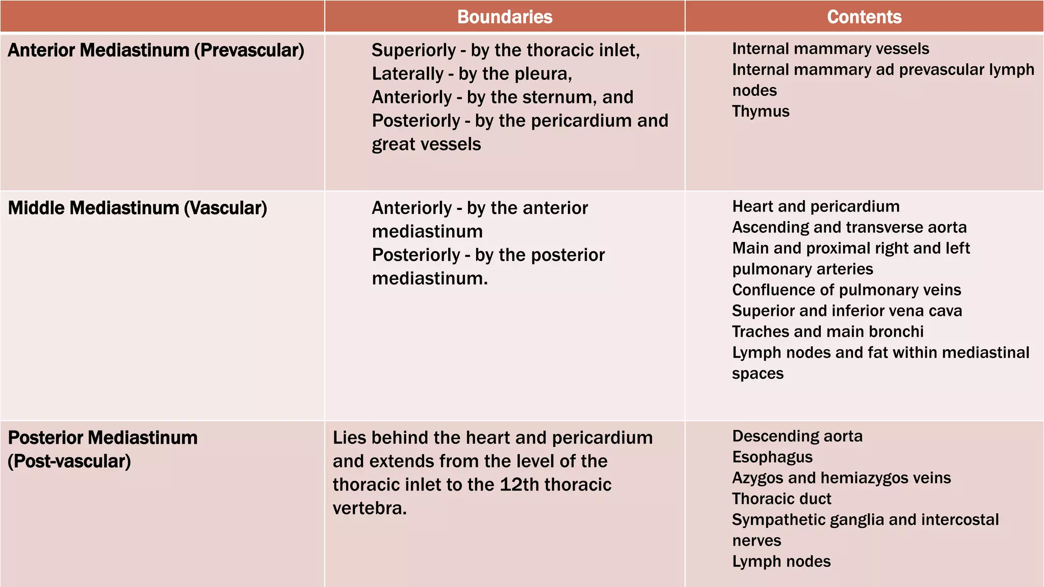
Johan Rey P. Mon
Post-Graduate Intern
MEDIASTINUM
NORMAL RADIOGRAPH
 Narrow, vertically oriented structure
that resides between the medial
parietal pleural layers of the lungs
 Extends from the sternum to the bodies
of the vertebrae; and from the superior
thoracic aperture to the diaphragm
 Contains the thymus gland, the
pericardial sac, the heart, the trachea
and the major arteries and veins
MEDIASTINUM
BOUNDARIES
 Boundaries
 Anterior – sternum and costal
cartilages
 Posterior – 12 thoracic vertebrae
 Superior – thoracic inlet
 Inferior – diaphragm
 Lateral – mediastinal pleura
BOUNDARIES OF THE MEDIASTINUM
DIVISIONS
 Superior mediastinum*
 Inferior mediastinum
 Anterior
- anterior to the pericardial sac and
posterior to the body of the sternum
 Middle
- contains the pericardial sac and its
contents
 Posterior
- posterior to the pericardial sac and
the diaphragm and anterior to the
bodies of the vertebrae
DIVISIONS OF THE MEDIASTINUM
Boundaries Contents
Anterior Mediastinum (Prevascular) Superiorly - by the thoracic inlet,
Laterally - by the pleura,
Anteriorly - by the sternum, and
Posteriorly - by the pericardium and
great vessels
Internal mammary vessels
Internal mammary ad prevascular lymph
nodes
Thymus
Middle Mediastinum (Vascular) Anteriorly - by the anterior
mediastinum
Posteriorly - by the posterior
mediastinum.
Heart and pericardium
Ascending and transverse aorta
Main and proximal right and left
pulmonary arteries
Confluence of pulmonary veins
Superior and inferior vena cava
Traches and main bronchi
Lymph nodes and fat within mediastinal
spaces
Posterior Mediastinum
(Post-vascular)
Lies behind the heart and pericardium
and extends from the level of the
thoracic inlet to the 12th thoracic
vertebra.
Descending aorta
Esophagus
Azygos and hemiazygos veins
Thoracic duct
Sympathetic ganglia and intercostal
nerves
Lymph nodes
MEDIASTINAL
CONTOUR
 Right paratracheal stripe
 seen in two thirds of normal films
 made up of right brachiocephalic vein and
SVC
 Arch of the azygous vein
 Ascending aorta
 Superior vena cava (SVC)
 Right atrium
 Inferior vena cava (IVC)
RIGHT CARDIOMEDIASTINAL CONTOUR
 Left paratracheal stripe
 made up of left common carotid artery,
left subclavian artery and the left
jugular vein
 Aortic arch +/- aortic nipple (left
superior intercostal vein)
 Pulmonary artery
 Auricle of left atrium
 Left ventricle
LEFT CARDIOMEDIASTINAL CONTOUR
 Superior mediastinum
 Great vessels
 Thymus
 Ascending aorta
 Right ventricular outflow track
 Right ventricle
ANTERIOR CARDIOMEDIASTINAL CONTOUR
 Left atrium and pulmonary veins
 Right atrium
 Inferior vena cava
POSTERIOR CARDIOMEDIASTINAL CONTOUR
 Consists of potential spaces used to describe the location of disease
processes
 Important diseases change the appearance of the aortic knuckle, the
aorto-pulmonary window and the right para-tracheal stripe
MEDIASTINAL CONTOURS
The aortic knuckle (red line)
represents the left lateral edge
of the aorta as it arches
backwards over the left main
bronchus, and pulmonary
vessels.
The contour of the descending
thoracic aorta (yellow line) can
be seen in continuation from
the aortic knuckle.
NORMAL AORTIC KNUCKLE
The aorto-pulmonary window
lies between the arch of the
aorta and the pulmonary
arteries.
Between the Aortic Knuckle (AK)
and the Left Pulmonary Artery
(LPA)
The descending aorta (DA)
marks its posterior boundary.
AORTO-PULMONARY WINDOW
 From the level of the clavicles to
the azygos vein the right edge of
the trachea is seen as a thin
white stripe.
 Normal: <3 mm
 Thickened: may represent
paratracheal mass or enlarged
lymph node
RIGHT PARA-TRACHEAL STRIPE
MEDIASTINAL MASSES
ANTERIOR MEDIASTINAL
MASSES
MIDDLE MEDIASTINAL MASSES
POSTERIOR MEDIASTINAL
MASSES
POSTERIOR MEDIASTINAL
MASSES
HILUM
• Junction of the lung with the
mediastinum and is composed of upper
lobe pulmonary veins and branches of
the pulmonary artery and corresponding
bronchi
• On chest radiographs, the term hilum
represents the composite shadow of the
bronchi, pulmonary arteries and veins,
and lymph nodes on the medial aspect of
each lung.
• Left hilar shadow is higher in the right in
90% of individuals
HILUM
 On a true lateral radiograph, the right
and left hilar shadows are not
completely superimposed and
comprise a combination of the right
and left pulmonary arteries and the
superior pulmonary veins
 Inferior hilar window
- An avascular aspect of the composite
hilar shadow, inferior to the shadow of
the right pulmonary artery and veins and
anterior to the descending left
pulmonary artery and left superior vein
LEFT LATERAL VIEW OF THE HILUM
HILAR MASS
 RUL bronchus is seen in
approximately 50% of
individuals as an end-on, round
lucency at the upper margin of
the composite hilar shadow
 Recognition of this bronchus,
when not visible on prior
radiographs, should suggest a
mass or lymph node
enlargement about the bronchus
LEFT LATERAL VIEW OF THE HILUM
HEART
• It lies primarily in the anterior left
hemithorax, with the LV lying on the
left hemidiaphragm
• The RA extends to the right of
midline as it receives systemic
blood from the SVC, IVC, and
coronary sinus
• The RA and RV lie primarily anterior
to the planes of the LA and LV
• The RV is the most anterior
chamber and abuts the sternum
• The LA is subcarinal and midline in
the thorax
HEART
HEART
CARDIOMEDIASTINAL
ANATOMY
(PA VIEW)
1. Superior vena cava
2. Right atrium
3. Inferior vena cava
4. Aortic arch
5. Main pulmonary artery
6. Left atrial appendage
7. Left ventricle
CARDIOMEDIASTINAL
ANATOMY
(LATERAL VIEW)
1. Left atrium
2. Left ventricle
3. Inferior vena cava
4. Right ventricle
5. Right pulmonary artery
6. Left pulmonary artery
7. Aorta
 Refer to the bulges of
the cardiomediastinal contour on
frontal chest radiographs.*
 Right cardiomediastinal border:
 the right atrium is the only
normal bulge
MOGULS OF THE
HEART
 Left cardiomediastinal border:
 1st mogul
- uppermost, located paratracheally above
the carina, and formed by the aortic arch
(aortic knuckle or knob)
 2nd mogul
- located just above the left main bronchus
and represents the main pulmonary artery
segment
 3rd mogul
- NEVER NORMAL
 if present, it lies below the left main
bronchus and is usually formed by
prominent left atrial appendage,
which is commonly seen in rheumatic
heart disease
MOGULS OF THE
HEART
 4th mogul
- bulge just above the diaphragm formed
by the left ventricular margin or cardiac
apex
 5th mogul
- bulge at the cardiophrenic angle
- may be caused by prominent pericardial
fat pad, pericardial cyst, or adenopathy
MOGULS OF THE
HEART
 Right cardiac borders:
1. Superior vena cava
2. Inferior vena cava
3. Right atrium
CARDIAC BORDERS
 Left cardiac borders:
1. Aortic knob
2. Main pulmonary trunk
3. Left ventricle
CARDIAC BORDERS
RA LV
RV
LA
LV
HEART CHAMBERS
 Heart size is assessed as the
cardiothoracic ratio (CTR)
 Determines the relation of the heart to
the width of the chest at its widest part
near the level of the diaphragm
 Cardiac size is measured by dropping
parallel lines down both sides of the
heart, at the most lateral points on
each side, and measuring between
them.
 Thoracic width is measured by
dropping parallel lines down the inner
aspect of the widest points of the rib
cage, and measuring between these.
 CTR =
Maximum cardiac diameter
Macimum intrathoracic diameter
CARDIOTHORACIC RATIO
Adult
PA view = 0.50
AP view = 0.55
Pedia
Age (wks) Range
0-3 0.60-0.50
4-7 0.64-0.52
8-15 0.62-0.51
16-23 0.62-0.51
24-31 0.61-0.50
32-39 0.61-0.51
40-47 0.60-0.49
48-55 0.57-0.49
Age (yrs)Range
0-1 0.65-0.39
1-2 0.60-0.39
2-3 0.50-0.39
3-4 0.52-0.40
4-5 0.52-0.40
5-6 0.50-0.40
7 0.49-0.43
8 0.49-0.42
9 0.49-0.41
10 0.49-0.43
11 0.49-0.43
12 0.46-0.40
CARDIOTHORACIC RATIO NORMAL VALUES
THANK YOU!!!

Normal mediastinum radiograph

  • 1.
    Johan Rey P.Mon Post-Graduate Intern MEDIASTINUM NORMAL RADIOGRAPH
  • 2.
     Narrow, verticallyoriented structure that resides between the medial parietal pleural layers of the lungs  Extends from the sternum to the bodies of the vertebrae; and from the superior thoracic aperture to the diaphragm  Contains the thymus gland, the pericardial sac, the heart, the trachea and the major arteries and veins MEDIASTINUM
  • 3.
  • 4.
     Boundaries  Anterior– sternum and costal cartilages  Posterior – 12 thoracic vertebrae  Superior – thoracic inlet  Inferior – diaphragm  Lateral – mediastinal pleura BOUNDARIES OF THE MEDIASTINUM
  • 5.
  • 6.
     Superior mediastinum* Inferior mediastinum  Anterior - anterior to the pericardial sac and posterior to the body of the sternum  Middle - contains the pericardial sac and its contents  Posterior - posterior to the pericardial sac and the diaphragm and anterior to the bodies of the vertebrae DIVISIONS OF THE MEDIASTINUM
  • 8.
    Boundaries Contents Anterior Mediastinum(Prevascular) Superiorly - by the thoracic inlet, Laterally - by the pleura, Anteriorly - by the sternum, and Posteriorly - by the pericardium and great vessels Internal mammary vessels Internal mammary ad prevascular lymph nodes Thymus Middle Mediastinum (Vascular) Anteriorly - by the anterior mediastinum Posteriorly - by the posterior mediastinum. Heart and pericardium Ascending and transverse aorta Main and proximal right and left pulmonary arteries Confluence of pulmonary veins Superior and inferior vena cava Traches and main bronchi Lymph nodes and fat within mediastinal spaces Posterior Mediastinum (Post-vascular) Lies behind the heart and pericardium and extends from the level of the thoracic inlet to the 12th thoracic vertebra. Descending aorta Esophagus Azygos and hemiazygos veins Thoracic duct Sympathetic ganglia and intercostal nerves Lymph nodes
  • 9.
  • 10.
     Right paratrachealstripe  seen in two thirds of normal films  made up of right brachiocephalic vein and SVC  Arch of the azygous vein  Ascending aorta  Superior vena cava (SVC)  Right atrium  Inferior vena cava (IVC) RIGHT CARDIOMEDIASTINAL CONTOUR
  • 11.
     Left paratrachealstripe  made up of left common carotid artery, left subclavian artery and the left jugular vein  Aortic arch +/- aortic nipple (left superior intercostal vein)  Pulmonary artery  Auricle of left atrium  Left ventricle LEFT CARDIOMEDIASTINAL CONTOUR
  • 13.
     Superior mediastinum Great vessels  Thymus  Ascending aorta  Right ventricular outflow track  Right ventricle ANTERIOR CARDIOMEDIASTINAL CONTOUR
  • 14.
     Left atriumand pulmonary veins  Right atrium  Inferior vena cava POSTERIOR CARDIOMEDIASTINAL CONTOUR
  • 15.
     Consists ofpotential spaces used to describe the location of disease processes  Important diseases change the appearance of the aortic knuckle, the aorto-pulmonary window and the right para-tracheal stripe MEDIASTINAL CONTOURS
  • 16.
    The aortic knuckle(red line) represents the left lateral edge of the aorta as it arches backwards over the left main bronchus, and pulmonary vessels. The contour of the descending thoracic aorta (yellow line) can be seen in continuation from the aortic knuckle. NORMAL AORTIC KNUCKLE
  • 18.
    The aorto-pulmonary window liesbetween the arch of the aorta and the pulmonary arteries. Between the Aortic Knuckle (AK) and the Left Pulmonary Artery (LPA) The descending aorta (DA) marks its posterior boundary. AORTO-PULMONARY WINDOW
  • 20.
     From thelevel of the clavicles to the azygos vein the right edge of the trachea is seen as a thin white stripe.  Normal: <3 mm  Thickened: may represent paratracheal mass or enlarged lymph node RIGHT PARA-TRACHEAL STRIPE
  • 22.
  • 23.
  • 24.
  • 25.
  • 26.
  • 27.
  • 28.
  • 29.
    • Junction ofthe lung with the mediastinum and is composed of upper lobe pulmonary veins and branches of the pulmonary artery and corresponding bronchi • On chest radiographs, the term hilum represents the composite shadow of the bronchi, pulmonary arteries and veins, and lymph nodes on the medial aspect of each lung. • Left hilar shadow is higher in the right in 90% of individuals HILUM
  • 31.
     On atrue lateral radiograph, the right and left hilar shadows are not completely superimposed and comprise a combination of the right and left pulmonary arteries and the superior pulmonary veins  Inferior hilar window - An avascular aspect of the composite hilar shadow, inferior to the shadow of the right pulmonary artery and veins and anterior to the descending left pulmonary artery and left superior vein LEFT LATERAL VIEW OF THE HILUM
  • 32.
  • 33.
     RUL bronchusis seen in approximately 50% of individuals as an end-on, round lucency at the upper margin of the composite hilar shadow  Recognition of this bronchus, when not visible on prior radiographs, should suggest a mass or lymph node enlargement about the bronchus LEFT LATERAL VIEW OF THE HILUM
  • 34.
  • 35.
    • It liesprimarily in the anterior left hemithorax, with the LV lying on the left hemidiaphragm • The RA extends to the right of midline as it receives systemic blood from the SVC, IVC, and coronary sinus • The RA and RV lie primarily anterior to the planes of the LA and LV • The RV is the most anterior chamber and abuts the sternum • The LA is subcarinal and midline in the thorax HEART
  • 36.
  • 37.
    CARDIOMEDIASTINAL ANATOMY (PA VIEW) 1. Superiorvena cava 2. Right atrium 3. Inferior vena cava 4. Aortic arch 5. Main pulmonary artery 6. Left atrial appendage 7. Left ventricle
  • 38.
    CARDIOMEDIASTINAL ANATOMY (LATERAL VIEW) 1. Leftatrium 2. Left ventricle 3. Inferior vena cava 4. Right ventricle 5. Right pulmonary artery 6. Left pulmonary artery 7. Aorta
  • 39.
     Refer tothe bulges of the cardiomediastinal contour on frontal chest radiographs.*  Right cardiomediastinal border:  the right atrium is the only normal bulge MOGULS OF THE HEART
  • 40.
     Left cardiomediastinalborder:  1st mogul - uppermost, located paratracheally above the carina, and formed by the aortic arch (aortic knuckle or knob)  2nd mogul - located just above the left main bronchus and represents the main pulmonary artery segment  3rd mogul - NEVER NORMAL  if present, it lies below the left main bronchus and is usually formed by prominent left atrial appendage, which is commonly seen in rheumatic heart disease MOGULS OF THE HEART
  • 42.
     4th mogul -bulge just above the diaphragm formed by the left ventricular margin or cardiac apex  5th mogul - bulge at the cardiophrenic angle - may be caused by prominent pericardial fat pad, pericardial cyst, or adenopathy MOGULS OF THE HEART
  • 44.
     Right cardiacborders: 1. Superior vena cava 2. Inferior vena cava 3. Right atrium CARDIAC BORDERS
  • 45.
     Left cardiacborders: 1. Aortic knob 2. Main pulmonary trunk 3. Left ventricle CARDIAC BORDERS
  • 46.
  • 47.
     Heart sizeis assessed as the cardiothoracic ratio (CTR)  Determines the relation of the heart to the width of the chest at its widest part near the level of the diaphragm  Cardiac size is measured by dropping parallel lines down both sides of the heart, at the most lateral points on each side, and measuring between them.  Thoracic width is measured by dropping parallel lines down the inner aspect of the widest points of the rib cage, and measuring between these.  CTR = Maximum cardiac diameter Macimum intrathoracic diameter CARDIOTHORACIC RATIO
  • 48.
    Adult PA view =0.50 AP view = 0.55 Pedia Age (wks) Range 0-3 0.60-0.50 4-7 0.64-0.52 8-15 0.62-0.51 16-23 0.62-0.51 24-31 0.61-0.50 32-39 0.61-0.51 40-47 0.60-0.49 48-55 0.57-0.49 Age (yrs)Range 0-1 0.65-0.39 1-2 0.60-0.39 2-3 0.50-0.39 3-4 0.52-0.40 4-5 0.52-0.40 5-6 0.50-0.40 7 0.49-0.43 8 0.49-0.42 9 0.49-0.41 10 0.49-0.43 11 0.49-0.43 12 0.46-0.40 CARDIOTHORACIC RATIO NORMAL VALUES
  • 49.

Editor's Notes

  • #2 The mediastinum occupies the portion of the chest surrounded by the right and left lung We must note the widening, either local (tumor), or diffuse (inflammation)
  • #6 This division of the mediastinum is purely arbitrary, as there are No true anatomic boundaries between the three compartments. However, by using the most easily recognizable mediastinal structure ”the heart” as the focal point, the relationship of mediastinal masses to the heart allows for simple and consistent compartmentalization. Furthermore, this division of the mediastinum corresponds to easily recognizable regions seen on the lateral chest radiograph.
  • #7 Mediastinum can be divided into the superior and inferior mediastinum by a transverse plane extending from the sternal angle to the intervertebral disc between T4 and T5 Superior mediastinum structures: Veins – SVC, R and L Brachiocephalic veins Arteries – Arch of the Aorta, Brachiocephalic Artery, L common carotid and L subclavian artery Note for obliteration of spaces Note for opacities Mediastinal width Upright: 8 cm Supine: 10 cm Thus, internal structures such as the chambers of the heart, the great vessels, etc. all blend into a single shadow except when they abut the lung
  • #8 Lateral view of chest showing divisions of the mediastinum. The superior mediastinum lies above the line extending from the sternal angle to the fourth dorsal vertebra. A: The anterior mediastinum; B: the middle mediastinum; and C: the posterior mediastinum. In the superior mediastinum the trachea is indicated by arrows. The lower pair of arrows indicate the region of the superimposed hila. Compartment margins on the film are not clear, because the mediastinum is a conglomeration of the structures. Inferior mediastinum Anterior - anterior to the pericardial sac and posterior to the body of the sternum Middle - contains the pericardial sac and its contents Posterior - posterior to the pericardial sac and the diaphragm and anterior to the bodies of the vertebrae
  • #10 The mediastinum itself contains the heart and great vessels (middle mediastinum) and potential spaces in front of the heart (anterior mediastinum), behind the heart (posterior mediastinum) and above the heart (superior mediastinum). These potential spaces are not defined on a normal chest X-ray, but an awareness of their position can help in describing the location of disease processes.
  • #17 Displacement or loss of definition of these lines can indicate disease, such as aneurysm or adjacent lung consolidation
  • #19 The right lateral edge of the Ascending Aorta (AA) is also marked. This is a potential space in the mediastinum where abnormal enlargement of lymph nodes can be seen on a chest X-ray.
  • #21 This appearance is created by air of low density (blacker) lying either side of the comparatively dense (whiter) tracheal wall. The left side of the trachea is not so well defined because of the position of the aortic arch and great vessels.
  • #31 Both hilar should be concave Both hilar should be of similar density The hilar points are the angle formed by the descending upper lobe veins, as they cross behind the lower lobe arteries The shape of the right hilum on frontal radiographs has been likened to a sideways V, with the opening pointing rightward (Fig. 12.19A, B). The upper portion of the V is composed primarily of the truncus anterior and the posterior division of the right superior pulmonary vein. The right interlobar artery forms the lower half of the V, as it descends lateral to the bronchus intermedius. The right inferior pulmonary vein crosses the lower right hilar shadow but does not contribute to its opacity (Fig. 12.19A). Left hilus is higher than the right because the left pulmo artery is higher than the right
  • #32 a right hilum that lies above the left suggests volume loss in the right upper or left lower lobe
  • #33 roughly triangular in shape, with its apex at the junction of the LUL and LLL bronchi and its base directed anteriorly and inferiorly.
  • #37 Lies approximately 2/3 to the left of midline and 1/3 to the right The LA is subcarinal and midline in the thorax, being supplied by the right and left superior and inferior pulmonary veins.
  • #41 More specifically, it refers to the left mediastinal outline beginning at the aortic knob.
  • #42 1st Mogul - A prominent knob is a clue to ectasia, aneurysm, or hypertension. Notching or a ‘Figure of 3’ sign of the aorta suggests coarctation(narrowing) 2ND Mogul – Excessive convexity is seen with poststenotic dilatation, chronic obstructive pulmonary disease, pulmonary artery hypertension, left-to-right shunts, and pericardial defects. Severe concavity suggests right-to-left shunts. 3rd Mogul – It is not usually seen with other causes of left atrial enlargement.
  • #44 4th Mogul – is a bulge just above the cardiophrenic angle, seen with infarction or ventricular aneurysm. 5th Mogul – fifth bulge at the cardiophrenic angle is caused by pericardial cysts, prominent fat pads, or adenopathy.
  • #46 *Right ventricle is more anteriorly located, better seen in lateral view
  • #47 *Right ventricle is more anteriorly located, better seen in lateral view
  • #48 Check for the aorta Note if the aortic notch is prominent and atherosclerotic
  • #49 The heart size should be considered on every chest X-ray, but the cardiothoracic ratio (CTR) can only be assessed confidently if a posterior - anterior (PA) view has been acquired. The rule is, if an anterior - posterior (AP) view has been taken, then the heart should not be called enlarged even if the CTR is >50%. This is because an AP view exaggerates the heart size. If the CTR is <50% on an AP view, then clearly the heart size is within normal.