Modern pharmaceutical analytical techniques (MPAT) electrophoresis techniques
•Download as DOCX, PDF•
6 likes•267 views

Paper electrophoresis gel electrophoresis capillary electrophoresis zone electrophoresis moving boundary electrophoresis iso electric focusing electrophoresis

Recommended
More Related Content
What's hot
What's hot (20)
Similar to Modern pharmaceutical analytical techniques (MPAT) electrophoresis techniques
Similar to Modern pharmaceutical analytical techniques (MPAT) electrophoresis techniques (20)
More from JayeshRajput7
More from JayeshRajput7 (20)
Recently uploaded
Recently uploaded (20)
Modern pharmaceutical analytical techniques (MPAT) electrophoresis techniques
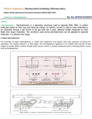
- 1. Masters of pharmacy, Pharmaceutical technology (Pharmaceutics) Subject- Modern pharmaceutical analytical techniques (MPAT) (MPH-101T) Lesion no- 5, Electrophoresis By- Drx JAYESH M.RAJPUT Points:- Electrophoresis: - Electrophoresis is a laboratory technique used to separate DNA, RNA, or protein molecules based on their size and electrical charge. An electric current is used to move molecules to be separated through a gel. Pores in the gel work like a sieve, allowing smaller molecules to move faster than larger molecules. The conditions used during electrophoresis can be adjusted to separate molecules in a desired size range. 1. Paper electrophoresis. The technique of paper electrophoresis is simple and inexpensive and requires only micro quantities of plasma for separation. The support medium is a filter paper. The electrophoresis apparatus in its simplest form consists of two troughs to contain buffer solution through which electric current is passed. Frequently used in isolating proteins, amino acids and oligopeptides.
- 2. Procedure:- 1. A long strip of filter paper is moistened with a suitable buffer solution of the desired pH and the sample is applied transversely across the central part of the strip 2. Ends are fixed to dip in buffer solutions in two troughs fitted with electrodes 3. Electric field of about 20 volts/cm is established 4. The charged particles of sample migrate along the strip towards respective electrodes of opposite polarity, according to net charges, sizes and interactions with the solid matrix 5. Homogeneous group of particles migrate as a separate band 6. The electrophoresis is carried out for 16-18 hours 7. Separated proteins are fixed to a solid support using a fixative such as acetone or methanol 8. Proteins are stained (bromophenol blue) to make them visible 9. The separated proteins appear as distinct bands 10. Drawback long time interval and blurring of margins `Observation:- The different fractions appear as blue colored bands across the filter paper starting from the moving boundary backwards. If a quantitative estimation is required for each fraction, the bands may be carefully cut and eluted, or the bands may be scanned optically in a densitometer. In human plasma five different bands can be identified on paper electrophoresis. Filter paper such as whatman no 1 and no 3mm in strip of 3 or 5 cm wide have been used to good effect, separation takes place in 12 to 14 hrs. Advantages: - it is economical, easy to use
- 3. Disadvantages:- certain compounds such as proteins, hydrophilic molecules cannot be resolved due to the adsorptive and inogenic properties of paper which results in tailing and distortion of component bands, electro osmosis is also a disadvantage. 2. Gel electrophoresis.
- 7. 4. Zone electrophoresis. Zone electrophoresis (ZE) is an electrophoretic separation technique typically used for analyzing proteins, nucleic acids, and biopolymers. During the process, different species in a sample are transported in a continuous electrolyte buffer system, subject to a potential gradient. Due to
- 8. differences in the mobilities, the species in the samples will eventually separate into different, well- resolved peaks.
- 9. 5. Moving boundary electrophoresis. Moving-boundary electrophoresis (MBE also free-boundary electrophoresis) is a technique for separation of chemical compounds by electrophoresis in a free solution. Apparatus The moving-boundary electrophoresis apparatus includes a U-shaped cell filled with buffer solution and electrodes immersed at its ends. The sample applied could be any mixture of charged components such as a protein mixture. On applying voltage, the compounds will migrate to the anode or cathode depending on their charges. The change in the refractive index at the boundary of the separated compounds is detected using Schlieren optics at both ends of the solution in the cell.
- 10. 6. Iso- electric focusing. Isoelectric focusing (IEF), also known as electrofocusing, is a technique for separating different molecules by differences in their isoelectric point (pI).[1][2] It is a type of zone electrophoresis usually performed on proteins in a gel that takes advantage of the fact that overall charge on the molecule of interest is a function of the pH of its surroundings. IEF involves adding an ampholyte solution into immobilized pH gradient (IPG) gels. IPGs are the acrylamide gel matrix co-polymerized with the pH gradient, which result in completely stable gradients except the most alkaline (>12) pH values. The immobilized pH gradient is obtained by the continuous change in the ratio of immobilines. An immobiline is a weak acid or base defined by its pK value.