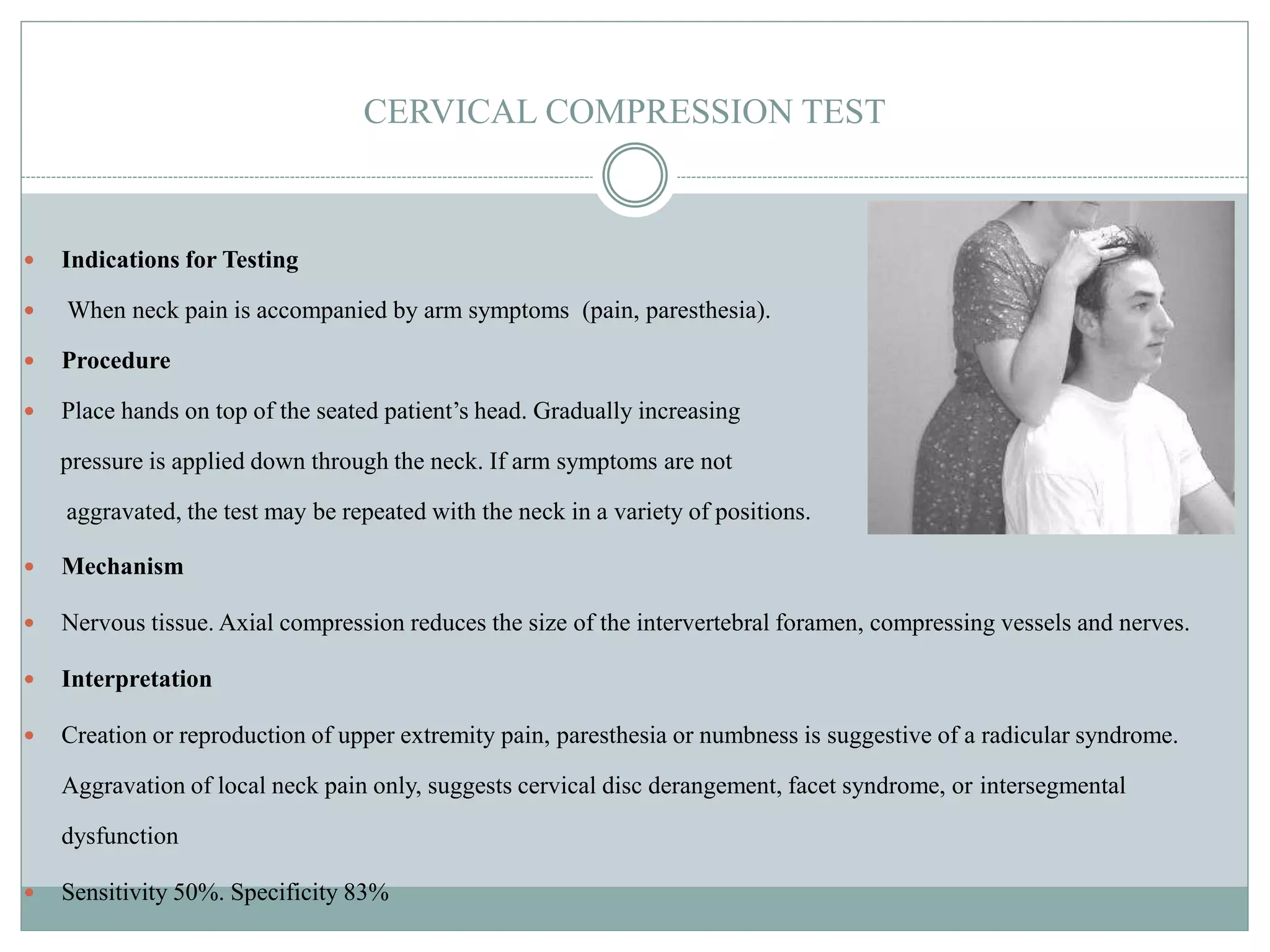
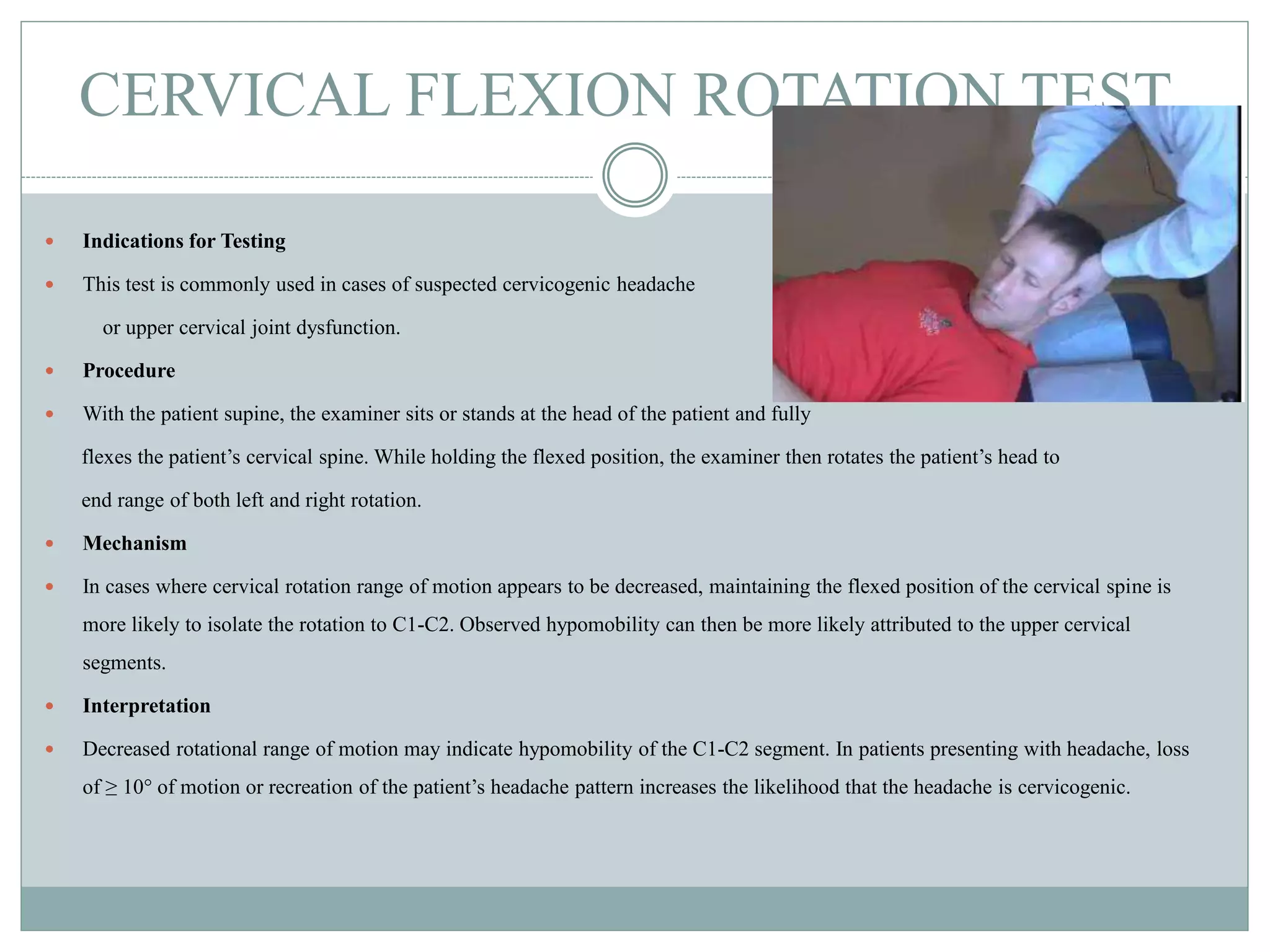
Call Girls Service in Bommanahalli - 7001305949 with real photos and phone nu...
cervical test .pptx
1. O R T H O P A E D I C U N I T P R E S E N T A T I O N B Y
C H U K W U M A , C H I N E N Y E A
Cervical Orthopaedic Test of yje
Cervical Spine
2. CERVICAL COMPRESSION TEST
Indications for Testing
When neck pain is accompanied by arm symptoms (pain, paresthesia).
Procedure
Place hands on top of the seated patient’s head. Gradually increasing
pressure is applied down through the neck. If arm symptoms are not
aggravated, the test may be repeated with the neck in a variety of positions.
Mechanism
Nervous tissue. Axial compression reduces the size of the intervertebral foramen, compressing vessels and nerves.
Interpretation
Creation or reproduction of upper extremity pain, paresthesia or numbness is suggestive of a radicular syndrome.
Aggravation of local neck pain only, suggests cervical disc derangement, facet syndrome, or intersegmental
dysfunction
Sensitivity 50%. Specificity 83%
3. CERVICAL FLEXION TEST
Indications for Testing
Cervical flexion can be used as a neurodynamic test for lesions of the meninges,
spinal cord or nerve roots. It is indicated in suspected cases of meningitis ,
spinal cord lesions, cervical or lumbar disc herniation or radiculopathy.
Procedure
Depending on the circumstances, the patient can be seated or supine. The practitioner supports the patient’s head
and moves the neck though the entire range of flexion to patient tolerance. Performing cervical flexion with
the patient in long leg sitting (i.e., sitting with the legs straight out on an adjusting table), rather than the
normal sitting position, creates more tension and may further sensitize the test.
4. Mechanism
With neck flexion, the dural sac, meninges, spinal cord, and nerve roots are elongated under a tensile
load. Stretching of an inflamed dural sac or injured spinal cord will cause pain at the level of
irritation.
Flexing the hips and knees reduces tension on the meninges (knee flexion decreasing tension on the
sciatic nerve and hip flexion decreasing tension on the femoral nerve).
Interpretation
In a suspected meningitis case, the patient can be tested in a supine position. Reactive flexion of the
patient’s
knees and hips is considered Brudzinski’s sign.
Whether passive neck flexion is performed with the patient seated or supine, sharp, shooting pain
down the spine and into the extremities is Lhermitte’s sign and suggests the presence of a cord tumor,
posterior column disease, meningeal adhesions, multiple sclerosis, or cervical spondylotic
myelopathy. This sign may take on other forms, such as increasing spastic rigidity in the lower
extremity.
A positive test that recreates symptoms along the nerve distribution in the lower extremity is
Lindner’s sign, suggesting radiculopathy. The patient can be tested either seated or supine..
5. CERVICAL DISTRACTION
Indications for Testing
Usually part of a basic orthopedic examination of the neck.
For patients with possible radicular syndromes.
To help document cervical sprain or facet capsular sprain.
Procedure
The patient is seated in a neutral posture. The PT places thumbs under
the patient’s occiput, thenar eminences under mastoid processes.
The examiner gradually adds force superiorly up to about 5 to 30 pounds of
lifting pressure. As an option
the distraction can be held for up to 30-60 seconds.
Mechanism
Nervous tissue. Distraction opens the IVF, potentially relieving pressure on the nerve roots.
Interpretation
A decrease in arm symptoms suggests IVF encroachment. Further, it also suggests that traction therapy may be
beneficial for this patient. An increase in local pain with distraction may suggest a ligamentous sprain, facet capsular
sprain, or may be aggravating a muscular strain.
Sensitivity 44% Specificity 90-97%
6. SOTO-HALL TEST
Indications for Testing
When suspecting a fracture or lesion of the
lower cervical or upper thoracic joints.
Procedure
Place one hand on the patient’s sternum to
prevent lumbar and thoracic regions from
flexing; the other hand under the patient’s
occiput. The neck is then passively flexed towards the chest while maintaining
gentle pressure on the sternum.
Mechanism
It places traction on the posterior elements of the cervical and upper thoracic spine and
compresses the anterior bodies and discs. It also causes traction of the nervous system .
In the case of vertebral fracture, when the supraspinous ligament pulls on the spinous
process of the fractured element, acute local pain will result.
Interpretation
Localized pain suggests a possible joint or bone injury or pathology (but not lower than
the T7 level).
7. CERVICAL RESISTED MUSCLE TESTS
(includes O’Donoghue maneuver)
Indications for Testing
Resisted muscle tests of the cervical spine are used to assess a possible muscle strain, particularly following trauma.
Procedure
The tests are performed to induce isometric contractions. In an acute setting, muscle testing is best performed with the
cervical spine in neutral. Appropriate muscle testing requires an adequate amount of force. The force can be initiated
either by the patient (“push into my hand”) or by the practitioner (“resist me”). Once the maximum force is achieved,
the contraction should be sustained for one to two seconds. In cases where the suspicion of a muscle strain is high but
the muscles test strong and do not elicit pain, the PT may choose to increase the challenge by extending the duration of
each contraction to 5 seconds or by repeating a one- to two-second contraction, ten times.
8. Mechanism
The isometric load imposed by the tests may cause pain due to an injured muscle. It may
also reveal weakness because of a tear in the muscle fibers.
Compression of the joints does occur and may be painful, especially during the initial phase
of the contraction. O’Donoghue maneuver When muscle testing is followed by passive
range of motion into each of the cardinal directions, this combination of procedures has
been referred to as the O’Donoghue maneuver and is performed to distinguish between
cervical muscle strain and ligament sprain. The passive tests place end range tensile loads
on the cervical ligaments and discs. Such tests also load the muscle at end range, without
eliciting an active contraction of that muscle..
9. CERVICAL FLEXION ROTATION TEST
Indications for Testing
This test is commonly used in cases of suspected cervicogenic headache
or upper cervical joint dysfunction.
Procedure
With the patient supine, the examiner sits or stands at the head of the patient and fully
flexes the patient’s cervical spine. While holding the flexed position, the examiner then rotates the patient’s head to
end range of both left and right rotation.
Mechanism
In cases where cervical rotation range of motion appears to be decreased, maintaining the flexed position of the cervical spine is
more likely to isolate the rotation to C1-C2. Observed hypomobility can then be more likely attributed to the upper cervical
segments.
Interpretation
Decreased rotational range of motion may indicate hypomobility of the C1-C2 segment. In patients presenting with headache, loss
of ≥ 10° of motion or recreation of the patient’s headache pattern increases the likelihood that the headache is cervicogenic.
10. DEEP NECK FLEXOR ENDURANCE
TEST and JULL’S TEST
Indications for Testing
In patients presenting with neck or upper back symptoms in which lack of strength endurance or
neuromuscular control in the deep cervical flexors is suspected.
Procedure
In the supine position, the patient is directed to maximally retract the chin and, while maintaining the
retraction, lift the head about one inch off the table. The examiner places a hand on the table under the
patient’s head and observes for the skin folds in the neck created by chin retraction. The test is
terminated when the patient’s head touches the examiners hand or separation of the skin folds occurs.
11. Jull’s Test : The physiotherapist places his hands under the head of a supine patient
who is instructed to retract the chin. The PT then passively raises their head about
an inch. They then instruct the patient to maintain the position of the head while the
PT slowly removes their hands.
Interpretation
If the patient is unable to maintain the beginning position or the PT observes a loss
of the skin folds created by chin retraction, chin jutting, or shaking, this would be
considered a failed test. The PT may also place their hand under the patient’s head
to be able to feel when the patient’s head lowers. Under normal circumstances the
patient should be able to hold the position for about 39 seconds.
12. SHOULDER ABDUCTION TEST
Indications for Testing
Used when evaluating a patient suffering from neck and arm
symptoms, especially if a radicular syndrome is suspected
Procedure
The patient is asked to actively raise (abduct) the symptomatic arm until it is near the head and to
report if there are any changes in arm symptoms.
Mechanism
Nervous tissue. Alleviation of arm pain or paresthesia with shoulder abduction over the head may
occur due to a reduction in nerve root tension, mainly C6-C8 or intraforaminal pressure
Interpretation
If shoulder abduction relieves the patient’s arm pain, this suggests there is a radicular syndrome of
the lower nerve roots. Patients may present in this posture to relieve radicular arm pain (Bakody’s
sign). It also suggests that abduction of the arm during cervical joint palpation and manipulation
may be helpful.
13. SHOULDER DEPRESSION
Indications for Testing
Used to indicate possible nerve root or brachial plexus irritation/inflammation in patients with
neck and arm symptoms.
Procedure
Patient may be seated or supine. PT first instructs patient to laterally bend the neck away from the
side to be tested. Then PT maintains the head in that amount of lateral flexion attained by the
patient and places the other hand on top of the patient’s shoulder. With gradually increasing
pressure, the examiner depresses the affected shoulder. Radicular pain produced on the side being
stretched constitutes a positive test. Confirmation of the positive test may be appreciated by
reducing downward pressure on the shoulder until symptoms just abate and then have the patient
rotate the head away from the affected shoulder. Exacerbation of radicular symptoms confirms a
positive test
14. REFRENCES
Magee DJ. (2014). Orthopedic Physical Assessment, sixth Ed. St. Louis, MO:
Elsevier;
Ronald LeFebvre, and Charles Novak, (2021) Cervical orthopedic tests
https://ftp.uws.edu/udocs/public/CSPE_Protocols_and_Care_Pathways/Pr
otocols/Cervical_Orthopedic_Tests.pdf