
Recommended
More Related Content
What's hot
What's hot (20)
Similar to DIC
Similar to DIC (20)
More from HIRENGEHLOTH
More from HIRENGEHLOTH (20)
Recently uploaded
Recently uploaded (20)
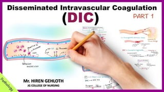
DIC
- 1. Mr. HIREN GEHLOTH JG COLLEGE OF NURSING
- 2. INTRODUCTION
- 3. • Disseminated intravascular coagulation (DIC) is a rare but serious condition that causes abnormal blood clotting throughout the body’s blood vessels. It is caused by another disease or condition, such as an infection or injury, that makes the body’s normal blood clotting process become overactive. • DIC is not a disease but a sign of an underlying condition. • DIC may be triggered by sepsis, trauma, cancer, shock, abruptio placenta, toxins, or allergic reactions. • The severity of DIC is variable, but it is potentially life-threatening.
- 5. “Disseminated intravascular coagulopathy is a syndrome in which either intrinsic or extrinsic or both pathways are activated to produce multiple fibrin clots in small blood vessels. The resultant reduction in the coagulation factors and platelets results in bleeding.” DEFINITION
- 8. • In DIC, the normal hemostatic mechanisms are altered so that tiny clots form within the microcirculation of the body. • These clots consume platelets and clotting factors, eventually causing coagulation to fail and bleeding to result. • This bleeding disorder is characterized by low platelet and fibrinogen levels; prolonged prothrombin time (PT), partial thromboplastin time (PTT), and thrombin time; and elevated fibrin degradation products (D-dimers). • The primary prognostic factor is the ability to treat the underlying condition that precipitated DIC. PATHOPHYSIOLOG Y
- 9. CLINICAL MANIFESTATION • Clinical manifestations of DIC are primarily reflected in compromised organ function or failure, usually a result of excessive clot formation (with resultant ischemia to all or part of the organ) or, less often, bleeding. • Patient may bleed from mucous membranes, venipuncture sites, and gastrointestinal and urinary tracts. • Bleeding can range from minimal occult internal bleeding to profuse hemorrhage from all orifices.
- 10. • Patients typically develop multiple organ dysfunction syndrome (MODS), and they may exhibit renal failure as well as pulmonary and multifocal central nervous system infarctions as a result of microthromboses, macrothromboses, or hemorrhages. • Initially, the only manifestation is a progressive decrease in the platelet count; then, progressively, the patient exhibits signs and symptoms of thrombosis in the organs involved. Eventually bleeding occurs (at first subtle, advancing to frank hemorrhage). Signs depend on the organs involved.
- 11. ASSESSMENT AND DAIGNOSTIC EVALUATION • Clinically, the diagnosis of DIC is often established by a drop in platelet count, an increase in PT and activated partial thromboplastin time (aPTT), an elevation in fibrin degradation products, and measurement of one or more clotting factors and inhibitors (eg, antithrombin [AT]). • The International Society on Thrombosis and Haemostasis has developed a highly sensitive and specific scoring system using the platelet count, fibrin degradation products, PT, and fibrinogen level to diagnose DIC.
- 12. MEDICAL MANAGEMENT • The most important management issue is treating the underlying cause of DIC. A second goal is to correct the secondary effects of tissue ischemia by improving oxygenation, replacing fluids, correcting electrolyte imbalances, and administering vasopressor medications. • If serious hemorrhage occurs, the depleted coagulation factors and platelets may be replaced (cryoprecipitate to replace fibrinogen and factors V and VII; fresh-frozen plasma to replace other coagulation factors). • A heparin infusion, which is a controversial management method, may be used to interrupt the thrombosis process.
- 13. NURSING MANAGEMENT Maintaining Hemodynamic Status • Avoid procedures and activities that can increase intracranial pressure, such as coughing and straining. • Closely monitor vital signs, including neurologic checks, and assess for the amount of external bleeding. • Avoid medications that interfere with platelet function, if possible (eg, beta-lactam antibiotics, acetylsalicylic acid, nonsteroidal anti- inflammatory drugs). • Avoid rectal probes and rectal or intramuscular injection medications. • Use low pressure with any suctioning.
- 14. • Administer oral hygiene carefully: use sponge-tipped swabs, salt or soda mouth rinses; avoid lemon-glycerine swabs, hydrogen peroxide, commercial mouthwashes. • Avoid dislodging any clots, including those around IV sites, injection sites, and so forth. Maintaining Skin Integrity • Assess skin, with particular attention to bony prominences and skin folds. • Reposition carefully; use pressure-reducing mattress and lamb’s wool between digits and around ears and soft absorbent material in skin folds, as needed. • Perform skin care every 2 hours; administer oral hygiene carefully. •Use prolonged pressure (5 minutes minimum) after essential injections.
- 15. Monitoring for Imbalanced Fluid Volume •Auscultate breath sounds every 2 to 4 hours. • Monitor extent of edema. • Monitor volume of IV medications and blood products; decrease volume of IV medications if possible. •Administer diuretics as prescribed. Assessing for Ineffective Tissue Perfusion Related to Micro thrombi • Assess neurologic, pulmonary, and skin systems. • Monitor response to heparin therapy; monitor fibrinogen levels. • Assess extent of bleeding. • Stop epsilon-aminocaproic acid if symptoms of thrombosis occur.
- 16. Reducing Fear and Anxiety • Identify previous coping mechanisms, if possible; encourage patient to use them as appropriate. • Explain all procedures and rationale in terms that the patient and family can understand. •Assist family in supporting patient. • Use services from behavioral medicine and clergy, if desired.