1. Anatomy of Skin
Dr. Syed Faseeh Hassan
Group :1
International School of Medicine
International University of Kyrgyzstan
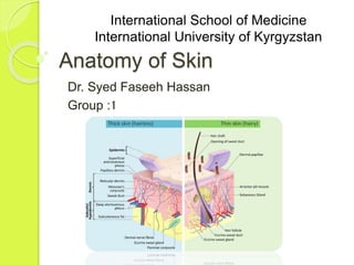
2. Anatomy of Skin
Skin is composed of three primary
layers (from outer to inner):
the Epidermis
the Dermis and
the Hypodermis / Subcutaneous Tissue
3. Epidermis
Epidermis, "epi" coming from the
Greek meaning "over" or "upon", is the
outermost layer of the skin.
It forms the waterproof, protective wrap
over the body's surface which also
serves as a barrier to infection and is
made up of stratified
squamous epithelium with an
underlying basal lamina.
The epidermis contains no blood
4. Nourishment of Epidermis
Cells in the deepest layers are
nourished almost exclusively by
diffused oxygen from the surrounding
air and to a far lesser degree by blood
capillaries extending to the outer
layers of the dermis.
5. Epidermis Layers(1)
The epidermis is divided into several
layers, where cells are formed
through mitosis at the innermost layers.
Cells move up the strata changing shape
and composition as they differentiate and
become filled with keratin. They eventually
reach the top layer called stratum
corneum and are sloughed off, or
desquamated. This process is
called keratinization and takes place
within weeks.
The outermost layer of the epidermis
consists of 25 to 30 layers of dead cells.
6. Epidermis Layers(2)
The epidermis is divided into the
following 5 sub layers or strata:
• Stratum corneum
• Stratum lucidum
• Stratum granulosum
• Stratum spinosum
• Stratum basale (also called "stratum
germinativum")
7.
8. Epidermis Layers(3)
The stratum corneum (Latin for 'horny
layer') is the outermost layer of
the epidermis
1. This layer is composed of 15–20 layers
of flattened cells with no nuclei or cell
organelles. Their cytoplasm shows
filamentous keratin.
2. performs protective and adaptive
physiological functions including
mechanical shear, impact resistance,
water flux and hydration regulation,
microbial proliferation and invasion
regulation, initiation of inflammation
through cytokine activation and dendritic
9. Epidermis Layers(4)
Desquamation, the process of cell
shedding from the surface of the stratum
corneum, balances proliferating
keratinocytes that form in the stratum
basale.
These cells migrate through the
epidermis towards the surface in a
journey that takes approximately
fourteen days.
10. Epidermis Layers(5)
The stratum lucidum (Latin for "clear
layer") is a thin, clear layer of dead skin
cells in the epidermis named for its
translucent appearance under
a microscope.
It is readily visible by light microscopy only
in areas of thick skin, which are found on
the palms of the hands and the soles of the
feet.
it is composed of 3 to 5 layers of dead,
flattened keratinocytes. The keratinocytes
of the stratum lucidum do not feature
distinct boundaries and are filled
11. Epidermis Layers(6)
The stratum granulosum (or granular
layer) is a thin layer of cells in
the epidermis.
Keratinocytes migrating from the
underlying stratum spinosum become
known as granular cells in this layer.
These cells contain keratohyalin granules,
which are filled with histidine- and cysteine-
rich proteins that appear to bind the keratin
filaments together. Therefore, the main
function of keratohyalin granules is to bind
12. Epidermis Layers(7)
The stratum spinosum (or spinous
layer/prickle cell layer)
Keratinization begins in the stratum
spinosum.
Their spiny (Latin, spinosum)
appearance is due to shrinking the of
microfilaments
between desmosomes that occurs when
stained with H&E.
13. Epidermis Layers(8)
The stratum basale (basal layer,
sometimes referred to as stratum
germinativum) is the deepest layer of the
five layers of the epidermis.
The stratum basale is a single layer
of columnar or cuboidal basal cells. The
cells are attached to each other and to the
overlying stratum spinosum cells
by desmosomes and hemidesmosomes
They divide to form the keratinocytes of the
stratum spinosum, which migrate
superficially. Other types of cells found
within the stratum basale are melanocytes
, Langerhans cells (immune cells),
14.
15. Histology of Epidermis(1)
The main type of cells that make up
the epidermis are Merkel
cells, keratinocytes,
with melanocytes and Langerhans
cells also present.
• Merkel Cells: Merkel cells, also known
as Merkel-Ranvier cells or tactile
epithelial cells, are oval-shaped
mechanoreceptors essential for light
touch sensation and found in the skin of
vertebrates.
16. Histology of Epidermis(2)
• Merkel Cells: They are abundant in
highly sensitive skin like that of the
fingertips in humans, and make synaptic
contacts with somatosensory afferent
nerve fibers. Although uncommon, these
cells may become malignant and form
a Merkel cell carcinoma—an aggressive
and difficult to treat skin cancer. They
are clear cells found in the stratum
basale (at the bottom of sweat duct
ridges) of the epidermis approximately
10 μm in diameter.
17. Histology of Epidermis(3)
• Keratinocytes constitute 90% of
the cells of the epidermis, the outermost
layer of the skin. Basal cells in the basal
layer (stratum basale) of the skin, are
sometimes referred to as basal
keratinocytes.
• The primary function of keratinocytes is
the formation of a barrier against
environmental damage by heat, UV
radiation, water
loss, pathogenic bacteria, fungi, parasite
s and viruses.
18. Histology of Epidermis(4)
Melanocytes are melanin-
producing neural crest-
derived cells located in the bottom layer
(the stratum basale) of the
skin's epidermis.
Once synthesized, melanin is contained
in
special organelles called melanosomes
which can be transported to
nearby keratinocytes to induce
pigmentation. Functionally, melanin
serves as protection against UV
19. Histology of Epidermis(5)
Langerhans cells (LC) are tissue-
resident macrophages of the skin, and
contain organelles called Birbeck
granules. They are present in all layers
of the epidermis and are most prominent
in the stratum spinosum.
They also occur in the papillary dermis,
particularly around blood vessels, as well
as in the mucosa of the mouth, foreskin,
and vaginal epithelium.
20. Dermis
The dermis is a layer of skin between
the epidermis and subcutaneous
tissues, that primarily consists of dense
irregular connective tissue and cushions
the body from stress and strain. The
dermis is tightly connected to the
epidermis through a basement
membrane.
Structural components of the dermis
are collagen, elastic fibers, and
extrafibrillar matrix.
21. Dermis
It also contains mechanoreceptors that
provide the sense
of touch and thermoreceptors that
provide the sense of heat.
In addition, hair follicles, sweat glands,
sebaceous glands (oil glands), apocrine
glands,
lymphatic vessels, nerves and blood
vessels are present in the dermis.
22. Layers of Dermis(1)
It is divided into two layers, the superficial
area adjacent to the epidermis called the
papillary region and a deep thicker area
known as the reticular dermis.
• The papillary region is composed of
loose areolar connective tissue. This is named
for its finger like projections called papillae, that
extend toward the epidermis and contain either
terminal networks of blood capillaries or
tactile Meissner's corpuscles.
• It is the uppermost layer of the dermis. It
intertwines with the rete ridges of the epidermis
and is composed of fine and loosely arranged
collagen fibers.
23. Layers of Dermis(2)
• The reticular dermis is the lower layer of the
dermis, found under the papillary dermis,
composed of dense irregular connective
tissue featuring densely packed collagen
fibers. It is the primary location of dermal
elastic fibers
• The reticular region is usually much thicker
than the overlying papillary dermis.
• Within the reticular region are the roots of the
hair, sebaceous glands, sweat
glands, receptors, nails, and blood vessels.
24. Hypodermis/ Sub-cutaneous
Tissue
• The subcutaneous tissue (meaning
'beneath the skin'), also called
the hypodermis, hypoderm (from Gre
ek, meaning 'beneath the
skin'), subcutis, or superficial fascia.
• The hypodermis is beneath the dermis
which is beneath the epidermis. It is
used mainly for fat storage.
• It consists of loose connective tissue,
adipose tissue and elastin.
25. Cells types in hypodermis
• The main cell types are fibroblasts,
macrophages
and adipocytes (subcutaneous tissue
contains 50% of body fat). Fat serves as
padding and insulation for the body.
26. Structures in Hypodermis
Fibrous bands anchoring the skin to the deep fascia
Collagen and elastin fibers attaching it to the dermis
Fat is absent from the eyelids, clitoris, penis, much
of pinna, and scrotum.
Blood vessels on route to the dermis
Lymphatic vessels on route from the dermis
The glandular part of some sweat glands; mammary
gland lie entirely within the subcutaneous tissue(which are
modified apocrine sweat glands)
Cutaneous nerves and free endings
Hair follicle roots
Ruffini and Pacinian corpuscles
Mast cells
Bursae, in the space overlying joints in order to facilitate
smooth passage of overlying skin